Dit protocol beschrijft de generatie en fenotypische karakterisering van een C57BL/6N-Cracdem1(c.538-83 to c.3352+255 del) muismodel met behulp van CRISPR/Cas9-technologie om de rol van CRACD bij cardiale remodellering na een myocardinfarct te bestuderen.
Methodenartikel
Generatie en fenotypische karakterisering van een CRISPR/Cas9-gemodificeerd Cracd-deficiënt muismodel voor studies naar remodellering na myocardinfarct
In dit artikel
Samenvatting
Samenvatting
Myocardinfarct (MI) blijft wereldwijd een belangrijke oorzaak van morbiditeit en mortaliteit. Dit protocol beschrijft een methode voor het genereren en karakteriseren van een Cracd-deficiënte muislijn op de C57BL/6N-achtergrond met behulp van CRISPR/Cas9-technologie. Zygoten werden co-geïnjecteerd met Cas9-mRNA, een gRNA-construct en een donor-template ontworpen om een genomische deletie van 3153-bp te genereren. Het bewerkte allel werd bevestigd via PCR-genotypering en Sanger-sequencing. Om de functionele rol van CRACD bij remodeling na een MI te beoordelen, ondergingen Cracd-deficiënte en wild-type (WT, C57BL/6N) muizen een MI, geïnduceerd door permanente ligatie van de linker anterior descending coronaire arterie. Op postoperatieve dag 7 werden de hartfunctie en de wandbeweging van de linker ventrikel beoordeeld met transthoracale echocardiografie en speckle-tracking strain imaging, gevolgd door histopathologische evaluatie met H&E- en Masson-trichroomkleuring. Representatieve resultaten toonden aan dat Cracd-deficiënte muizen een verminderde ventriculaire dilatatie en een behouden systolische functie vertoonden in vergelijking met WT-controles. Dit protocol biedt een betrouwbaar experimenteel platform voor mechanistische studies naar CRACD in de cardiale pathofysiologie.
Inleiding
Myocardinfarct (MI) blijft wereldwijd een leidende oorzaak van morbiditeit en mortaliteit, wat bijdraagt aan een aanzienlijke last voor de mondiale cardiovasculaire gezondheid1. De langetermijnprognose na een MI wordt primair bepaald door het proces van remodellering van de linker ventrikel (LV)2, wat gepaard gaat met ventriculaire dilatatie, wandverdunning en interstitiële fibrose3,4, welke belangrijke factoren zijn bij de progressie naar hartfalen (HF). De wereldwijde incidentie van HF als gevolg van cardiale remodellering na MI verergert de last van cardiovasculaire aandoeningen verder5. Ondanks deze goed onderbouwde klinische observaties ontbreken systematische studies die gebruikmaken van sensitieve, reproduceerbare in vivo-modellen om de rol van specifieke genen bij remodellering na MI te beoordelen. Geen enkel protocol combineert momenteel een met CRISPR/Cas9 gemanipuleerd knockout-model met geavanceerde strain-imaging om een cytoskeletregulator zoals CRACD te evalueren. Daarom is er een dringende behoefte aan een methode die nauwkeurige genetische manipulatie en cardiale fenotypering met hoge resolutie mogelijk maakt.
Capping protein inhibiting regulator of actin dynamics (CRACD), ook bekend als KIAA1211, is een regulator van actinpolymerisatie die hoofdzakelijk in het cytosol gelokaliseerd is en een cruciale rol speelt in de dynamiek van het cytoskelet6Het reguleert de actinpolymerisatie positief door te binden aan actin-capping-eiwitten (CAPZA1, CAPZA2, CAPZB) en te voorkomen dat deze de barbed ends van actinfilamenten afdekken.7In epitheelweefsels speelt CRACD een kritische rol bij het behouden van de celmorfologie en signaaltransductie door de integriteit van het actinecytoskelet te waarborgen.8,9In tumorcellen is de dysregulatie ervan geassocieerd met versterkte celmetastase en celproliferatie.6,10De rol van CRACD in het hart is nog niet volledig begrepen. Transcriptoomstudies van het hart en experimentele interventiestudies laten zien dat Cracd genexpressie reageert op pathologische stress en kan genen reguleren die verband houden met de cytoskeletdynamiek tijdens ventriculaire remodellering9,11Bovendien is het inhiberende eiwit CapZ nauw verbonden met cardiale pathologie: de fosforylering van CapZ reguleert de groei van myofibrillen tijdens hypertrofie geïnduceerd door fenylefrine.12Ons vroege onderzoek wijst uit dat een matige downregulatie van CRACD bescherming biedt tegen myocardiale schade en de contractiliteit van myofilamenten behoudt na acute ischemie.13Deze bevindingen wijzen op de mogelijkheid dat CRACD een rol speelt bij maladaptieve ventriculaire remodellering na een MI. Systematische in vivo loss-of-function-studies om echter te testen of Cracd tekortaanbod moduleert de remodellering na een myocardinfarct (MI) ontbraken, grotendeels door de afwezigheid van een gevalideerd stapsgewijs protocol voor het genereren en fenotyperen van een Cracd‑deficiënte muis onder ischemische stress.
Er bestaan verschillende benaderingen voor het genereren van constitutieve knockout-muizen. Traditionele homologe recombinatie in embryonale stamcellen (ES-cellen) is nauwkeurig, maar kostbaar, tijdrovend (12–18 maanden) en beperkt tot bepaalde muizenstammen14. ENU (N-ethyl-N-nitrosourea) mutagenese produceert willekeurige puntmutaties, waarvoor uitgebreide terugkruising en sequencing vereist zijn15. In contrast hiermee biedt CRISPR/Cas9 duidelijke praktische voordelen voor deze studie: (i) directe injectie in de zygote verkort de tijdlijn naar 4–6 maanden; (ii) het maakt de deletie van een groot genomisch fragment (3153 bp) mogelijk om volledig verlies van functie te garanderen; (iii) het werkt direct op de C57BL/6N-achtergrond zonder stamconversie; en (iv) het vermijdt een selecteerbare marker, waardoor onbedoelde transcriptionele interferentie wordt geminimaliseerd. Daarom is CRISPR/Cas9 de meest efficiënte keuze voor het genereren van een Cracd-deficiënte lijn die geschikt is voor routinematige cardiovasculaire fenotypering. Deze technologie is op grote schaal gebruikt voor het uitvoeren van nauwkeurige in vivo loss-of-function studies16,17.
Speckle-tracking strain-imaging is een post-processingtechniek die de beweging van natuurlijke akoestische speckles binnen het myocard frame voor frame volgt, waardoor het mogelijk is om myocardiale deformatieparameters te berekenen zoals strain (fractionele verandering in lengte) en strain rate (snelheid van deformatie)18. In tegenstelling tot conventionele M-mode of tweedimensionale echocardiografie, die globale functionele indices verschaft (ejacctiefractie, fractionele verkorting, interne diameter van het linker ventrikel) en relatief ongevoelig is voor regionale wandbewegingsstoornissen, kan strain-imaging segmentale contractiele functie kwantificeren. Dit is bijzonder belangrijk in de vroege post-infarctperiode, waar compensatoire hyperkinesie van niet-geïnfarceerde segmenten de globale ejectiefractie kan behouden ondanks significante regionale dysfunctie. Speckle-tracking biedt verschillende voordelen ten opzichte van standaard eindpunten: het levert segmentale strain en strain rate, verplaatsing en snelheid op, waardoor subtiele subklinische veranderingen kunnen worden gedetecteerd19; het kan dyssynchronie en vroege contractiele tekorten identificeren voordat onomkeerbare remodellering optreedt; en het onderzoekt direct het subendocard, de laag die het meest kwetsbaar is voor ischemie20. In deze studie is CRACD een cytoskelet-regulator die de contractiliteit van myofibrillen op microscopisch niveau kan beïnvloeden. Conventionele metingen op basis van de holte zouden dergelijke lokale mechanische effecten gemakkelijk kunnen missen. Daarom vergroot de integratie van speckle-tracking-analyse in dit protocol het vermogen om vroege beschermende effecten van Cracd-deficiëntie te detecteren. Bijgevolg biedt de integratie van speckle-tracking in dit protocol een gevoelig en kwantitatief instrument om post-infarct remodellering te evalueren, vooral tijdens de eerste week na een MI wanneer vroege dilatatie en contractiele dysfunctie ontstaan21,22.
Dit protocol is ontworpen voor onderzoekers die de functionele impact van een specifieke gen deletie op acute (7-daagse) remodeling na een MI onderzoeken, gebruikmakend van een combinatie van constitutieve knockout en hoge-resolutie beeldvorming. Het is geschikt voor hypothesevormende studies waarbij het globale verlies van het doelgen acceptabel is en het belangrijkste eindpunt de vroege systolische functie en structurele veranderingen zijn. Er moeten echter enkele beperkingen in overweging worden genomen. Ten eerste is de Cracd-deficiënte muis een globale knockout; daarom kunnen de waargenomen cardioprotectieve effecten niet uitsluitend worden toegeschreven aan het verlies van CRACD in cardiomyocyten, aangezien systemische bijdragen ook een rol kunnen spelen. Toekomstige studies met conditionele knockout-strategieën zullen nodig zijn om hartspecifieke deletie te bereiken. Ten tweede richt het protocol zich op één enkel tijdstip (dag 7) en behandelt het geen chronische remodeling of de progressie van HF. Longitudinale studies voorbij de acute fase zullen vereist zijn. Ten derde vereist speckle-tracking analyse beelden van hoge kwaliteit (duidelijke endocardiale grenzen) en gespecialiseerde software, die mogelijk niet in alle kernfaciliteiten beschikbaar is. Ondanks deze beperkingen biedt het protocol een robuust, reproduceerbaar platform voor de initiële functionele karakterisering van genen die betrokken zijn bij cardiale remodeling na een MI.
Deze studie had tot doel het CRISPR/Cas9-systeem te gebruiken om een Cracd-deficiënt muismodel (C57BL/6N-Cracdem1 (c.538-83 to c.3352+255 del)) te genereren en te valideren voor het onderzoeken van de impact van Cracd-deficiëntie op cardiale remodellering en ventrikelwandbeweging na een myocardinfarct (MI). MI werd geïnduceerd in zowel wild-type (WT, C57BL/6N) als Cracd-deficiënte muizen, en één week na het MI werden uitgebreide evaluaties uitgevoerd. De beoordelingen omvatten conventionele echocardiografische parameters, speckle-tracking strain-analyse en histopathologisch onderzoek. We hebben ons specifiek gericht op de vroege fase na het MI (dag 7) om initiële remodelleringsgebeurtenissen vast te leggen en te bepalen of Cracd-deficiëntie de ventriculaire dilatatie en systolische functie tijdens de acute fase van het MI beïnvloedt. De resultaten worden geacht waardevolle inzichten te verschaffen in de rol van CRACD bij het moduleren van nadelige cardiale remodellering na een MI.
Protocol
All animal experiments were approved by the Institutional Animal Care and Use Committee of the Guangdong Institute of Biotechnology (Approval No. IACUC2025179). The study utilized 20 SPF Cracd-deficient and 20 age- and weight-matched WT mice. All mice were 10–12 weeks old and weighed 20–25 g. All male animals were housed in an AAALAC-accredited facility (License No. SYXK (Yue) 2016-0122) at the Guangdong Institute of Biotechnology (Guangdong Laboratory Animals Monitoring Institute). The housing environment was maintained at 22–25 °C with 50–70% relative humidity under a 12 h light/12 h dark cycle.
1. Generation of Cracd-deficient mice using CRISPR/Cas9
- Use CRISPR/Cas9 gene editing technology to generate Cracd-deficient mice on a C57BL/6N background. Delete the genomic sequence from c.538-83 to c.3352+255 (partial intron 5 to intron 6) of Cracd by homology-directed repair.
NOTE: The mouse Cracd gene (NCBI RefSeq: NM_001163793.1; Ensembl: ENSMUSG00000036377) is located on chromosome 5 and contains 9 exons, with the ATG start codon in exon 2 and the TAA stop codon in exon 9. - Design the sequence of gRNA targeting vector and donor oligonucleotide (with targeting sequence flanked by 120 bp homologous sequences combined on both sides).
- Log in to the NCBI website, select the Primer-BLAST function, and design PCR primers for genotyping.
- Paste the NCBI reference sequence number of Cracd (NM_001163793.1) into the PCR template field, specify the exon range for amplification, and click “Get Primers” to retrieve multiple pairs of candidate primers for PCR-based detection of the deletion and wild-type alleles.
- Design single-guide RNAs (gRNAs) to target sites flanking the intended deletion interval in the Cracd gene.
- Select four gRNA sequences (20 nt upstream of the PAM) using online CRISPR design tools.
- To minimize potential off-target effects, score the candidate gRNAs with CRISPRater and select the two optimal sequences (including the NGG PAM).
NOTE: Example selected sequences:
gRNA-A1: CGGCGCTCTTAACATCCAGAGGG (score: 0.71)
gRNA-A2: CCCTTCAGTCTGGGTAGTAGAGG (score: 0.71)
Accept gRNAs with a CRISPRater score ≥ 0.5. For in vitro validation, accept gRNAs with > 50% cleavage efficiency in a Cas9/gRNA target efficiency detection kit.
- Design a single-stranded donor oligonucleotide to promote efficient joining of the intended breakpoints. Example donor sequence: TTAACCATCAACGGGCCGCTGCAAGGGCTGCTGT
GTCTATA CTCAGGACCACCTGCCCTCTAGAGGCAGGAAGGT
AGAGTTCCAAGCTCTGCTGTGCTAGGTTAAATGCAACTTAACATC. - Co-inject Cas9 mRNA, gRNA and donor oligonucleotide into fertilized eggs to generate Cracd-deficient mice. Prepare Cas9 mRNA and gRNA by in vitro transcription.
- Preparation of gRNA.
- Transcribe gRNAs in vitro from DNA templates using the MEGAshortscript™ T7 transcription kit (see Table of Materials) following the manufacturer's protocol.
- Purify the transcribed gRNAs using an RNA purification kit (see Table of Materials) and elute them in RNase-free water.
- Assess gRNA cleavage activity using an in vitro Cas9/gRNA target efficiency detection kit (see Table of Materials).
- Perform all RNA preparation steps on ice using RNase‑free consumables (pipette tips, tubes, water). Keep RNA samples on ice at all times to prevent degradation.
- To perform the cleavage assay: mix the transcribed gRNA (25 ng/µL) with recombinant Cas9 protein (50 ng/µL) and a PCR-amplified DNA fragment containing the target sequence (50 ng).
- Incubate the reaction at 37 °C for 30 min.
- Run the products on a 2% agarose gel to visualize uncleaved and cleaved bands.
- Calculate cleavage efficiency as the ratio of cleaved band intensity to total (cleaved + uncleaved) band intensity.
- Accept gRNAs with > 50% cleavage efficiency.
- Preparation of Cas9 mRNA.
- Thaw the reagents from the T7 ARCA mRNA Kit (see Table of Materials).
- At room temperature, combine 10 µL of 2× ARCA/NTP mix, 1 µL of template DNA, 2 µL of T7 RNA polymerase mix, and nuclease-free water to 20 µL.
- Mix thoroughly, pulse-spin in a microfuge (5 s at 5000 × g), and incubate at 37 °C for 30 min.
- Remove the template DNA by adding 2 µL of DNase I (1 U/µL), mix well, and incubate at 37 °C for 15 min to obtain Cas9 mRNA. Optionally add 1 µL of RNase inhibitor (40 U/µL) (see Table of Materials) to the reaction to prevent RNA degradation.
- Perform all steps on ice with RNase‑free consumables. Keep the Cas9 mRNA on ice until use.
- Superovulate 4-week-old C57BL/6N female mice by intraperitoneal injection of 5 IU of pregnant mare serum gonadotropin (PMSG), followed 48 h later by 5 IU of human chorionic gonadotropin (hCG).
CAUTION: PMSG and hCG are hormones that may cause skin sensitization. Wear gloves and dispose of needles in a sharps container. - Approximately 14–18 h after hCG injection, euthanize the C57BL/6N female mice and one 8–12-week-old C57BL/6N male mouse by cervical dislocation. Collect oocytes from the oviducts of the females and sperm from the cauda epididymis of the male.
CAUTION: Euthanasia must be performed in accordance with institutional guidelines. Cervical dislocation should only be performed by trained personnel. - Capacitate the sperm in capacitation medium (see Table of Materials).
- Add a small volume of the sperm suspension to the oocytes for in vitro fertilization and co-culture for 3–4 h at 37 °C to obtain fertilized zygotes.
- Dilute the transcribed gRNA and Cas9 mRNA with RNase-free water to working concentrations (typically 25 ng/µL for gRNA and 50 ng/µL for Cas9 mRNA) and donor oligonucleotide at 10 ng/µL.
- Microinject 1–2 pL of the mixture into the cytoplasm of fertilized zygotes using a microinjection system (see Table of Materials).
- Identify healthy zygotes by the presence of two distinct pronuclei and an intact polar body under a stereomicroscope.
- Transplant 300 healthy fertilized zygotes into the oviducts of pseudopregnant female mice that have been mated with vasectomized males.
NOTE: This results in the generation of 2 positive founder mice (F0). - Use RNase‑free water and ice‑cold conditions during dilution to maintain RNA integrity.
- Identify F0 mice by PCR genotyping of tail biopsies (see section 1.6).
- Breed F0 founders with WT mice to generate F1 heterozygous offspring, to obtain germline transmission of the deletion.
- Intercross heterozygous F1 mice to produce F2 homozygous Cracd-deficient mice.
- Preparation of gRNA.
- Genotype by Polymerase Chain Reaction (PCR)
NOTE: The following protocol uses a commercial TIANamp Genomic DNA Kit (see Table of Materials). Buffer GL is the lysis buffer, Buffer GB is the binding buffer, and Buffer WA and Buffer WB are wash buffers.- Place approximately 2–5 mm of mouse tail tissue in a microcentrifuge tube containing 180 µL of Buffer GL, 20 µL of Proteinase K (20 mg/mL stock solution), and 10 µL of RNase A (10 mg/mL stock solution).
- Incubate the sample at 56 °C overnight to digest the tissue.
- Centrifuge the tube at 13800 × g for 2 min at room temperature to pellet tissue debris and transfer the supernatant to a new tube.
- Add 200 µL of Buffer GB and 200 µL of 100% ethanol to the supernatant, mix thoroughly, and apply the mixture to a spin column placed in a collection tube.
- Centrifuge at 13800 × g for 2 min at room temperature and discard the flow-through.
- Add 500 µL of Buffer WA to the column, centrifuge at 13800 × g for 1 min at room temperature and discard the flow-through.
- Carefully add 700 µL of Buffer WB (wash buffer containing ethanol, pre-mixed with 100% ethanol) to the spin column, centrifuge at 13800 × g for 1 min at room temperature and discard the flow-through.
- Apply Buffer WB to the inner wall of the column to wash residual salts.
- Repeat step 1.6.5 once, then transfer the column into a new collection tube and centrifuge at 13800 × g for 2 min at room temperature to dry the membrane and remove residual ethanol.
- Place the column into a new 1.5 mL microcentrifuge tube. Add 50–200 µL of sterilized water or elution buffer (preheated to 65 °C) to the center of the membrane, incubate for 5 min.
- Centrifuge the column at 13800 × g for 2 min at room temperature to elute the DNA.
- To increase DNA yield, either reapply the flow-through to the membrane or add an additional 50–200 µL of sterilized water, incubate for 5 min.
- Perform a final centrifugation at 13800 × g for 2 min.
NOTE: Expect DNA yield of 50–200 ng/µL from tail tissue; A260/A280 ratio of 1.8–2.0 indicates pure DNA.
- Place approximately 2–5 mm of mouse tail tissue in a microcentrifuge tube containing 180 µL of Buffer GL, 20 µL of Proteinase K (20 mg/mL stock solution), and 10 µL of RNase A (10 mg/mL stock solution).
- Genotyping by multiplex PCR using three primers in a single reaction.
- Design three primers to distinguish WT, heterozygous, and homozygous genotypes in one PCR reaction: Forward primer F1 (5’-CAGAAGTCATAGCAGCAAGGCAG-3’, binds upstream of the deletion), Forward primer F2 (5’-GAGGGAATTGAGAGGGAGCC-3’, binds within the deleted region, present only in the WT allele), and Reverse primer R1 (5’-GCCAATTTAGCTGCTTCAAATGTTC-3’, common reverse primer downstream of the deletion).
NOTE: The targeted allele generates a 421 bp amplicon (F1 + R1) only when the deletion is present; the WT allele generates a 357 bp amplicon (F2 + R1) only when the intact allele is present. - Set up the PCR reaction in a 25 µL total volume containing 50–100 ng genomic DNA, 0.2 µM of each primer (F1, F2, and R1), 200 µM dNTPs, 1.5 mM MgCl₂, and 1 U Taq DNA polymerase (see Table of Materials).
- Use the following thermocycling program: initial denaturation at 95 °C for 3 min; 35 cycles of denaturation at 95 °C for 15 s, annealing at 60°C for 15 s, and extension at 72 °C for 25 s; and a final extension at 72 °C for 5 min.
- Separate the PCR products by 1–2% agarose gel electrophoresis (120 V, 30 min) in 1× TAE buffer.
- Genotype interpretation based on band pattern: WT shows only a 357 bp band (F2+R1); Heterozygous shows both 421 bp (F1+R1) and 357 bp (F2+R1) bands; Homozygous shows only a 421 bp band (F1+R1).
- Design three primers to distinguish WT, heterozygous, and homozygous genotypes in one PCR reaction: Forward primer F1 (5’-CAGAAGTCATAGCAGCAAGGCAG-3’, binds upstream of the deletion), Forward primer F2 (5’-GAGGGAATTGAGAGGGAGCC-3’, binds within the deleted region, present only in the WT allele), and Reverse primer R1 (5’-GCCAATTTAGCTGCTTCAAATGTTC-3’, common reverse primer downstream of the deletion).
2. Myocardial infarction model establishment
- Anesthetize the mice via inhalation of 3% isoflurane (delivered in 100% oxygen at 1 L/min), place them in a supine position on a surgical platform, and confirm deep anesthesia by the absence of pedal withdrawal reflex.
CAUTION: Isoflurane is a volatile anesthetic. Use only in a well-ventilated area or under a fume hood to avoid inhalation. - Perform orotracheal intubation using a 20-gauge intravenous catheter sheath. Apply ophthalmic ointment to both eyes to prevent corneal drying during surgery.
- Connect the tube to a mechanical ventilator upon successful intubation to maintain ventilation and anesthesia with 2% isoflurane.
NOTE: Successful intubation is confirmed by visible chest movements synchronized with the ventilator.
- Connect the tube to a mechanical ventilator upon successful intubation to maintain ventilation and anesthesia with 2% isoflurane.
- Remove hair from the anterior chest with depilatory cream. Disinfect the surgical area by applying povidone-iodine and 70% alcohol in a circular motion for multiple times, starting from the center and moving outward.
- After fully exposing the skin, make a 1–1.5 cm incision in the left chest wall between the 2nd and 3rd intercostal spaces to open the thoracic cavity.
- Incise the pericardium to fully expose the heart.
CAUTION: Surgical procedures must be performed under sterile conditions. Use appropriate personal protective equipment (PPE), including gloves and a surgical mask.
- Using the inferior border of the left auricle as a landmark, pass a 7–0 atraumatic suture needle beneath the left anterior descending coronary artery (LAD) approximately 2–3 mm distal to its origin.
- Pass the needle to a depth of approximately 0.5–1 mm and tighten the ligature slowly to avoid myocardial tearing.
- Confirm successful occlusion by observing pallor (whitening) of the left ventricular anterior wall distal to the ligation site within 30 s.
- In sham-operated controls, open the chest and pass the suture beneath the coronary artery without tying the knot.
- After establishing the model, sequentially suture the ribs (4-0 absorbable suture, simple interrupted pattern) and skin (4-0 absorbable suture, continuous pattern) to close the thoracic cavity. Remove the endotracheal tube once the mouse resumes stable spontaneous breathing.
- Place mice on a heating pad (37 °C) until fully conscious.
- Administer postoperative analgesia (e.g., buprenorphine 0.05–0.1 mg/kg, subcutaneously, every 12 h for 48 h) according to institutional IACUC guidelines and closely monitor the postoperative health status for several days.
NOTE: Monitor for signs of distress (hunched posture, ruffled fur, reduced mobility, weight loss > 20%).
- Administer postoperative analgesia (e.g., buprenorphine 0.05–0.1 mg/kg, subcutaneously, every 12 h for 48 h) according to institutional IACUC guidelines and closely monitor the postoperative health status for several days.
- Mortality and exclusion criteria.
- Record the number of mice that die within the first 7 days after MI surgery.
NOTE: In a typical cohort, expect a mortality rate of approximately 10–20% by postoperative day 7, with no significant difference between WT and Cracd‑deficient mice. Common causes of death include extensive infarction leading to acute heart failure, surgical bleeding, or pneumothorax. Exclude any mouse with persistent arrhythmia (heart rate < 350 bpm) or signs of severe distress (e.g., hunched posture, ruffled fur, weight loss > 20%) from subsequent echocardiographic and histological analyses. For sham‑operated mice, mortality should be < 5%.
- Record the number of mice that die within the first 7 days after MI surgery.
3. Echocardiographic assessment
- Animal Preparation and Anesthesia
- Power on the high-resolution small animal ultrasound imaging system (see Table of Materials) and its integrated heated stage.
- Ensure that the appropriate high-frequency transducer is securely connected to the mounting apparatus following the manufacturer’s instructions. Set the temperature of the heated stage to 37 °C and allow it to stabilize.
- Place mice in an induction chamber and anesthetize them with 3% isoflurane in oxygen (1–2 L/min) until the pedal withdrawal reflex is absent. Transfer anesthetized mice to the heated platform in a supine position.
- Maintain anesthesia with 1–1.5% isoflurane delivered via a nose cone, adjusting to maintain heart rate between 450–550 beats per min.
- Apply ophthalmic ointment to both eyes to prevent corneal drying.
- Shave the thoracic region, evenly apply depilatory cream for no longer than 3 min, then remove the depilatory cream thoroughly with wet gauze.
- Rinse the skin gently with warm saline if necessary and dry the area. Attach surface ECG electrodes to the limbs using electrode gel at the contact sites and secure them with tape.
NOTE: Confirm stable ECG and heart rate within 450–550 bpm before proceeding to ultrasound imaging.
- Power on the high-resolution small animal ultrasound imaging system (see Table of Materials) and its integrated heated stage.
- Conventional Echocardiography Image Acquisition
- Position the mouse in a horizontal, supine posture on the heated stage, with a slight leftward tilt if needed to optimize cardiac imaging.
- Apply a generous amount of pre-warmed ultrasound coupling gel to the hairless left precordial region, avoiding air bubbles.
- Place the transducer gently on the chest wall at the left parasternal position. Adjust its orientation so that the index mark points toward the mouse’s right shoulder and fine-tune the angle and pressure to optimize image quality.
- Switch to B-mode (two-dimensional) imaging, adjust depth, focus zone, and frame rate to obtain a clear parasternal long-axis view of the left ventricle. Acceptable image quality requires clear visualization of the endocardial border with no dropouts.
- Once the heart rate stabilizes (450–550 bpm), acquire three separate parasternal long-axis B-mode cine loops, each containing at least 3–5 consecutive cardiac cycles.
- Save each loop by pressing “Cine Store”, and store representative still frames using “Frame Store”.
- From the long‑axis view, rotate the transducer 90° clockwise to obtain a short‑axis view at the mid‑papillary level.
- Acquire three separate short‑axis B‑mode cine loops (each with ≥ 3–5 cycles).
- Activate M‑mode, position the cursor perpendicular to the interventricular septum and posterior wall, and record three separate M‑mode tracings.
NOTE: Acceptable M‑mode tracings require clear endocardial borders throughout the cardiac cycle.
- Use the VevoLab 3.2.0 software (see Table of Materials) to measure the following parameters from M‑mode tracings according to the American Society of Echocardiography leading‑edge method: interventricular septum thickness at diastole and systole (IVSd, IVSs), left ventricular internal diameter at diastole and systole (LVIDd, LVIDs), and left ventricular posterior wall thickness at diastole and systole (LVPWd, LVPWs).
- Calculate left ventricular ejection fraction (EF) and fractional shortening (FS) using the Teichholz formula: EF (%) = [(LV Vold - LV Vols) / LV Vold] × 100, LV Vold = ((7 / (2.4 + LVIDd)) × LVIDd3), LV Vol;s = ((7 / (2.4 + LVIDs)) × LVIDs3), FS (%) = [(LVIDd – LVIDs) / LVIDd] × 100.
NOTE: For each mouse, measurements were averaged from three consecutive cardiac cycles per M‑mode recording, and the average was taken across three separate recordings, resulting in a total of 9 measurements per parameter per mouse.
- Calculate left ventricular ejection fraction (EF) and fractional shortening (FS) using the Teichholz formula: EF (%) = [(LV Vold - LV Vols) / LV Vold] × 100, LV Vold = ((7 / (2.4 + LVIDd)) × LVIDd3), LV Vol;s = ((7 / (2.4 + LVIDs)) × LVIDs3), FS (%) = [(LVIDd – LVIDs) / LVIDd] × 100.
- Position the mouse in a horizontal, supine posture on the heated stage, with a slight leftward tilt if needed to optimize cardiac imaging.
- Speckle-Tracking Strain Analysis
- Perform speckle-tracking analysis using the dedicated analysis software (VevoStrain module within VevoLab 3.2.0).
- Open a clear parasternal long-axis B-mode cine loop, select "Vevo Strain" to enter the strain analysis mode, retain only complete cardiac cycles, and choose "Long Axis".
- Manually trace the endocardial border at the end‑diastolic frame. After the endocardial border is drawn, the software automatically generates an epicardial border (adjust as needed). Then start the strain analysis and activate the "toggle contour/vector/orbit line/B mode" function to visualize the motion trajectory.
- Assess tracking quality via the colour‑coded mesh: it should appear green, closely follow the endocardial and epicardial borders without jerking, and move synchronously with myocardial contraction/relaxation. If not, manually refine the borders and re‑analyze.
- Activate "Time‑to‑peak analysis" to divide the left ventricle into six segments (basal anterior, mid anterior, apical anterior, apical posterior, mid posterior, basal posterior), with each colour representing an individual segment. Once satisfactory tracking is achieved, export data for wall motion parameters: velocity (cm/s), displacement (cm), strain (%), strain rate (1/s) for both longitudinal and radial.
NOTE: If tracking quality remains poor after three acquisition attempts (e.g., erratic strain curves, or inability to visualise clear motion trajectories), exclude the mouse from strain analysis.
- Perform speckle-tracking analysis using the dedicated analysis software (VevoStrain module within VevoLab 3.2.0).
4. Histopathological analysis
- After echocardiography, administer a lethal dose of isoflurane to deeply anesthetized mice and apply a secondary physical method (e.g., cervical dislocation) according to institutional guidelines.
- Open the thoracic cavity, rapidly harvest the hearts and rinse the hearts gently in cold phosphate-buffered saline (PBS).
CAUTION: Euthanasia must be performed according to institutionally approved protocols.
- Open the thoracic cavity, rapidly harvest the hearts and rinse the hearts gently in cold phosphate-buffered saline (PBS).
- Trim the hearts, remove the great vessels and cut transversely from apex to base into 2–3 slices (approximately 2–3 mm thick).
- Immerse the tissue slices in 4% paraformaldehyde in PBS and fix them at 4 °C for 12–24 h.
NOTE: Use a volume ratio of fixative to tissue of at least 10:1.
CAUTION: Paraformaldehyde is toxic and a suspected carcinogen. Handle in a fume hood and wear appropriate PPE (gloves, lab coat, safety goggles). Dispose of paraformaldehyde waste as hazardous chemical waste according to institutional guidelines.
- Immerse the tissue slices in 4% paraformaldehyde in PBS and fix them at 4 °C for 12–24 h.
- Dehydrate tissue through a graded ethanol series (50%, 70%, 80%, 95%, 100% ethanol, each for 1 h), clear them in xylene (2 changes, 30 min each), embed them in paraffin, and section them at 4–5 µm thickness using a microtome.
NOTE: Sections should be free of folds and tears.
CAUTION: Xylene is flammable and toxic. Use only in a fume hood and avoid skin contact. Collect xylene waste in a designated container and dispose of as hazardous chemical waste. - Deparaffinize the sections in xylene (2 changes, 5–10 min each), rehydrate them through descending ethanol series (100%, 95%, 80%, 70%, each for 3 min) to water.
- Stain them using standard Hematoxylin and Eosin (H&E) (see Table of Materials) and Masson’s trichrome protocols (see Table of Materials).
CAUTION: Xylene is toxic. Use only in a fume hood and avoid skin contact. Dispose of xylene waste as hazardous material.
- Stain them using standard Hematoxylin and Eosin (H&E) (see Table of Materials) and Masson’s trichrome protocols (see Table of Materials).
- Examine the stained sections under a light microscope (see Table of Materials).
- For quantitative analysis, capture digital images at 4×, 20×, and 40× magnification using a digital camera attached to the microscope. Measure infarct size and fibrotic area using ImageJ.
NOTE: Infarct size (%) = (infarct area / total LV area) × 100; collagen content (%) = (blue-stained area / total myocardial area) × 100.
- For quantitative analysis, capture digital images at 4×, 20×, and 40× magnification using a digital camera attached to the microscope. Measure infarct size and fibrotic area using ImageJ.
Resultaten
Generation of Cracd-deficient mice
A heritable mouse model carrying a targeted deficiency in the Cracd gene was generated using CRISPR/Cas9-mediated genome editing (Figure 1A, Figure 1B). To distinguish WT, heterozygous, and homozygous genotypes, a multiplex PCR was performed using three primers (F1, F2, R1) in a single reaction. Mice homozygous for the deletion (Cracd‑deficient) displayed only the 421 bp band, WT mice showed only the 357 bp band (Figure 1C). Sanger sequencing of genomic DNA from Cracd-deficient mice confirmed the predicted deletion in the target gene compared to WT controls (Figure 1D). Western blot analysis further demonstrated a marked reduction in CRACD protein levels in cardiac tissues from Cracd-deficient mice compared with WT controls (Figure 1E). Together, these data confirm the successful establishment of the Cracd-deficient mouse line.
Echocardiography
Echocardiographic assessment on postoperative day 7 revealed pronounced pathological cardiac remodeling in WT mice after MI (WT-MI) compared with sham-operated controls (Figure 2A). The WT-MI group showed thinning of the interventricular septum (Figure 2B, C), significant left ventricular (LV) dilation as reflected by increased LVIDd (Figure 2D) and LVIDs (Figure 2E), and thinning of the posterior wall (Figure 2F, G). These structural alterations were associated with a pronounced decline in global systolic function, as demonstrated by significantly reduced EF (Figure 2H) and FS (Figure 2I). In contrast, Cracd-deficient mice displayed a cardioprotective phenotype following MI. LV dilation was significantly attenuated, with notably smaller LVIDd and LVIDs compared with the WT-MI group. Furthermore, these mice exhibited improved systolic thickening of the posterior wall and maintained significantly higher EF and FS values. These findings indicate that Cracd deficiency effectively attenuates post-infarction LV dilation and preserves global systolic function.
Speckle-tracking strain analysis
Left ventricular myocardial motion and deformation were further evaluated by speckle‑tracking echocardiography using only parasternal long‑axis views (Figure 3A). To ensure data reliability, we defined pragmatic criteria for acceptable output based on the capabilities of the VevoStrain software. Acceptable strain analysis required: (i) a clear parasternal long‑axis view with endocardial borders visible throughout the cardiac cycle; (ii) a stable heart rate between 450–550 bpm during acquisition; (iii) after automatic tracking, the colour‑coded mesh (activated via “toggle contour/vector/orbit line/B mode”) had to closely follow the endocardial and epicardial borders without jerking or losing contact; and (iv) strain curves had to be smooth, show consistent waveforms across segments, and lack sudden spikes or arrhythmic interruptions. Suboptimal output—most commonly erratic strain curves or inability to visualise clear motion trajectories—led to manual adjustment of endocardial or epicardial contours and re‑analysis. If quality remained poor after three attempts, the mouse was excluded. Using these criteria, we obtained reliable strain data in WT‑MI mice and Cracd‑deficient MI mice.
Compared with the sham group, WT-MI mice exhibited significantly reduced radial velocity (Figure 3B) and longitudinal velocity (Figure 3C), as well as radial displacement (Figure 3D) and longitudinal displacement (Figure 3E), indicating impaired global myocardial motion. The absolute values of radial strain (Figure 3F), radial strain rate (Figure 3G), longitudinal strain (Figure 3H), and longitudinal strain rate (Figure 3I) were also markedly decreased, reflecting severely compromised regional myocardial deformation and function. In contrast, Cracd-deficient mice showed significant improvement in these parameters. Compared with the WT-MI group, they displayed better recovery of radial and longitudinal displacements. Furthermore, radial velocity, radial strain, and radial strain rate were maintained at higher levels, while longitudinal strain and longitudinal strain rate were significantly improved. These results suggest that Cracd deficiency not only preserves global systolic function but also enhances regional LV myocardial deformation after MI.
The speckle‑tracking imaging analysis also included visualization of segmental strain distribution and cardiac motion trajectories. Parasternal long‑axis B‑mode images with segmented six regions (BA, MA, AA, AP, MP, BP) and color gradient mapping are displayed in Figure 4A. Cardiac motion trajectories, time‑to‑peak curves for radial and longitudinal strain, parametric distribution maps of strain synchrony, and three‑dimensional reconstructions are presented in Figure 4B. These data further support the functional improvements observed in Cracd‑deficient mice.
Histological examination
To assess the protective effect of Cracd deficiency against MI, histopathological examination was performed on heart sections from WT and Cracd-deficient mice at 7 days post-MI using H&E and Masson's trichrome staining. Whole‑heart longitudinal sections are shown in Figure 5A, with quantitative analysis of infarct size and collagen content presented in Figure 5B and Figure 5C, respectively. High‑magnification views of the infarct zone, border zone, and remote zone are displayed in Figure 5D. In WT-MI hearts, H&E staining showed pronounced left ventricular wall thinning and a well-demarcated infarct zone. Within the infarct region, extensive inflammatory cell infiltration, myocardial necrosis, and marked fibrosis with scar formation were observed. In the border zone, inflammatory infiltration, myocardial necrosis, fibrosis and cardiomyocyte hypertrophy were evident, while the remote zone displayed prominent hypertrophic changes. Corresponding Masson's trichrome staining further demonstrated extensive interstitial fibrosis within both the infarct and border zones of WT hearts following MI. In contrast, Cracd-deficient mice exhibited substantially attenuated pathological alterations, including reduced wall thinning, diminished inflammatory infiltration and myocardial necrosis, less pronounced cardiomyocyte hypertrophy, and markedly decreased interstitial fibrosis compared with WT-MI hearts. These histological findings indicate that Cracd deficiency mitigates post-infarction myocardial damage and adverse remodeling.
Successful protocol execution should yield: (i) a well‑demarcated infarct zone with extensive collagen deposition in WT-MI hearts; (ii) clear cellular morphology without staining artifacts; and (iii) significantly attenuated pathology in Cracd‑deficient MI hearts. Although infarct size may vary with ligation precision, the genotype‑dependent difference is a reliable indicator of successful performance.
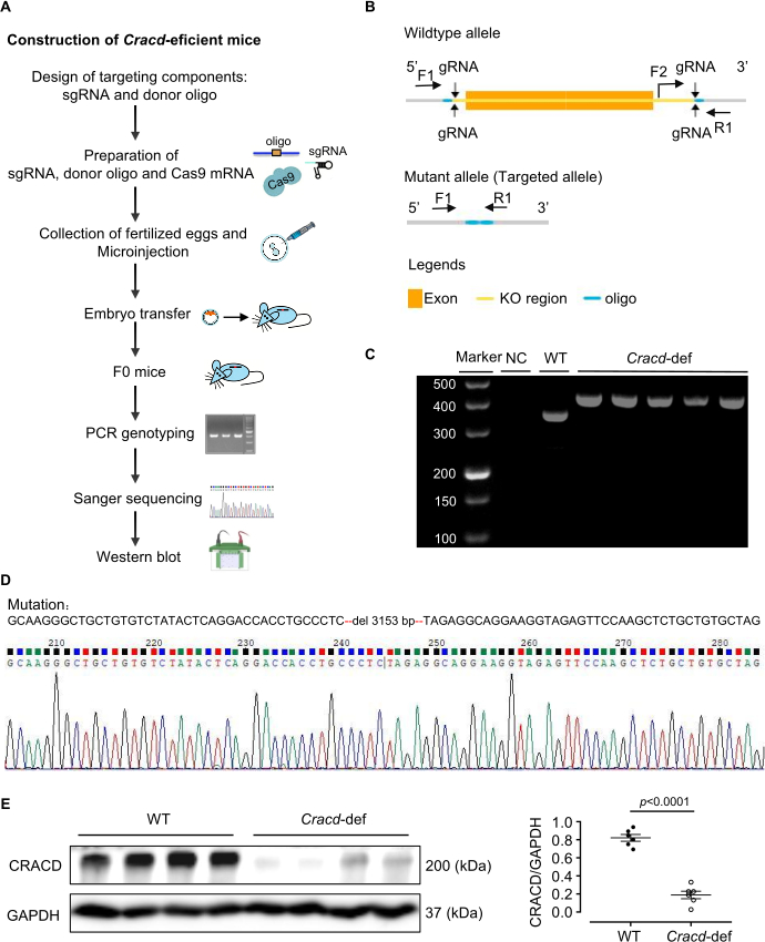
Figure 1: Generation of Cracd-deficient mice. (A) Construction of Cracd-deficient mice. (B) Schematic of the Cracd gene modification strategy. (C) PCR genotyping of tail genomic DNA. WT mice show a single 357 bp band, while homozygous (Cracd‑deficient) mice show a single 421 bp band. (D) Sanger sequencing of genomic DNA confirmed the predicted deletion in the Cracd locus of deficient mice compared to WT controls. (E) Western blot analysis showed significantly reduced CRACD protein levels in the hearts of Cracd-deficient mice compared to WT controls. n = 6 mice per group. Data are presented as mean ± SEM. Statistical significance was calculated with a two-tailed unpaired Student's t-test. Please click here to view a larger version of this figure.
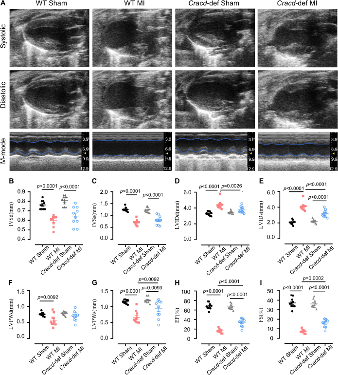
Figure 2: Echocardiographic assessment of cardiac function. (A) Representative B-mode and M-mode echocardiographic images of mouse hearts at diastole and systole. Quantitative analysis of (B) interventricular septum thickness at diastole (IVSd) and (C) systole (IVSs), (D) left ventricular internal diameter at diastole (LVIDd) and (E) systole (LVIDs), (F) left ventricular posterior wall thickness at diastole (LVPWd) and (G) systole (LVPWs), (H) ejection fraction (EF), and (I) fractional shortening (FS). n = 10 mice per group. Data are presented as mean ± SEM. Statistical significance was determined by one-way ANOVA followed by Tukey's multiple comparisons test. Please click here to view a larger version of this figure.
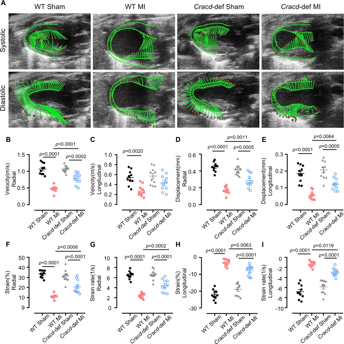
Figure 3: Speckle-tracking strain analysis. (A) Representative images of left ventricular wall motion and deformation at diastole and systole. Quantitative analyses of the following parameters: (B) radial velocity, (C) longitudinal velocity, (D) radial displacement, (E) longitudinal displacement, (F) radial strain, (G) radial strain rate, (H) longitudinal strain, and (I) longitudinal strain rate. n = 10 mice per group. Data are presented as mean ± SEM. Statistical significance was determined by one-way ANOVA followed by Tukey's multiple comparisons test. Please click here to view a larger version of this figure.
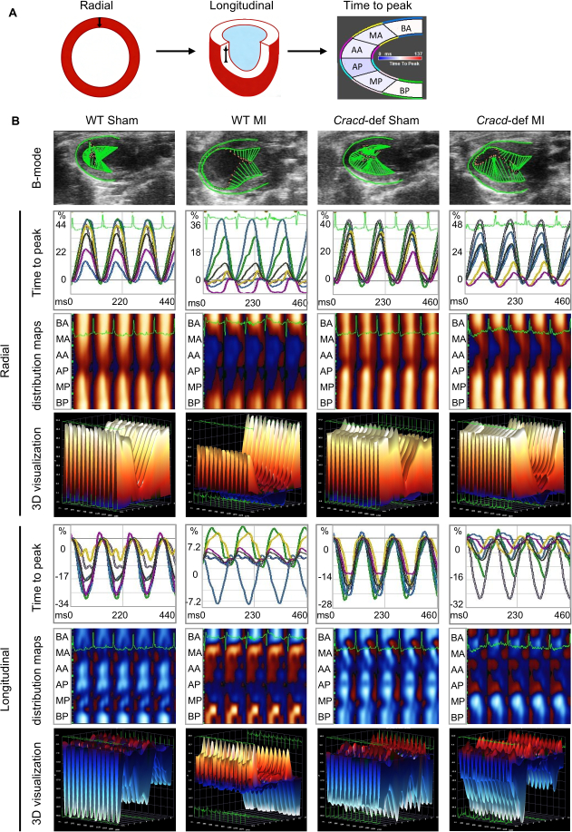
Figure 4: Speckle-tracking imaging in WT-Sham, WT-MI, Cracd-deficient Sham, and Cracd-deficient MI groups at 7 days post-MI. (A) Parasternal long‑axis B‑mode images. Left ventricular wall was segmented into six regions: BA (basal anterior), MA (mid anterior), AA (apical anterior), AP (apical posterior), MP (mid posterior), and BP (basal posterior). Color gradient from blue to red represents increasing strain magnitude. (B) Cardiac motion trajectories; time-to-peak curves for radial and longitudinal strain values along with corresponding peak times. Parametric distribution maps of strain synchrony (blue/red indicate direction: radial – away from/toward center; longitudinal – away from/toward apex). White dots = highest velocity regions, black dots = lowest velocity. Three-dimensional reconstructions (X: time, Y: spatial position, Z: strain magnitude). Please click here to view a larger version of this figure.
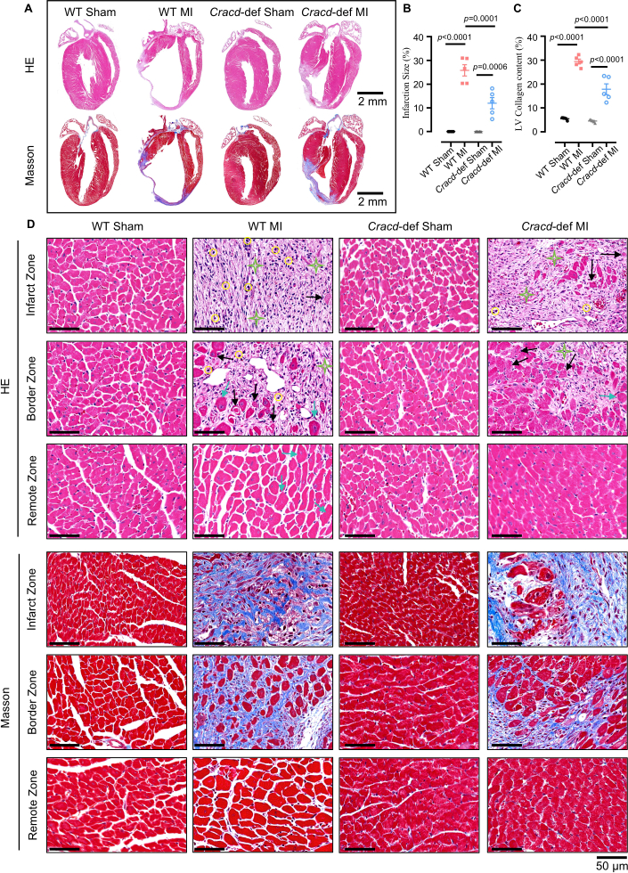
Figure 5: Histopathological analysis of cardiac remodeling after MI. (A) Whole-heart longitudinal sections with H&E and Masson's trichrome staining. Scale bars: 2 mm. (B) Quantitative analysis of infarct size and (C) collagen content. (D) High‑magnification views of infarct zone (IZ), border zone (BZ), and remote zone (RZ). H&E: yellow circles = inflammatory infiltration; black arrows = myocardial necrosis; green stars = fibrosis; blue arrows = hypertrophy. Masson’s trichrome: blue indicates collagen deposition. Scale bar: 50 μm. n = 5 mice per group. Data are presented as mean ± SEM. Statistical significance was determined by one-way ANOVA followed by Tukey's multiple comparisons test. Please click here to view a larger version of this figure.
Discussie
In deze studie hebben we een CRISPR/Cas9-gemodificeerde Cracd-deficiënte muis gegenereerd en een workflow opgezet om deze na een MI te fenotyperen met behulp van conventionele echocardiografie, speckle-tracking strain imaging en histologie. Het protocol leverde knock-outs op met een bevestigd verlies van het CRACD-eiwit. Cracd-deficiënte muizen vertoonden na een MI minder ventriculaire dilatatie, een betere EF en verminderde fibrose dan de WT-controles, wat het nut van het model voor het bestuderen van cardiale remodeling bevestigt.
Het succes van dit model hangt af van verschillende kritische stappen. Het ontwerp van het gRNA en de in vitro validatie beïnvloeden direct de efficiëntie van het bewerken van de zygote23. Wij gebruiken CRISPick of Benchling om gRNA's te selecteren met CRISPRater-scores ≥ 0,5 en testen vervolgens de splitsingsefficiëntie met een in vitro Cas9/gRNA-assay. Alleen gRNA's die een splitsing van > 50% bereiken, worden gemicro-injecteerd. Een correcte ligatie van de LAD is even cruciaal: de 7‑0 hechtdraad moet 2–3 mm vanaf de oorsprong van de LAD passeren op een diepte van 0,5–1 mm om te voorkomen dat de rechterventrikel wordt geperforeerd (te diep) of dat het vat niet wordt afgesloten (te ondiep); onmiddellijke bleking van de voorwand bevestigt de occlusie. Hartslagcontrole tijdens echocardiografie is ook essentieel24; het handhaven van 450–550 bpm door het aanpassen van isofluraan (doorgaans 1–2%) verbetert de reproduceerbaarheid van conventionele metingen en strain-metingen. Ten slotte is voor speckle‑tracking-analyse een visuele beoordeling van het kleurgecodeerde raster essentieel25. Het raster moet de myocardiale grenzen volgen zonder schokken, anders is handmatige verfijning nodig; als de kwaliteit na drie pogingen slecht blijft, wordt de muis uitgesloten.
Zelfs wanneer het protocol nauwgezet wordt gevolgd, kunnen er bepaalde technische moeilijkheden optreden. Systematische probleemoplossing kan de meeste hiervan verhelpen. Lage kiembaantransmissiesnelheden (onder de 10%) wijzen meestal op een slechte integratie van het donor-oligonucleotide of een zwakke gRNA-activiteit26. Bijgevolg kan het verhogen van de donor-oligo naar 20 ng/µL, het verlengen van de homologiearmen naar 120–200 bp en het opnieuw controleren van de gRNA-splitsing (> 50%) de transmissie verbeteren. Hoge mortaliteit na MI (boven de 30%) wordt vaak veroorzaakt door pneumothorax, bloedingen en een groot infarct27. Daarom vermindert het houden van de muizen op continue zuurstof tot ze ontwaken en het gebruik van een warmtemat om de lichaamstemperatuur te handhaven de sterftecijfers aanzienlijk. Als de voorwand na ligatie niet bleek wordt, is de LAD waarschijnlijk verkeerd geïdentificeerd of is de hechting te oppervlakkig. In dat geval kan het opnieuw lokaliseren van de LAD (een helderrood diagonaal vat) en het opnieuw doorvoeren van de naald tot een diepte van 0,5–1 mm, soms met een dissectiemicroscoop, dit probleem oplossen. Bij een slechte kwaliteit van de speckle-tracking zijn een lage framerate, wazige randen of bewegingsartefacten de gebruikelijke oorzaken28. Dienovereenkomstig helpt het verhogen van de framerate, het nauwkeurig afstellen van de gain, het handmatig corrigeren van het endocardiale spoor en het weggooien van cycli met aritmieën.
Naast deze operationele overwegingen heeft de methode duidelijke beperkingen. De Cracd knockout is globaal, waardoor CRACD in alle weefsels ontbreekt. Bijgevolg kan de waargenomen bescherming niet uitsluitend worden toegeschreven aan cardiomyocyten, aangezien systemische bijdragen mogelijk ook een rol spelen. Een conditionele knockout zou nodig zijn om celtype-specifieke rollen te onderscheiden. Een andere beperking is het enkele tijdstip op dag 7; we zien niets over chronische remodellering. Daarom zouden latere tijdstippen (4 of 8 weken) moeten worden toegevoegd voor langetermijnstudies. De reproduceerbaarheid lijdt bovendien onder de afhankelijkheid van de beeldkwaliteit: niet elk echografiesysteem kan scherpe endocardiale randen leveren, en de ervaring van de operator is van belang. Daarom wordt aanbevolen dat alle operaties en metingen door een professioneel getrainde persoon worden uitgevoerd.
Gezien deze beperkingen is het nuttig om onze aanpak te vergelijken met alternatieve strategieën voor het bestuderen van genfunctie bij remodellering na een myocardinfarct (MI). AAV-gemedieerde genmanipulatie en antisense-oligonucleotiden (ASO's) zijn twee gangbare alternatieven29,30. AAV9 kan hartspecifieke, transiënte overexpressie of knockdown bewerkstelligen zonder dat fokken nodig is, maar de verpakkingslimiet (~4,7 kb), de variabele transductie-efficiëntie en off-target effecten zijn nadelen31. In tegenstelling hiermee biedt ons CRISPR/Cas9-knockoutmodel permanent en volledig verlies van functie, wat voordelig is voor langetermijnstudies. ASO's zorgen voor een omkeerbare, dosisafhankelijke knockdown met een snelle werking, waardoor ze geschikt zijn voor acute interventies; ze vereisen echter herhaalde dosering en missen weefselspecificiteit32. Daarom is de hier beschreven constitutieve knockout het meest geschikt voor initiële ontdekking en chronische fenotypering.
Ten slotte kan het protocol ruim verder worden uitgebreid dan CRACD. Dezelfde workflow — CRISPR/Cas9-generatie van knockout-muizen gecombineerd met echocardiografie, strain-imaging en histologie — kan elk kandidaatgen in post-MI remodellering evalueren. Het fenotyperingskader is ook geschikt voor andere cardiovasculaire ziektemodellen, zoals transversale aortacontrictie (drukoverbelasting) of ischemie-reperfusieschade. Bovendien kunnen de weefselmonsters die met dit protocol zijn verkregen, worden gebruikt voor multi-omics-analyses (transcriptomics, proteomics, metabolomics) om moleculaire mechanismen te ontrafelen. Het Cracd-deficiënte model kan dienen als instrument voor het testen van geneesmiddelen: verbindingen waarvan wordt vermoed dat ze via de CRACD-route werken, kunnen worden geëvalueerd. Zo biedt dit protocol een platform dat aanpasbaar is aan een breed scala aan mechanistische en translationele studies binnen het cardiovasculaire onderzoek.
Openbaarmakingen
De auteurs verklaren dat zij geen concurrerende belangen hebben.
Dankbetuigingen
Dit werk werd ondersteund door de Guangdong Basic and Applied Basic Research Foundation (2023A1515011278), het Guangdong Provincial Biotechnology Research Institute Self-Funded Project (GDBRI-ZL202602) en de China-Canada Academic Exchange and Cooperation on Cardiovascular Disease Models and Pathogenesis (MS202500058).
Materialen
| Naam | Bedrijf | Catalogusnummer | Opmerkingen |
|---|---|---|---|
| CRISPR/Cas9 | |||
| Trizma Hydrochloride-oplossing | Sigma-Aldrich, USA | T2663 | Gebruikt voor het bereiden van DNA-extractiebuffer |
| Proteïnase K | Sigma-Aldrich, USA | 539480 | Gebruikt voor digestie van muizenstaarten |
| Green Taq Mix | Vazyme, China | P131 | Gebruikt voor PCR-amplificatie |
| Agarose | BioFroxx, Germany | 1110GR500 | Gebruikt voor DNA-gelelektroforese bij een concentratie van 1–2% |
| DNA-marker | Thermo Scientific, USA | SM0242 | Gebruikt als DNA-ladder voor het bepalen van de grootte van PCR-producten |
| TIANamp Genomic DNA Kit | Tiangen Biotech, China | DP304 | Gebruikt voor het extraheren van genomisch DNA uit muizenstaartweefsel |
| MEGAshortscript™ T7 transcriptiekit | Thermo Scientific, USA | AM1354 | Gebruikt voor in vitro transcriptie van gRNA |
| RNA-zuiveringskit | Zymo Research, USA | R1016 | Gebruikt voor het zuiveren van getranscribeerd gRNA |
| BeyoCRISPR™ sgRNA Screening Kit | Beyotime, China | D8412S | Gebruikt voor in vitro evaluatie van de splitsingsactiviteit van gRNA |
| RNase-inhibitor | NEB, USA | M0314 | Gebruikt om RNA-degradatie tijdens in vitro transcriptie te voorkomen |
| TaKaRa Taq | Takara Bio, Japan | R001A | Gebruikt voor PCR-genotypering van muizenstaart-DNA |
| FERTIUP® Mouse Sperm Preincubation Medium: PM | Cosmo Bio, Japan | KYD-002-EX | Gebruikt voor capacitatie van sperma voorafgaand aan in vitro fertilisatie |
| T7 ARCA mRNA-kit | NEB, USA | E2060 | Gebruikt voor in vitro transcriptie van Cas9-mRNA |
| Drachtig merrie serum gonadotropine | MCE, USA | 9002-70-4 | Gebruikt voor superovulatie, toegediend via intraperitoneale injectie |
| Humaan choriongonadotropine | ProSpec, Israel | HOR-250 | Gebruikt voor inductie van ovulatie, toegediend via intraperitoneale injectie |
| RNase-vrij water | Aladdin, China | R665526 | Gebruikt voor RNA-oplossing en PCR-opstelling |
| Primers | Sangon Biotech, China | / | Gebruikt voor genotypering |
| Chirurgie | |||
| Isofluraan | RWD Life Science, China | R510-22-10 | Gebruikt voor inductie en instandhouding van anesthesie tijdens de operatie |
| Ontharingscrème | Veet, France | 1000207 | Aangebracht op het borstgebied om haar te verwijderen voorafgaand aan chirurgie en echografie |
| 7-0 Hechtmateriaal | Lingqiao, China | LQ7022380206 | Gebruikt voor permanente ligatie van de linker anterior descending (LAD) kransslagader |
| 4-0 Hechtmateriaal | Lingqiao, China | LQ4022380412 | Gebruikt voor het sluiten van de huidincisie na de operatie |
| Anesthesieventilator | Harvard Apparatus, USA | Tabletop | Gebruikt voor het toedienen van isofluraan en ondersteuning van de ademhaling |
| Kleine dieren ventilator | Taimeng, China | HX-101E | Gebruikt voor het bieden van positieve drukventilatie tijdens openhartoperaties |
| Warmtemat met constante temperatuur | TigerGene, USA | TG-TP-GS | Gebruikt voor het behouden van de lichaamstemperatuur van de muis tijdens operatie en herstel |
| Ribspreider | Shenzhen Huayon Biotech, China | LN-18-4101 | Gebruikt voor het spreiden van de ribben en het blootleggen van het hart voor LAD-ligatie |
| Scharen | Shenzhen Huayon Biotech, China | 18-0551 | Gebruikt voor het maken van huidincisies en het knippen van weefsel |
| Stereomicroscoop | Nikon, Japan | SMZ 745 | Gebruikt voor het bieden van een vergroot beeld tijdens de LAD-slagaderligatie-operatie |
| Microscopische naaldvoerder | Shenzhen Huayon Biotech, China | 18-2211 | Gebruikt voor het manipuleren van de kleine 7-0 hechtnald onder de microscoop |
| Chemicaliën en reagentia | |||
| Ultrasone koppelingsmiddelen | Tianjin Jinya Technology Development, China | TM-100 | Aangebracht op de borst voor echocardiografie, waarborgt correct contact van de probe |
| Paraformaldehyde | Sigma-Aldrich, USA | V900894 | Gebruikt voor fixatie van hartweefsel na oogst |
| Xyleen | Macklin, China | 1330-20-7 | Gebruikt voor paraffineverwijdering en weefselklaring voor kleuring |
| Hematoxyline | Sigma-Aldrich, USA | H3136 | Kernkleuring, gebruikt bij HE-kleuring, |
| Eosine | Sigma-Aldrich, USA | E4009 | Cytoplasmakleuring, gebruikt bij HE-kleuring, |
| Ponceau S | Sigma-Aldrich, USA | P3504 | Gebruikt bij Masson-kleuring voor cytoplasmakleuring |
| Fosfomolybdeenzuur | Sigma-Aldrich, USA | 221856 | Gebruikt bij Masson-kleuring als beitsmiddel en voor differentiatie |
| Anilineblauw-oplossing | Sigma-Aldrich, USA | 415049 | Gebruikt bij Masson-kleuring voor kleuring van collageenvezels (blauw) |
| CRACD-antilichaam | Absea, China | KC-35356 | Primair antilichaam voor detectie van CRACD-eiwit via Western Blot, verdunning 1:2000 |
| GAPDH-antilichaam | CST, USA | 2118S | Loading control antilichaam voor Western Blot, verdunning 1:5000 |
| Ethanol | Macklin, China | 64-17-5 | Gebruikt voor dehydratie en bereiding van reagentia (graden 70%, 80%, 95%, 100%) |
| Laboratoriumapparatuur | |||
| Sorvall™ Legend™ Micro 17 Centrifuge | Thermo Scientific, USA | 75002541 | Gebruikt voor routinematige monstervoorbereiding |
| PCR Thermal Cycler | Bio-Rad Laboratories, USA | 1861096 | Gebruikt voor DNA-amplificatie |
| Micro-injectiesysteem | Eppendorf, Germany | 5193000020 | Gebruikt voor micro-injectie van CRISPR/Cas9-reagentia in zygoten |
| Vevo 2100 Imaging System | VisualSonics, Canada | Vevo2100 | Hoogfrequent beeldsysteem voor niet-invasieve beoordeling van hartfunctie en -structuur na MI |
| Lichtmicroscoop | Leica, Germany | DM2500 | Gebruikt voor het observeren van HE- en Masson-gekleurde hartcoupes |
| Analysesoftware | |||
| ImageJ | National Institutes of Health, USA | / | Gebruikt voor beeldanalyse, inclusief oppervlaktemeting, signaalquantificatie en verwerking van histologische beelden (HE- en Masson-kleuring) |
| VevoLab 3.2.0 / VevoStrain | VisualSonics, Canada | / | VevoLab-software voor analyse van de hartfunctie; VevoStrain-module gebruikt voor myocardiale strainanalyse om de hartprestaties na MI te beoordelen |
| GraphPad Prism 8 | GraphPad Software, USA | / | Gebruikt voor statistische analyse en het maken van grafieken |
Referenties
- Fauconnier, J., Meli, A. C., Thireau, J., Roberge, S., Shan, J., et al. Ryanodine receptor leak mediated by caspase-8 activation leads to left ventricular injury after myocardial ischemia-reperfusion. Proc Natl Acad Sci U S A. 108 (32), 13258-13263 (2011).
- Cohn, J. N., Ferrari, R., Sharpe, N. Cardiac remodeling-concepts and clinical implications: a consensus paper from an international forum on cardiac remodeling. J Am Coll Cardiol. , (2000).
- Liu, D., Tian, X., Liu, Y., Song, H., Cheng, X., et al. CREG ameliorates the phenotypic switching of cardiac fibroblasts after myocardial infarction via modulation of CDC42. Cell Death Dis. 12 (4), (2021).
- Contessotto, P., Pandit, A. Therapies to prevent post-infarction remodelling: from repair to regeneration. Biomaterials. 275, (2021).
- Zhu, R., Sun, H., Yu, K., Zhong, Y., Shi, H., et al. Interleukin-37 and dendritic cells treated with interleukin-37 plus troponin I ameliorate cardiac remodeling after myocardial infarction. J Am Heart Assoc. 5 (12), (2016).
- Chang, Y., Zhang, J., Huo, X., Qu, X., Xia, C., et al. Substrate rigidity dictates colorectal tumorigenic cell stemness and metastasis via CRAD-dependent mechanotransduction. Cell Rep. 38 (7), (2022).
- Seo, Y., Jang, J., Ko, K. P., Zou, G., Huang, Y., et al. Actin dysregulation induces immune evasion via oxidative stress-activated PD-L1 in gastric cancer. bioRxiv. , (2024).
- Kim, B., Zhang, S., Huang, Y., Ko, K. P., Jung, Y. S., et al. CRACD loss induces neuroendocrine cell plasticity of lung adenocarcinoma. Cell Rep. 43 (6), (2024).
- Jung, Y. S., Wang, W., Jun, S., Zhang, J., Srivastava, M., et al. Deregulation of CRAD-controlled cytoskeleton initiates mucinous colorectal cancer via β-catenin. Nat Cell Biol. 20 (11), 1303-1314 (2018).
- Liu, Z., Cao, H., Shi, Y., Yang, R. KIAA1211 plays an oncogenic role in human non-small cell lung cancer. J Cancer. 10 (26), 6747-6753 (2019).
- Snider, P. L., Snider, E., Simmons, O., Lilly, B., Conway, S. J. Analysis of uncharacterized mKiaa1211 expression during mouse development and cardiovascular morphogenesis. J Cardiovasc Dev Dis. 6 (2), (2019).
- Lin, Y. H., Warren, C. M., Li, J., McKinsey, T. A., Russell, B. Myofibril growth during cardiac hypertrophy is regulated through dual phosphorylation and acetylation of the actin capping protein CapZ. Cell Signal. 28 (8), 1015-1024 (2016).
- Yang, F. H., Pyle, W. G. Reduced cardiac CapZ protein protects hearts against acute ischemia-reperfusion injury and enhances preconditioning. J Mol Cell Cardiol. 52 (3), 761-772 (2012).
- Tong, C., Huang, G., Ashton, C., Wu, H., Yan, H., et al. Rapid and cost-effective gene targeting in rat embryonic stem cells by TALENs. J Genet Genomics. 39 (6), 275-280 (2012).
- Cordes, S. P. N-ethyl-N-nitrosourea mutagenesis: boarding the mouse mutant express. Microbiol Mol Biol Rev. 69 (3), 426-439 (2005).
- Cong, L., Ran, F. A., Cox, D., Lin, S., Barretto, R., et al. Multiplex genome engineering using CRISPR/Cas systems. Science. 339 (6121), 819-823 (2013).
- Kanke, K. L., Rayner, R. E., Bozik, J., Abel, E., Venugopalan, A., et al. Single-stranded DNA with internal base modifications mediates highly efficient knock-in in primary cells using CRISPR-Cas9. Nucleic Acids Res. 52 (22), 13561-13576 (2024).
- Mihos, C. G., Liu, J. E., Anderson, K. M., Pernetz, M. A., O'Driscoll, J. M., et al. Speckle-tracking strain echocardiography for the assessment of left ventricular structure and function: a scientific statement from the American Heart Association. Circulation. 152 (10), (2025).
- Garg, P. K., Biggs, M. L., Kizer, J. R., Shah, S. J., Psaty, B., et al. Glucose dysregulation and subclinical cardiac dysfunction in older adults: the Cardiovascular Health Study. Cardiovasc Diabetol. 21 (1), (2022).
- Asanuma, T., Fukuta, Y., Masuda, K., Hioki, A., Iwasaki, M., et al. Assessment of myocardial ischemic memory using speckle tracking echocardiography. JACC Cardiovasc Imaging. 5 (1), 1-11 (2012).
- Chong, S. Y., Zharkova, O., Yatim, S. M. J. M., Wang, X., Lim, X. C., et al. Tissue factor cytoplasmic domain exacerbates post-infarct left ventricular remodeling via orchestrating cardiac inflammation and angiogenesis. Theranostics. 11 (19), 9243-9261 (2021).
- Hung, C. L., Verma, A., Uno, H., Shin, S. H., Bourgoun, M., et al. Longitudinal and circumferential strain rate, left ventricular remodeling, and prognosis after myocardial infarction. J Am Coll Cardiol. 56 (22), 1812-1822 (2010).
- Labuhn, M., Adams, F. F., Ng, M., Knoess, S., Schambach, A., et al. Refined sgRNA efficacy prediction improves large- and small-scale CRISPR-Cas9 applications. Nucleic Acids Res. 46 (3), 1375-1385 (2018).
- Wu, J., Bu, L., Gong, H., Jiang, G., Li, L., et al. Effects of heart rate and anesthetic timing on high-resolution echocardiographic assessment under isoflurane anesthesia in mice. J Ultrasound Med. 29 (12), (2010).
- Voigt, J. U., Pedrizzetti, G., Lysyansky, P., Marwick, T. H., Houle, H., et al. Definitions for a common standard for 2D speckle tracking echocardiography: consensus document of the EACVI/ASE/Industry Task Force to standardize deformation imaging. J Am Soc Echocardiogr. 28 (2), 183-193 (2015).
- Port, F., Chen, H. M., Lee, T., Bullock, S. L. Optimized CRISPR/Cas tools for efficient germline and somatic genome engineering in Drosophila. Proc Natl Acad Sci U S A. 111 (29), (2014).
- Gao, E., Lei, Y. H., Shang, X., Huang, Z. M., Zuo, L., et al. A novel and efficient model of coronary artery ligation and myocardial infarction in the mouse. Circ Res. 107 (12), 1445-1453 (2010).
- Rösner, A., Barbosa, D., Aarsæther, E., Kjønås, D., Schirmer, H., et al. The influence of frame rate on two-dimensional speckle-tracking strain measurements: a study on silico-simulated models and images recorded in patients. Eur Heart J Cardiovasc Imaging. 16 (10), 1137-1147 (2015).
- Argiro, A., Bui, Q., Hong, K. N., Ammirati, E., Olivotto, I., et al. Applications of gene therapy in cardiomyopathies. JACC Heart Fail. 12 (2), 248-260 (2024).
- Micheletti, R., Plaisance, I., Abraham, B. J., Sarre, A., Ting, C. C., et al. The long noncoding RNA Wisper controls cardiac fibrosis and remodeling. Sci Transl Med. 9 (395), (2017).
- Wu, Z., Yang, H., Colosi, P. Effect of genome size on AAV vector packaging. Mol Ther. 18 (1), 80-86 (2010).
- Juliano, R. L. The delivery of therapeutic oligonucleotides. Nucleic Acids Res. 44 (14), 6518-6548 (2016).
Herprints en machtigingen
