The video demonstrates the procedure for a growth competition assay to measure the relative replication fitness of two viral strains by comparing their replication in human immune cells.
Subskrypcja JoVE jest wymagana do oglądania tego materiału. Zaloguj się lub rozpocznij bezpłatny okres próbny.
Artykuł metodologiczny
An Assay for Measuring the Relative Replication Fitness of Viral Strains
181 wyświetleń
⸱
31 lipca 2026
W tym artykule
Streszczenie
Protokół
1. Construction of Chimeric HIV-1 NL4-3 Molecular Clones
- Amplify Insert DNA Fragment
- Design chimeric primers. The 5’ halves of both forward and reverse primers contain an HIV-1 vector sequence, at which the fragment will be inserted. The 3’ half of the primers must contain the end of the insert sequence (Figure 1). Make sure that the chimeric primer sequence retains the original open reading frames.
- Use primers at least 20 bases in length, with a melting temperature greater than or equal to 60 °C, ~50% GC content, and a low tendency to form primer dimers, heterodimers, and/or hairpin structures. Assess these properties using the OligoAnalyzer web tool (https://www.idtdna.com/analyzer/Applications/OligoAnalyzer/).
- Use polymerase chain reaction (PCR) and the chimeric primers to amplify the insert DNA (Figure 1). For each PCR reaction, use 1× high fidelity buffer, 0.2 mM dNTPs, 1 U of high fidelity DNA polymerase, 0.5 µM of forward chimeric primer, 0.5 µM of reverse chimeric primer, and 1 pg0 ng of DNA sample carrying the insert region. Add dH2O to a final volume of 50 µl.
- Set thermal cycling steps as follows: Perform an initial DNA denaturation step at 98 °C for 10 sec. Amplify with 30 cycles of DNA denaturation at 98 °C for 10 sec and DNA annealing at 3 °C above the lowest melting temperature of the two primers for 20 sec. Perform a final extension at 72 °C for 10 min. Store PCR products at 4 °C.
- Take 5 µl of the PCR products from the previous step and run agarose gel electrophoresis.
- Use a 0.7% agarose gel, 1× TAE buffer (40 mM Tris-acetate, 1 mM EDTA), 0.5 μg/ml ethidium bromide (EtBr) final concentration, and 1 kb ladder as the DNA size marker. Set the power source voltage to 5 V/cmthe the distance between electrodes. Stop the electrophoresis when the loading dye migrates through about 2/3 of the gel length. Visualize the gel using a gel documentation system.
NOTE: EtBr is a suspected carcinogen and must be properly disposed of, per institution regulations. Gloves should always be worn when handling gels containing EtBr. To prevent cross-contamination, change to new gloves after finishing handling EtBr-containing material and before handling other materials or equipment.
- Use a 0.7% agarose gel, 1× TAE buffer (40 mM Tris-acetate, 1 mM EDTA), 0.5 μg/ml ethidium bromide (EtBr) final concentration, and 1 kb ladder as the DNA size marker. Set the power source voltage to 5 V/cmthe the distance between electrodes. Stop the electrophoresis when the loading dye migrates through about 2/3 of the gel length. Visualize the gel using a gel documentation system.
- If only one DNA band with a size corresponding to the desired PCR product is detected, purify the rest of the PCR product using a commercial kit such as QIAquick PCR Purification Kit according to the manufacturer’s protocols.
- If other, non-specific bands are also present, use the rest of the PCR product to run preparative gel electrophoresis. Use the same parameters and conditions specified in step 1.1.4. Ensure that the gel well is large enough to load ~45 µl of PCR products. Cut out the band of interest and extract the DNA from the gel using the QIAquick gel extraction kit according to the manufacturer’s protocols.
- Design chimeric primers. The 5’ halves of both forward and reverse primers contain an HIV-1 vector sequence, at which the fragment will be inserted. The 3’ half of the primers must contain the end of the insert sequence (Figure 1). Make sure that the chimeric primer sequence retains the original open reading frames.
- Introduce the Insert Fragment into Full-length Infectious HIV-1 Subtype B Vector (pNL4-3)
- Use purified PCR products from step 1.1.5 as PCR primers. Use pNL4-3VifA as template DNA in one PCR reaction and use pNL4-3VifB as template in the other reaction (Figure 1). For each PCR reaction, use 1× high-fidelity buffer, 0.2 mM dNTPs, 2 U of high-fidelity DNA polymerase, 500 ng of primer DNA, and 50 ng of template DNA in a final volume of 50 µl. Set the thermal cycling parameters to: 98 °C for 30 sec, 35 cycles at 98 °C for 10 sec, 48 °C for 1 min, and 72 °C for 10 min, followed by 72 °C for 10 min.
- Add 10 U of DpnI to 50 µl of the PCR reaction and incubate at 37 °C for 1 hr to digest the template DNA. Ensure that the plasmid DNA is isolated from a methylation-competent bacterial strain, e.g., TOP10 chemically competent Escherichia coli.
- Use the DpnI-digested product to transform competent bacterial cells. Use heat-shock transformation with TOP10 chemically competent E. coli, according to the manufacturer’s protocol. To select for bacterial cells containing the recombinant plasmid, use Luria Broth (LB) culture plates containing 100 mg/L carbenicillin.
- Pick ~10 well-separated colonies and grow each separately in 3 ml LB liquid medium containing 100 mg/L carbenicillin and incubate at 30 °C in a shaker overnight (O/N).
- Use the QIAprep Spin Miniprep kit to isolate plasmid DNA from the bacterial liquid culture, according to the manufacturer’s protocol.
- Use double restriction digestion to determine whether the plasmid DNA contains the proper insert. Ensure that one of the restriction sites exists only within the insert region and the other restriction site exists only once in the HIV-1 vector, outside of the insert region.
- Digest at least 300 ng of plasmid DNA in a 10 µl final reaction volume. Select restriction buffers, incubation temperature, and incubation time according to the manufacturer’s protocol of the selected restriction enzymes. Take 9 µl of the digested DNA and run gel electrophoresis as described in step 1.1.4. Select recombinant plasmids that have DNA bands of the predicted sizes.
- Confirm sequence integrity of the recombinant plasmids by Sanger sequencing. While rare, unwanted mutation(s) can be introduced during the PCR reactions.
- Sequence both strands of the plasmid DNA. Follow instructions in step 1.1.1.i. to design sequencing primers. In addition, ensure that the forward and reverse sequencing primers anneal at least 50 bp upstream and downstream of the insert region in the recombinant plasmid, respectively.
- Submit plasmid DNA and sequencing primers to a commercial DNA sequencing service provider. Prepare the DNA sample and primers as specified by the service provider.
- Make an endotoxin-free stock of the mutated plasmid DNA using an Endotoxin free plasmid DNA kit according to manufacturer’s protocol. Prepare at least 1 µg of endotoxin-free plasmid DNA for transfection in the following step.
- Introduce Small-scale Mutations Via Site-directed Mutagenesis
- Design mutagenic primers with overlapping forward and reverse primers containing the desired mutation(s). Position the base(s) to be substituted, inserted, or deleted in the middle of the primers, flanked by 10-15 homologous bases. Follow instructions in step 1.1.1.i.
- Use PCR to synthesize mutant plasmids. For each PCR reaction, use 1× high-fidelity buffer, 10 mM dNTPs, 2 U of high-fidelity DNA polymerase, 0.5 µM of forward mutagenic primer, 0.5 µM of reverse mutagenic primer, and 50 ng of chimeric pNL4-3VifB, from step 1.2.6, in a final volume of 50 µl. Set the thermal cycling parameters to: 98 °C for 30 sec, 25 cycles at 98 °C for 10 sec, 48 °C for 1 min, and 72 °C for 10 min, followed by 72 °C for 10 min.
- Repeat steps 1.2.2 to 1.2.6.
2. Generation of Viral Stock Using Transfection
- Calculate the amount of viral stock desired and plasmid DNA required. With a viral titer of 104 infectious units (IU)/ml or higher, 1.8 ml of viral stock is sufficient for two sets of growth competition assays, including monoinfections, each done in triplicate. For a transfection done in a 6-well plate, about 1.8 mL supernatant is harvested per well. One µg of plasmid DNA is needed for each transfection done in a 6-well plate.
- For each well of a 6-well plate, prepare 100 µl of transfection mixture, e.g., consisting of 1 µl X-tremeGENE 9 transfection reagent (or comparable product), 1 µg of plasmid DNA, and serum-free Dulbecco’s Modified Eagle Medium (DMEM).
- Determine the volume of plasmid DNA needed, using 1 µg plasmid DNA per well. Ensure that the final concentration of the plasmid DNA is at least 50 ng/µl.
- Determine how much serum-free medium (DMEM) is needed per well using the formula: Total volume of DMEM in µl = 100 µl - DNA volume in µl.
- Add 106 human embryonic kidney or HEK 293T-17 (ATCC) cells/well in 2 ml of propagation medium (DMEM + 10% fetal bovine serum (FBS)) into a 6-well plate. Incubate for 1 hr at 37 °C in a 5% CO2 atmosphere. Seed as many wells as needed (determined in step 2.1).
- To prepare the transfection mixture, aliquot the appropriate volume of serum-free DMEM, as calculated above, into a 1.8 ml polypropylene microcentrifuge tube, and then add the transfection reagent.
- Pipette reagent directly into the media solution, do not add it to the plastic surface of the microcentrifuge tube. Add plasmid DNA last. Pipette up and down gently to mix the solution. Incubate for 15 min at room temperature or RT (15 °C to 25 °C) to allow the formation of transfection complexes.
- Add the mixture in a drop-wise manner to cells seeded in the 6-well plate. Gently shake or swirl the wells to ensure even distribution of transfection complexes.
- Seal plates with plastic wrap.
- Incubate cultures at 37 °C in a 5% CO2 atmosphere for 48 hr.
- Use a pipette to carefully collect and transfer supernatant to a 15 ml tube through a 0.22 µm filter top.
- Use a pipette to transfer 250 µl or more of the filtered supernatant to 1.8 ml microfuge tubes with rubber gaskets in the lids.
- Store filtered supernatants at -80 °C until use.
3. Establish Viral Growth Kinetics
- Monoinfection
- Seed 3 × 10⁵ PHA-stimulated peripheral blood mononuclear cells (PBMC)/well in 48-well plates in a total volume of 500 µl/well. Keep the culture plates at 37 °C in a 5% CO2 atmosphere until inoculation.
- For each virus, prepare an inoculum containing 6,000 IU in 2 ml of complete Iscove’s Modified Dulbecco’s Medium (cIMDM).
- Inoculate wells in triplicate by adding 500 µl of the inoculum (1,500 IU) to the seeded cells. The final volume of the infected cell culture is 1 ml/well, and the multiplicity of infection (MOI) is 0.005.
- Aliquot 200 µl of the remaining inoculum to each of two 96-well plates for RNA isolation, one of which is saved as a backup.
- Incubate cultures at 37 °C in a 5% CO2 atmosphere for 16-24 hr.
- Wash cultures 16-24 hr after inoculation.
- Remove and discard 750 µl of culture supernatant.
- Add 750 µl of fresh cIMDM. Wrap plates in plastic wrap and spin for 10 minutes at 300 × g. Remove and discard 750 µl supernatant.
- Add 750 µl of fresh cIMDM. Incubate at 37 °C with a 5% CO2 atmosphere (day 1).
- Sample cultures daily from day 2 to day 7.
- Transfer 500 µl of culture supernatant to a 1.8 ml centrifuge tube. Spin for 1 min at 3,000 × g.
- Transfer 200 µl of the cell-free supernatant to the two 96-well sample plates for RNA isolation, again saving one plate as backup. Store supernatants at -80 °C until RNA isolation.
- Add 500 µl fresh cIMDM to each culture. Incubate at 37 °C in a 5% CO2 atmosphere.
- Discard cultures into Wescodyne at the end of the experiment.
- Isolate RNA from 200 µl of supernatant (use commercial kits such as QIAamp Viral RNA Mini Kit) following the manufacturer’s standard protocol. For a large number of samples, use the Qiagen QIAxtractor.
- Store RNA samples at -80 °C until cDNA synthesis.
- cDNA Synthesis (Reverse Transcription)
- For each RNA sample, add 1.2 nmol of dNTP and 1.2 pmol of cDNA synthesis primer (5’-GTTGATCCTTTAGGTATCTTTCCACAGC-3’, HXB2 nucleotide 7968 to 7995) to 10 µl of viral RNA. Add water to a final volume of 14 µl. Flick the tube to mix and spin briefly to collect liquid at the bottom of the tube.
- Incubate mixture for 5 min at 65 °C, then hold at 4 °C until the master mix is prepared.
- Prepare master mix using 5× first-strand buffer (250 mM Tris-HCl, pH 8.3, 375 mM KCl, 15 mM MgCl2), 5 mM dithiothreitol (DTT), 120 U of SuperScriptIII, and 240 U of RNase inhibitor. Add water to the final volume of 10 µl.
- Add 10 µl of master mix to RNA mixture, flick to mix, and spin to collect.
- Incubate mixture for 90 min at 50 °C to allow synthesis of cDNA. Incubate for 15 min at 70 °C to inactivate reverse transcriptase. Hold at 4 °C as needed.
- Add 2 U of RNase H, flick to mix, and then spin to collect.
- Incubate 20 min at 37 °C. Store cDNA at -20 °C.
4. Growth Competition Assay
- Seed 3 × 10⁵ PHA-stimulated PBMCs (or 1 × 105 CEM×174 cells) in 500 µl total volume per well in a 48-well flat-bottomed plate.
- Keep the plate at 37 °C in a 5% CO2 atmosphere until inoculation.
- For each virus, prepare 3 ml of inoculum containing 6,000 IU.
- Transfer 1.5 ml of each viral inoculum to a sterile tube to create the dual infection inoculum. Add 500 µl of the dual inoculum (1,500 IU) to 3 × 10⁵ cells in a 48-well plate. The final culture volume is 1 ml/well. Aliquot 200 µl of the inoculum to two 96-well plates for RNA isolation; save one plate as a backup.
- Incubate inoculated cells at 37 °C with a 5% CO2 atmosphere for 16-24 hr.
- Wash cultures 16-24 hr after inoculation.
- Remove and discard 750 µl of culture supernatant.
- Add 750 µl of fresh cIMDM. Wrap plates in plastic wrap and spin for 10 min at 300 × g. Remove and discard 750 µl supernatant.
- Add 750 µl of fresh cIMDM. Incubate at 37 °C with a 5% CO2 atmosphere (day 1).
- Select sampling times to include at least 3 time points within the exponential growth phase observed in step 3.4.1.
- For each sampling, follow step 3.1.7.
- Perform cDNA synthesis as described in section 3.2.
Dostęp ograniczony. Zaloguj się lub rozpocznij wersję próbną, aby wyświetlić tę treść.
Wyniki
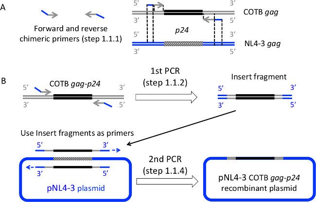
Figure 1: Construction of HIV-1 NL4-3 COTB Gag-p24 recombinant molecular clones using overlap extension PCR. (A) Design the chimeric primers. The 5’ halves of the primers contain the NL4-3 vector sequence and the 3&#x...
Dostęp ograniczony. Zaloguj się lub rozpocznij wersję próbną, aby wyświetlić tę treść.
Materiały
| Nazwa | Firma | Numer katalogowy | Komentarze |
|---|---|---|---|
| Construction of recombinant clones | |||
| Chimeric primer/Mutagenic/Sequencing primers | IDT | Custom DNA oligos | |
| pNL4-3VifA and pNL4-3VifB plasmid | |||
| High-Fidelity DNA polymerase | Thermo Scientific | F-549S | |
| High-fidelity buffer | Thermo Scientific | F-549S | |
| Deoxynucleotide triphosphate (dNTP) | Bioline | Bio-39026 | |
| Thermal cycler | Life Technologies | Applied Biosystems® GeneAmp® PCR System 9700 | |
| DNA loading dye | Thermo Scientific | R0631 | |
| 1 kb Plus DNA ladder | Invitrogen | 10787-018 | |
| QIAquick PCR Purification Kit | Qiagen | 28104 | |
| QIAquick gel extraction kit | Qiagen | 28704 | |
| DpnI enzyme | New England Biolabs | R0176S | |
| TOP10 chemically competent Escherichia coli | Invitrogen | C4040-10 | |
| Luria Broth base powder | Invitrogen | 12795-084 | |
| Carbenicillin | Research Products International | C46000-25.0 | |
| QIAprep Spin Miniprep kit | Qiagen | 27104 | |
| EndoFree Plasmid Maxi Kit | Qiagen | 12362 | |
| Transfection | |||
| X-tremeGENE 9 DNA transfection reagent | Roche | 6365787001 | |
| HEK 293T-17 | ATCC | CRL-11268 | http://www.atcc.org/ |
| 0.22 μm filter top tube | VWR International | 89220-716 | |
| Cell culture | |||
| Dulbecco's Modified Eagle Medium (DMEM) | Life Technologies | 10566016 | |
| Iscove′s Modified Dulbecco′s Medium (IMDM) | Life Technologies | 31980-030 | |
| RPMI-1640 media | Life Technologies | 61870-036 | |
| Phytohemagglutinin (PHA) | Thermo Scientific | R30852801 | |
| Human Interleukin-2 | Roche | 11147528001 | |
| Fetal bovine serum | JR Scientific | 43640 | |
| Penicillin and Streptomycin | Corning Cellgro | 30-001-CI | |
| 6 well plate | VWR Scientific | 73520-906 | |
| 48 well plate | VWR Scientific | 62407-338 | |
| 96 well flat-bottomed plate | ISC Bioexpress | T-3015-4 | |
| 96 well round-bottomed plate | BD Falcon | 353077 | |
| 1.5 ml Microcentrifuge Tube | Mt. Baker Bio | MBD-1500 | |
| 1.5 ml tubes w/ O-ring | VWR Scientific | 89004-290 | |
| 50 ml conical tube | ISC Bioexpress | C-3317-6 |
