PCR stał się powszechną techniką w wielu laboratoriach biologii molekularnej. Niniejszym przedstawiamy krótki przewodnik po kilku konwencjonalnych protokołach PCR. Ponieważ każda reakcja jest unikalnym eksperymentem, optymalne warunki niezbędne do otrzymania produktu są zmienne. Zrozumienie zmiennych w reakcji znacznie zwiększy efektywność rozwiązywania problemów, zwiększając tym samym szansę na uzyskanie pożądanego wyniku.
Artykuł metodologiczny
Reakcja łańcuchowa polimerazy: podstawowy protokół oraz strategie rozwiązywania problemów i optymalizacji
W tym artykule
Podsumowanie
Streszczenie
W naukach biologicznych nastąpiły postępy technologiczne, które wprowadziły tę dyscyplinę w złote ery odkryć. Na przykład dziedzina mikrobiologii została przekształcona dzięki wynalezieniu mikroskopu przez Antona van Leeuwenhoeka, co po raz pierwszy pozwoliło naukowcom na wizualizację prokariontów. Opracowanie reakcji łańcuchowej polimerazy (PCR) jest jedną z tych innowacji, które zmieniły bieg nauk molekularnych, a ich wpływ objął niezliczone poddyscypliny biologii. Proces teoretyczny został opisany przez Keppego i współpracowników w 1971 roku; jednak minęło kolejne 14 lat, zanim pełna procedura PCR została opisana i zastosowana eksperymentalnie przez Kary'ego Mullisa w 1985 roku podczas pracy w Cetus Corporation. Automatyzacja i udoskonalenie tej techniki postępowały wraz z wprowadzeniem termostabilnej polimerazy DNA z bakterii Thermus aquaticus, co zaowocowało nazwą polimeraza DNA Taq.
PCR jest potężną techniką amplifikacji, która pozwala na wygenerowanie znacznej ilości konkretnego fragmentu DNA (tj. amplikonu) z niewielkiej ilości materiału wyjściowego (tj. matrycy DNA lub sekwencji docelowej). Choć metoda ta jest prosta i zazwyczaj bezproblemowa, istnieją pułapki, które mogą skomplikować reakcję i doprowadzić do błędnych wyników. Niepowodzenie PCR może skutkować powstaniem wielu niespecyficznych produktów DNA o różnych rozmiarach, które na żelach agarozowych widoczne są jako drabinka lub rozmycie prążków. Czasami nie powstają żadne produkty. Inny potencjalny problem pojawia się, gdy w amplikonach nieumyślnie wprowadzone zostaną mutacje, co prowadzi do powstania heterogenicznej populacji produktów PCR. Niepowodzenia w PCR mogą być frustrujące, chyba że zastosuje się cierpliwość i staranną analizę błędów w celu zidentyfikowania i rozwiązania problemów. Niniejszy protokół przedstawia podstawowe zasady PCR, opisuje metodologię pozwalającą na amplifikację większości sekwencji docelowych oraz prezentuje strategie optymalizacji reakcji. Podążając za tym przewodnikiem po PCR, studenci powinni być w stanie:
● Przygotować reakcje i warunki cykli termicznych dla konwencjonalnego eksperymentu PCR
● Zrozumieć funkcję poszczególnych składników reakcji oraz ich ogólny wpływ na przebieg eksperymentu PCR
● Zaprojektować i zoptymalizować eksperyment PCR dla dowolnej matrycy DNA
● Rozwiązywać problemy w przypadku nieudanych eksperymentów PCR
Protokół
1. Projektowanie starterów
Projektowanie odpowiednich starterów jest kluczowe dla pomyślnego wyniku eksperymentu PCR. Podczas projektowania zestawu starterów do konkretnego regionu DNA przeznaczonego do amplifikacji, jeden starter powinien przyłączać się do nici dodatniej, która zgodnie z konwencją jest zorientowana w kierunku 5' → 3' (znana również jako nić sensowna lub nietematyczna), a drugi starter powinien być komplementarny do nici ujemnej, zorientowanej w kierunku 3' → 5' (nić antysensowna lub tematyczna). Podczas projektowania starterów pojawia się kilka typowych problemów: 1) samozszywanie się starterów, prowadzące do powstania struktur drugorzędowych, takich jak pętle spinek do włosów (Figura 1a); 2) przyłączanie się starterów do siebie nawzajem, zamiast do matrycy DNA, co prowadzi do powstania dimerów starterów (Figura 1b); 3) drastycznie różne temperatury topnienia (Tm) dla każdego z starterów, co utrudnia wybór temperatury przyłączania umożliwiającej obu starterom efektywne wiązanie się z sekwencją docelową podczas cyklicznego ogrzewania i chłodzenia (Figura 1c) (Więcej informacji na temat Tm znajduje się w sekcjach OBLICZANIE TEMPERATURY TOPNIENIA (Tm) oraz MODYFIKACJE WARUNKÓW CYKLA).
- Poniżej znajduje się lista cech, które należy wziąć pod uwagę podczas projektowania starterów.
- Długość startera powinna wynosić od 15 do 30 nukleotydów (zasad).
- Optymalna zawartość G-C powinna mieścić się w przedziale 40-60%.
- Końcówka 3' starterów powinna zawierać G lub C, aby zakotwiczyć starter i zapobiec „oddychaniu” końców, co zwiększa wydajność inicjacji syntezy. „Oddychanie” DNA występuje, gdy końce nie pozostają zdenaturowane, lecz rozwarstwiają się lub rozdzielają. Trzy wiązania wodorowe w parach GC pomagają zapobiegać oddychaniu, ale zwiększają również temperaturę topnienia starterów.
- Końcówki 3' zestawu starterów, składającego się ze startera nici plus i startera nici minus, nie powinny być komplementarne względem siebie; podobnie końcówka 3' pojedynczego startera nie może być komplementarna do innych sekwencji w obrębie tego samego startera. Te dwa scenariusze prowadzą odpowiednio do tworzenia dimerów starterów oraz struktur spinek (hairpins).
- Optymalne temperatury topnienia (Tm) dla starterów mieszczą się w przedziale 52-58 °C, choć zakres ten może zostać rozszerzony do 45-65 °C. Końcowa Tm dla obu starterów nie powinna różnić się o więcej niż 5 °C.
- Należy unikać powtórzeń dinukleotydowych (np. GCGCGCGCGC lub ATATATATAT) oraz ciągów pojedynczych zasad (np. AAAAA lub CCCCC), ponieważ mogą one powodować poślizg wzdłuż zaprimowanego segmentu DNA lub prowadzić do tworzenia się struktur spinek. Jeśli jest to nieuniknione ze względu na naturę matrycy DNA, należy dopuszczać powtórzenia lub ciągi pojedynczych zasad o maksymalnej długości 4 zasad.
Uwagi:
- Istnieje wiele programów komputerowych wspomagających projektowanie par starterów. Rekomendowanymi stronami internetowymi do tego celu są narzędzie do projektowania starterów NCBI http://www.ncbi.nlm.nih.gov/tools/primer-blast/ oraz Primer3 http://frodo.wi.mit.edu/primer3/.
- W celu uniknięcia amplifikacji pokrewnych pseudogenów lub homologów warto przeprowadzić analizę BLAST w bazie NCBI, aby sprawdzić specyficzność starterów względem celu.
2. Materiały i odczynniki
- Podczas przygotowywania eksperymentu PCR ważne jest odpowiednie przygotowanie. Należy założyć rękawiczki, aby uniknąć zanieczyszczenia mieszaniny reakcyjnej lub odczynników. Należy uwzględnić kontrolę negatywną, a jeśli to możliwe, również kontrolę pozytywną.
- Wszystkie odczynniki potrzebne do eksperymentu PCR należy umieścić w świeżo napełnionym wiaderku z lodem i pozwolić im całkowicie odmarznąć przed przygotowaniem reakcji (Rycina 2). Odczynniki należy przechowywać w lodzie przez cały czas trwania eksperymentu.
- Standardowe odczynniki do PCR obejmują zestaw odpowiednich starterów dla pożądanego genu docelowego lub segmentu DNA do amplifikacji, polimerazę DNA, bufor dla konkretnej polimerazy DNA, doksy nukleotydy (dNTPs), matrycę DNA oraz wodę sterylną.
- Dodatkowe odczynniki mogą obejmować sól magnezu Mg2+ (w stężeniu końcowym od 0.5 do 5.0 mM), sól potasu K+ (w stężeniu końcowym od 35 do 100 mM), dimetylosulfotlenek (DMSO; w stężeniu końcowym 1-10%), formamid (w stężeniu końcowym 1.25-10%), albuminę surowiczą wołu (w stężeniu końcowym 10-100 μg/ml) oraz betainę (w stężeniu końcowym od 0.5 M do 2.5 M). Dodatki są omówione szerzej w sekcji rozwiązywania problemów.
- Należy zorganizować sprzęt laboratoryjny na blacie roboczym.
- Materiały obejmują probówki PCR i nakrętki, statyw na probówki PCR, marker odporny na etanol oraz zestaw mikropipet dozujących od 1 - 10 μl (P10), 2 - 20 μl (P20), 20 - 200 μl (P200) i 200 - 1000 μl (P1000), a także termocykler.
- Podczas przygotowywania kilku eksperymentów PCR wykorzystujących te same odczynniki, można je odpowiednio przeskalować i połączyć w jednej mieszaninie głównej (Master Mix). Ten krok można wykonać w sterylnej probówce do mikrocentryfugi o pojemności 1.8 ml (patrz Uwagi).
- Do analizy amplikonów powstałych w wyniku eksperymentu PCR wymagane są odczynniki i sprzęt do elektroforezy żelowej w agarozie. W celu przybliżenia wielkości produktu PCR niezbędny jest odpowiedni, dostępny komercyjnie marker wielkości molekularnej.
3. Przygotowanie mieszaniny reakcyjnej
- Rozpocznij od sporządzenia tabeli odczynników, które zostaną dodane do mieszaniny reakcyjnej (patrz Tabela 1).
- Następnie opisz probówki do PCR markerem odpornym na etanol.
- Objętości reakcji będą się różnić w zależności od stężeń roztworów zapasowych odczynników. Stężenia końcowe (CF) dla typowej reakcji o objętości 50 μl są następujące:
- Bufor X (zazwyczaj dostarczany przez producenta polimerazy DNA; może zawierać 15 mM MgCl2). Dodaj 5 μl buforu 10X na każdą reakcję.
- 200 μM dNTP (50 μM każdego z czterech nukleotydów). Dodaj 1 μl dNTP o stężeniu 10 mM na każdą reakcję (dATP, dCTP, dTTP i dGTP znajdują się w stężeniu 2,5 mM każdy).
- 1,5 mM Mg2+. Dodaj tylko wtedy, gdy nie jest on obecny w buforze 10X lub w zależności od potrzeb optymalizacji PCR. Na przykład, aby uzyskać stężenie 4,0 mM Mg2+ wymagane do optymalnej produkcji amplikonu konserwowanego 566 bp segmentu DNA znalezionego w nieopisowanym bakteriofagu mykobakterii, dodaj 8 μl MgCl2 o stężeniu 25 mM do reakcji (Rysunek 3).
- od 20 do 50 pmol każdego primera. Dodaj 1 μl każdego primera o stężeniu 20 μM.
- Dodaj od 104 do 107 cząsteczek (lub około od 1 do 1000 ng) matrycy DNA. Dodaj 0,5 μl genomowego DNA bakteriofaga mykobakterii o stężeniu 2ng/μl.
- Dodaj od 0,5 do 2,5 jednostek polimerazy DNA na 50 μl reakcji (patrz zalecenia producenta). Na przykład dodaj 0,5 μl polimerazy DNA Taq Sigma o stężeniu 0,5 jednostek/μl.
- Dodaj odpowiednią ilość (Q.S.) sterylnej wody destylowanej, aby uzyskać końcową objętość 50 μl na reakcję, zgodnie z wcześniejszymi ustaleniami w tabeli odczynników (Q.S. to łaciński skrót od quantum satis, oznaczający ilość potrzebną). Zatem wymagane jest 33 μl na reakcję, aby zwiększyć objętość do 50 μl. Należy jednak zauważyć, że wodę dodaje się jako pierwszą, ale wymaga to uprzedniego sporządzenia tabeli odczynników i ustalenia objętości wszystkich pozostałych składników dodawanych do reakcji.
4. Podstawowy protokół PCR
- Umieść płytkę 96-dołkową w pojemniku z lodem, aby służyła jako uchwyt dla cienkościennych probówek PCR 0,2 ml. Dodawanie odczynników PCR do schłodzonych cienkościennych probówek PCR 0,2 ml pomoże zapobiec aktywności nukleaz i niespecyficznemu przyłączaniu starterów.
- Do cienkościennej probówki PCR 0,2 ml (Rysunek 4) wprowadź pipetą następujące odczynniki PCR w podanej kolejności: wodę sterylną, bufor PCR 10X, dNTP, MgCl2, startery oraz matrycę DNA (patrz Tabela 1). Ponieważ eksperymenty powinny obejmować co najmniej kontrolę negatywną, a opcjonalnie także kontrolę pozytywną, zaleca się przygotowanie mieszaniny Master Mix w probówce do mikrocentryfugi o objętości 1,8 ml (patrz wyjaśnienie w Notatkach).
- W osobnych cienkościennych probówkach PCR 0,2 ml (Rysunek 4) dodaj wszystkie odczynniki z wyjątkiem matrycy DNA dla kontroli negatywnej (zwiększ ilość wody, aby zrekompensować brakującą objętość). Dodatkowo kolejna reakcja (jeśli dostępne są odczynniki) powinna zawierać kontrolę pozytywną z wykorzystaniem matrycy DNA i/lub starterów, o których wiadomo, że ulegają amplifikacji w tych samych warunkach co badane probówki PCR.
- Polimeraza DNA Taq jest zazwyczaj przechowywana w 50% roztworze glicerolu i w celu jej pełnego rozprowadzenia w mieszaninie reakcyjnej wymagane jest delikatne wymieszanie odczynników PCR poprzez co najmniej 20-krotne pipetowanie w górę i w dół. Podczas mieszania mikropipetę należy ustawić na około połowę objętości reakcji Master Mix, dbając o to, aby nie wprowadzić pęcherzyków powietrza.
- Zakręć cienkościenne probówki PCR 0,2 ml i umieść je w termocyklerze (Rysunek 5). Po szczelnym zamknięciu pokrywy termocyklera uruchom program (patrz Tabela 2).
- Po zakończeniu programu cienkościenne probówki PCR 0,2 ml można wyjąć i przechowywać w temperaturze 4 °C. Produkty PCR można wykryć, ładując alikwoty z każdej reakcji do dołków żelu agarozowego, a następnie barwiąc bromkiem etydium DNA, który przemieścił się w głąb żelu podczas elektroforezy. Jeśli produkt PCR jest obecny, bromek etydium wbuduje się między zasady nici DNA, umożliwiając wizualizację pręg w iluminatorze UV.
Uwagi:
- Przy przygotowywaniu wielu eksperymentów PCR korzystne jest sporządzenie mieszaniny odczynników wspólnych dla wszystkich reakcji (tj. Master Mix). Zazwyczaj koktajl ten zawiera roztwór polimerazy DNA, dNTP, bufor reakcyjny i wodę, zgromadzone w probówce do mikrowirówki o pojemności 1,8 ml. Ilość każdego odczynnika dodanego do Master Mix odpowiada całkowitej liczbie reakcji powiększonej o 10%, zaokrąglone w górę do pełnej reakcji. Na przykład, jeśli mamy 10 reakcji, to (10 + 1) x 5 μl buforu 10X daje 55 μl buforu 10X dla Master Mix. Odczynniki w Master Mix należy dokładnie wymieszać, delikatnie pompując tłok mikropipety w górę i w dół około 20 razy, zgodnie z wcześniejszym opisem. Do każdej probówki PCR dodaje się alikwot Master Mix, do którego następnie dodaje się matrycę DNA, wymagane startery oraz odczynniki specyficzne dla danego eksperymentu (patrz Tabele 1 i 7).
- Na poniższej stronie internetowej znajduje się kalkulator służący do wyznaczania liczby kopii matrycy DNA (http://www.uri.edu/research/gsc/resources/cndna.html). Całkowitą liczbę kopii dwuniciowego DNA można obliczyć za pomocą następującego równania:
Liczba kopii DNA = (ilość DNA (ng) x 6,022x1023) / (długość DNA x 1x10⁹ ng/ml x 650 Daltonów)
Obliczanie liczby kopii DNA służy do określenia, ile matrycy jest potrzebne na jedną reakcję. - Wyniki fałszywie dodatnie mogą wystąpić w następstwie kontaminacji z innej reakcji PCR, co objawiłoby się jako wiele niepożądanych produktów na żelu agarozowym po elektroforezie. Dlatego zaleca się stosowanie właściwej techniki oraz uwzględnienie kontroli negatywnej (oraz kontroli pozytywnej, jeśli to możliwe).
- Choć bromek etydyny jest najczęstszym barwnikiem do kwasów nukleinowych, istnieje kilka bezpieczniejszych i mniej toksycznych alternatyw. Poniższa strona opisuje kilka z nich, w tym błękit metylenowy, fiolet krystaliczny, SYBR Safe oraz Gel Red, wraz z opisami sposobu ich użycia i wykrywania produktu końcowego (http://bitesizebio.com/articles/ethidium-bromide-the-alternatives/).
- Choć większość nowoczesnych urządzeń do PCR wykorzystuje probówki o pojemności 0,2 ml, niektóre modele mogą wymagać prowadzenia reakcji w probówkach 0,5 ml. Należy sprawdzić instrukcję obsługi termocyklera, aby dobrać odpowiedni rozmiar probówki.
5. Obliczanie temperatury topnienia (Tm)
- Znajomość temperatury topnienia (Tm) starterów jest niezbędna dla powodzenia eksperymentu PCR. Chociaż dostępnych jest kilka kalkulatorów Tm, należy pamiętać, że obliczenia te stanowią jedynie szacunek rzeczywistej Tm ze względu na brak szczegółowych informacji o konkretnej reakcji oraz założenia przyjęte w samych algorytmach kalkulatorów Tm. Niemniej jednak, modelom termodynamicznym najbliższych sąsiadów preferuje się nad bardziej konwencjonalnym obliczeniem: Tm ≈ 4(G-C) + 2(A-T). Pierwszy z nich pozwala na dokładniejszą estymację Tm, ponieważ uwzględnia energię stosowania sąsiednich par zasad. Ten drugi jest stosowany częściej, ponieważ obliczenia są proste i można je szybko wykonać ręcznie. Informacje o tym, jak poszczególne warunki PCR i dodatki wpływają na temperaturę topnienia, znajdują się w sekcji Rozwiązywanie problemów.
Do obliczania wartości Tm za pomocą modeli termodynamicznych najbliższych sąsiadów zaleca się jeden z poniższych kalkulatorów:
http://www6.appliedbiosystems.com/support/techtools/calc/
http://www.cnr.berkeley.edu/~zimmer/oligoTMcalc.html
http://www.idtdna.com/analyzer/Applications/OligoAnalyzer/
http://www.finnzymes.com/tm_determination.html
http://mobyle.pasteur.fr/cgi-bin/portal.py?#forms::melting
6. Konfiguracja warunków cyklu termicznego
- Termocyklery do PCR szybko ogrzewają i chłodzą mieszaninę reakcyjną, umożliwiając indukowaną ciepłem denaturację dwuniciowego DNA (rozdzielenie nici), przyłączanie starterów do nici dodatniej i ujemnej matrycy DNA oraz elongację produktu PCR. Czasy cykli są obliczane na podstawie wielkości matrycy i zawartości par G-C w DNA. Ogólny schemat rozpoczyna się od wstępnego etapu denaturacji w temperaturze od 94 °C do 98 °C, zależnie od optymalnej temperatury aktywności polimerazy DNA i zawartości G-C w matrycowym DNA. Typowa reakcja rozpoczyna się od jednominutowej denaturacji w 94 °C. Czas przekraczający 3 minuty może doprowadzić do inaktywacji polimerazy DNA, niszcząc jej aktywność enzymatyczną. Jedna z metod, znana jako PCR z gorącym startem (hot-start PCR), drastycznie wydłuża czas wstępnej denaturacji z 3 minut do nawet 9 minut. W przypadku hot-start PCR polimerazę DNA dodaje się po zakończeniu wstępnego, wydłużonego etapu denaturacji. Ta modyfikacja protokołu zapobiega prawdopodobnej inaktywacji enzymu polimerazy DNA. Więcej informacji na temat hot-start PCR i innych alternatywnych metod znajduje się w sekcji rozwiązywania problemów niniejszego protokołu.
- Kolejnym krokiem jest ustawienie termocyklera tak, aby rozpoczął pierwszą z 25 do 35 rund trzystopniowego cyklu temperaturowego (Tabela 2). Choć zwiększenie liczby cykli powyżej 35 spowoduje uzyskanie większej ilości produktów PCR, zbyt wiele rund często prowadzi do wzbogacenia o niepożądane produkty wtórne. Trzy etapy temperaturowe w pojedynczym cyklu realizują trzy zadania: pierwszy etap denaturuje matrycę (a w późniejszych cyklach również amplikony), drugi etap umożliwia optymalne przyłączenie starterów, a trzeci etap pozwala polimerazie DNA związać się z matrycą DNA i zsyntetyzować produkt PCR. Czas trwania i temperatura każdego etapu w cyklu mogą być modyfikowane w celu optymalizacji produkcji pożądanego amplikonu.
Czas etapu denaturacji utrzymuje się tak krótko, jak to możliwe. Zazwyczaj od 10 do 60 sekund jest wystarczające dla większości matryc DNA. Czas i temperatura denaturacji mogą się różnić w zależności od zawartości G-C w matrycowym DNA, a także od szybkości zmiany temperatury (ramp rate), czyli czasu, w jakim termocykler zmienia temperaturę z jednej na drugą. Temperatura w tym etapie jest zazwyczaj taka sama, jak w fazie wstępnej denaturacji (krok nr 1 powyżej; np. 94 °C).
W cyklu następuje 30-sekundowy etap przyłączania starterów w temperaturze ustawionej na około 5 °C poniżej teoretycznej Tm starterów (idealnie od 52 °C do 58 °C).
Cykl kończy się etapem elongacji. Temperatura zależy od polimerazy DNA wybranej do eksperymentu. Na przykład polimeraza DNA Taq ma optymalną temperaturę elongacji od 70 °C do 80 °C i wymaga 1 minuty na elongację pierwszych 2 kb, a następnie dodatkowej minuty na każde kolejne 1 kb amplifikatu. Polimeraza DNA Pfu to inny enzym termostabilny, który ma optymalną temperaturę elongacji 75 °C. Polimeraza DNA Pfu jest zalecana do stosowania w reakcjach PCR i rozszerzania starterów wymagających wysokiej wierności i wymaga 2 minut na każde 1 kb do amplifikacji. Dokładne temperatury i czasy elongacji wskazane dla konkretnej polimerazy DNA należy sprawdzić w zaleceniach producenta. - Końcowa faza cykli temperaturowych obejmuje wydłużony okres elongacji trwający 5 minut lub dłużej. Ten ostatni krok pozwala na dokończenie syntezy wielu niekompletnych amplikonów, a w przypadku polimerazy DNA Taq umożliwia dodanie reszty adeniny do końców 3' wszystkich produktów PCR. Modyfikacja ta jest pośredniczona przez aktywność terminalowej transferazy polimerazy DNA Taq i jest użyteczna w późniejszych procedurach klonowania molekularnego wymagających nawisu 3'.
- Zakończenie reakcji osiąga się poprzez schłodzenie mieszaniny do 4 °C i/lub dodanie EDTA do końcowego stężenia 10 mM.
7. Ważne uwagi podczas rozwiązywania problemów z PCR
Jeśli standardowe warunki PCR nie pozwalają na uzyskanie pożądanego amplikonu, konieczna jest optymalizacja PCR w celu osiągnięcia lepszych wyników. Rygorystyczność reakcji można modulować w taki sposób, aby dostosować specyficzność poprzez zmianę zmiennych (np. stężeń reagentów, warunków cykli), które wpływają na profil amplikonu. Na przykład, jeśli reakcja nie jest wystarczająco rygorystyczna, powstanie wiele niespecyficznych amplikonów o zmiennej długości. Jeśli natomiast reakcja jest zbyt rygorystyczna, produkt nie powstanie. Rozwiązywanie problemów z reakcjami PCR może czasami być frustrujące. Jednak dokładna analiza i dobre zrozumienie reagentów użytych w eksperymencie PCR mogą skrócić czas i liczbę prób potrzebnych do uzyskania pożądanych wyników. spośród wszystkich czynników wpływających na rygorystyczność PCR, miareczkowanie Mg2+ i/lub manipulacja temperaturami przyłączania prawdopodobnie rozwiążą większość problemów. Zanim jednak cokolwiek zmienisz, upewnij się, że błędny wynik nie wynikał z błędu ludzkiego. Zacznij od potwierdzenia, że wszystkie reagenty zostały dodane do danej reakcji i że nie były one zanieczyszczone. Zwróć również uwagę na błędny wynik i zadaj następujące pytania: Czy na żelu po elektroforezie widoczne są dimery starterów (przesuwają się one jako małe prążki <100 b w pobliżu dołu ścieżki)? Czy występują produkty niespecyficzne (prążki migrujące w innym rozmiarze niż produkt pożądany)? Czy wystąpił brak jakiegokolwiek produktu? Czy docelowe DNA znajduje się na plazmidzie, czy w ekstrakcie DNA genomicznego? Warto również przeanalizować zawartość G-C pożądanego amplikonu.
- W pierwszej kolejności należy ustalić, czy któryś z odczynników do PCR całkowicie blokuje reakcję. Można to osiągnąć poprzez przygotowanie nowych odczynników (np. świeżych roztworów roboczych, nowych rozcieńczeń), a następnie systematyczne dodawanie jednego nowego odczynnika na raz do mieszanin reakcyjnych. Proces ten pozwoli zidentyfikować, który odczynnik był przyczyną niepowodzenia eksperymentu PCR. W przypadku bardzo starego DNA, w którym często kumulują się inhibitory, wykazano, że dodatek albuminy surowicy bydlęcej może pomóc w rozwiązaniu tego problemu.
- Dimery starterów mogą powstawać, gdy startery preferencyjnie hybrydyzują ze sobą nawzajem lub z drugim starterem w reakcji. Jeśli tak się stanie, na żelu agarozowym pojawi się niewielki produkt o długości mniej niż 100 bp. Należy zacząć od zmiany stosunku matrycy do startera; jeśli stężenie starterów jest w znacznym nadmiarze względem stężenia matrycy, startery będą bardziej skłonne do hybrydyzacji z samymi sobą lub ze sobą nawzajem niż z matrycą DNA. Dodanie DMSO i/lub zastosowanie metody hot start w cyklu termicznym może rozwiązać ten problem. W ostateczności konieczne może być zaprojektowanie nowych starterów.
- Produkty niespecyficzne powstają, gdy rygorystyczność (stringency) PCR jest zbyt niska, co skutkuje pojawieniem się niespecyficznych prążków PCR o zmiennych długościach. Powoduje to efekt drabinki na żelu agarozowym. W takim przypadku zaleca się dobranie warunków PCR, które zwiększą rygorystyczność. Rozmycie (smear) produktów o różnych rozmiarach może również wynikać ze zastosowania starterów zaprojektowanych dla sekwencji silnie powtórzonych podczas amplifikacji DNA genomowego. Jednak te same startery mogą amplifikować sekwencję docelową na plazmidzie, nie wywołując tego problemu.
- Brak produktów PCR wynika prawdopodobnie z zastosowania zbyt rygorystycznych warunków reakcji. Dimery starterów oraz struktury spinek do włosów, które tworzą się z starterów lub w zdenaturowanym DNA matrycowym, mogą również uniemożliwić amplifikację produktów PCR, ponieważ cząsteczki te nie mogą już tworzyć par zasad z pożądanym odpowiednikiem DNA.
- Jeśli zawartość par G-C nie została przeanalizowana, należy to zrobić. PCR regionów bogatych w G-C (zawartość GC >60%) stanowi jedno z największych wyzwań w tej metodzie. Istnieje jednak wiele dodatków, które są stosowane w celu złagodzenia tych trudności.
8. Manipulacja odczynnikami do PCR
Zrozumienie funkcji odczynników stosowanych w konwencjonalnej PCR jest kluczowe przy podejmowaniu decyzji o tym, jak najlepiej zmodyfikować warunki reakcji w celu uzyskania pożądanego produktu. Sukces może zależeć po prostu od zmiany stężenia MgCl2, KCl, dNTP, starterów, matrycowego DNA lub polimerazy DNA. Jednak niewłaściwe stężenie tych odczynników może prowadzić do błędnych wyników poprzez zmniejszenie rygorystyczności reakcji. Podczas rozwiązywania problemów z PCR należy manipulować tylko jednym odczynnikiem jednocześnie. Może być jednak zasadne przeprowadzenie miareczkowania odpowiedniego odczynnika.
- Sól magnezu Mg2+ (końcowe stężenie w reakcji od 0,5 do 5,0 mM)
Termostabilne polimerazy DNA wymagają obecności magnezu, który pełni rolę kofaktora podczas procesu reakcji. Zmiana stężenia magnezu jest jednym z najprostszych do manipulacji odczynników, a jednocześnie może mieć największy wpływ na rygorystyczność PCR. Zasadniczo wydajność produktu PCR wzrasta wraz z dodaniem większych stężeń Mg2+. Jednak zwiększone stężenia Mg2+ obniżają również specyficzność i wierność polimerazy DNA. Większość producentów dołącza roztwór chlorku magnezu (MgCl2) wraz z polimerazą DNA i 10X roztworem buforu do PCR. Roztwory 10X buforu do PCR mogą zawierać 15 mM MgCl2, co jest wystarczające dla typowej reakcji PCR, lub może on być dodawany oddzielnie w stężeniu zoptymalizowanym dla konkretnej reakcji. Mg2+ nie jest właściwie zużywany w reakcji, ale reakcja nie może zajść bez jego obecności. Zbyt duża ilość Mg2+ może zapobiegać całkowitej denaturacji matrycy DNA poprzez stabilizację nici duplex. Nadmiar Mg2+ może również stabilizować niespecyficzne przyłączanie starterów do niewłaściwych miejsc matrycy i obniżać specyficzność, co prowadzi do powstania niepożądanych produktów PCR. Gdy Mg2+ jest niewystarczające, reakcja nie zajdzie, w wyniku czego nie powstanie produkt PCR. - Sól potasu K+ (końcowe stężenie w reakcji od 35 do 100 mM)
W przypadku dłuższych produktów PCR (10 do 40 kb) korzystne jest zmniejszenie ilości soli potasu (KCl) z normalnego stężenia reakcyjnego 50 mM, często w połączeniu z dodatkiem DMSO i/lub glicerolu. Jeśli pożądany amplikon ma mniej niż 1000 bp i powstają długie produkty niespecyficzne, specyficzność można poprawić poprzez miareczkowanie KCl, zwiększając stężenie w przyrostach 10 mM aż do 100 mM. Zwiększenie stężenia soli pozwala na preferencyjną denaturację krótszych cząsteczek DNA w stosunku do dłuższych cząsteczek DNA. - Deoksynukleotydowe 5'-trifosforany (końcowe stężenie w reakcji od 20 do 200 μM dla każdego)
Deoksynukleotydowe 5'-trifosforany (dNTP) mogą powodować problemy w PCR, jeśli nie znajdują się w odpowiednich stężeniach ekwiwalentnych (tzn. [A] = [T] = [C] = [G]) i/lub ze względu na ich niestabilność wynikającą z wielokrotnego zamrażania i rozmrażania. Zwyczajne stężenie dNTP wynosi 50 μM dla KAŻDEGO z czterech dNTP. PCR może jednak tolerować stężenia od 20 do 200 μM dla każdego. Niższe stężenia dNTP mogą zwiększyć zarówno specyficzność, jak i wierność reakcji, podczas gdy nadmierne stężenia dNTP mogą w rzeczywistości hamować PCR. Jednak w przypadku dłuższych fragmentów PCR może być wymagane wyższe stężenie dNTP. Znaczna zmiana stężenia dNTP może wymagać odpowiadającej jej zmiany stężenia Mg2+. - Termostabilne polimerazy DNA
Enzymy PCR i powiązane z nimi bufory poczyniły ogromne postępy od czasu pierwszego zastosowania polimerazy DNA Taq. Dlatego wybór odpowiedniego enzymu może pomóc w uzyskaniu pożądanych produktów amplikonowych. Na przykład zastosowanie polimerazy DNA Taq może być preferowane nad polimerazą DNA Pfu, jeśli pożądana jest procesywność i/lub dodanie reszty adeniny do końców 3' produktu PCR. Dodanie adeniny 3' stało się użyteczną strategią klonowania produktów PCR do wektorów TA z nawisami tyminy 3'. Jeśli jednak ważniejsza jest wierność, lepszym wyborem może być enzym taki jak Pfu. Kilku producentów oferuje szereg specyficznych polimeraz DNA zaprojektowanych do specjalistycznych potrzeb. Należy przeanalizować warunki reakcji i charakterystykę pożądanego amplikonu, a następnie dopasować eksperyment PCR do odpowiedniej polimerazy DNA. Większość producentów dysponuje tabelami ułatwiającymi wybór polimerazy DNA, wymieniając cechy takie jak wierność, wydajność, szybkość, optymalne długości celu oraz to, czy enzym jest użyteczny do amplifikacji sekwencji bogatych w G-C lub do PCR z gorącym startem (hot start). - Matryca DNA
Jakość i czystość DNA będą miały istotny wpływ na prawdopodobieństwo powodzenia eksperymentu PCR. Stężenia DNA i RNA można określić za pomocą pomiarów gęstości optycznej przy 260 nm (OD260). Masę oczyszczonych kwasów nukleinowych w roztworze oblicza się przyjmując 50 μg/ml dla dwuniciowego DNA lub 40 μg/ml dla RNA lub jednoniciowego DNA przy OD260 = 1,0. Zanieczyszczenia z ekstrakcji DNA są częstymi inhibitorami w PCR i należy ich starannie unikać. Typowe inhibitory ekstrakcji DNA w PCR to białka, RNA, rozpuszczalniki organiczne i detergenty. Porównując maksymalną absorpcję kwasów nukleinowych OD260 do absorpcji białek OD280 (OD260/280), można określić szacunkową czystość ekstrahowanego DNA. Idealnie stosunek OD260/280 mieści się między 1,8 a 2,0. Niższy stosunek OD260/280 wskazuje na zanieczyszczenie białkami i/lub rozpuszczalnikami, co z dużym prawdopodobieństwem będzie problematyczne dla PCR.
Oprócz jakości matrycy DNA, optymalizacja ilości DNA może znacznie wpłynąć na wynik eksperymentu PCR. Choć wygodne jest określanie ilości w ng/μl, co często jest wynikiem nowoczesnych nanospektrofotometrów, odpowiednią jednostką dla udanego eksperymentu PCR jest liczba cząsteczek. Chodzi o to, ile kopii matrycy DNA zawiera sekwencję komplementarną do starterów PCR? Optymalna liczba cząsteczek docelowych mieści się w przedziale od 104 do 107 cząsteczek i można ją obliczyć zgodnie z opisem w powyższych uwagach.
9. Odczynniki dodatkowe
Odczynniki dodatkowe mogą przynieść rezultaty w sytuacjach, gdy inne metody zawiodą. Zrozumienie charakterystyki tych odczynników oraz celu ich zastosowania jest kluczowe dla określenia, które z nich będą najskuteczniejsze w uzyskaniu pożądanego produktu PCR. Dodawanie odczynników do reakcji jest utrudnione przez fakt, że modyfikacja jednego z nich może wpłynąć na stężenie robocze innego odczynnika. Oprócz wymienionych poniżej odczynników, wiele firm biotechnologicznych oferuje zastrzeżone, dostępne komercyjnie dodatki.
10. Dodatki wspomagające matryce bogate w G-C
- Dimetylosulfotlenek (końcowe stężenie w reakcji 1-10% DMSO)
W eksperymentach PCR, w których matrycowe DNA jest szczególnie bogate w pary G-C (zawartość GC >60%), dodanie DMSO może usprawnić reakcję poprzez zaburzanie parowania zasad i efektywne obniżenie Tm. Niektóre kalkulatory Tm zawierają pole wprowadzania pożądanego stężenia DMSO w eksperymencie PCR. Należy jednak pamiętać, że dodanie więcej niż 2% DMSO może wymagać zastosowania większej ilości polimerazy DNA, ponieważ wykazano, że DMSO hamuje działanie polimerazy DNA Taq. - Formamid (końcowe stężenie w reakcji 1,25-10%)
Podobnie jak DMSO, formamid również zaburza parowanie zasad, zwiększając jednocześnie rygorystyczność przyłączania starterów, co skutkuje mniejszą liczbą nieswoistych miejsc przyłączania i zwiększoną wydajnością amplifikacji. Wykazano również, że formamid jest wzmacniaczem w przypadku matryc bogatych w pary G-C. - 7-deaza-2'-deoksyguanozyno-5'-trifosforan (końcowe stężenie w reakcji dc7GTP; 3 dc7GTP:1 dGTP 50 μM)
Zastosowanie 3 części (czyli 37,5 μM) analogu zasady guanozynowej dc7GTP wraz z 1 częścią (czyli 12,5 μM) dGTP zdestabilizuje tworzenie struktur drugorzędowych w produkcie. Podczas denaturacji amplikonu lub matrycowego DNA często powstają struktury drugorzędowe, takie jak pętle spinek. Wbudowanie dc7GTP do amplikonu DNA zapobiegnie tworzeniu tych aberracyjnych struktur.
Uwaga:
dc7GTP osłabia sygnał barwienia bromkiem etydyny, dlatego stosuje się go w stosunku 3:1 z dGTP.
- Betaina (końcowe stężenie w reakcji od 0,5M do 2,5M)
Betaina (N,N,N-trimetyloglicyna) jest zwitterionowym analogiem aminokwasu, który zmniejsza, a nawet może całkowicie niwelować zależność temperatury topnienia DNA od składu nukleotydowego. Jest stosowana jako dodatek wspomagający amplifikację PCR sekwencji bogatych w G-C. Betainę często stosuje się w połączeniu z DMSO, co może znacznie zwiększyć szanse na amplifikację docelowego DNA o wysokiej zawartości G-C.
11. Dodatki wspomagające PCR w obecności inhibitorów
- Detergenty niejonowe pełnią funkcję tłumienia tworzenia się struktur drugorzędowych i pomagają stabilizować polimerazę DNA. W celu zwiększenia produkcji amplikonów można stosować detergenty niejonowe, takie jak Triton X-100, Tween 20 lub NP-40, w stężeniach reakcyjnych od 0,1 do 1%. Jednak stężenia powyżej 1% mogą hamować reakcję PCR. Obecność detergentów niejonowych zmniejsza rygorystyczność PCR, co może prowadzić do powstania produktów niespecyficznych. Z drugiej strony, ich zastosowanie pozwala zneutralizować inhibicyjny wpływ SDS, który bywa zanieczyszczeniem w protokołach ekstrakcji DNA.
- Dodatek specyficznych białek, takich jak albumina surowicy bydlęcej (BSA) w stężeniu 400 ng/μl i/lub białko T4 gene 32 w stężeniu 150 ng/μl, wspomaga PCR w obecności inhibitorów, takich jak FeCl3, hemina, kwas fulwowy, kwas huminowy, garbniki lub ekstrakty z kału, wody słodkiej i wody morskiej. Jednakże niektóre inhibitory PCR, w tym sole żółciowe, bilirubina, EDTA, NaCl, SDS lub Triton X-100, nie są neutralizowane przez dodatek BSA ani białka T4 gene 32.
12. Modyfikacje warunków cyklicznych
- Optymalizacja temperatury przyłączania zwiększy wydajność każdej reakcji PCR i powinna być rozważana w połączeniu z innymi dodatkami i/lub modyfikacjami warunków cyklicznych. W celu obliczenia optymalnej temperatury przyłączania stosuje się następujące równanie:
TaOPT = 0,3 TmPrimer + 0,7 TmProduct -14,9
TmPrimer oblicza się jako Tm mniej stabilnej pary, korzystając z równania:
TmPrimer = ((ΔH/(ΔS+R x ln(c/4)))-273,15 + 16,6 log[K+] gdzie ΔH jest sumą zmian entalpii najbliższych sąsiadów dla hybryd; ΔS jest sumą zmian entropii najbliższych sąsiadów; R to stała gazowa (1,99 cal K-1 mol-1); C to stężenie primera; a [K+] to stężenie potasu.
Ostatnie równanie można obliczyć za pomocą jednego z kalkulatorów Tm wymienionych na poniższej stronie internetowej:
http://protein.bio.puc.cl/cardex/servers/melting/sup_mat/servers_list.html
TmProduct oblicza się w następujący sposób:
TmProduct = 0,41(%G-C) + 16,6 log [K+] - 675/długość produktu
W większości reakcji PCR stężenie potasu ([K+]) wynosi 50 mM. - Hot start PCR jest wszechstronną modyfikacją, w której czas początkowej denaturacji jest znacznie zwiększony (Tabela 4). Modyfikację tę można wprowadzić z innymi modyfikacjami warunków cyklicznych lub bez nich. Ponadto jest ona często stosowana w połączeniu z dodatkami w przypadku trudnej amplifikacji amplikonów. W rzeczywistości hot start PCR jest coraz częściej włączany jako standardowy element ogólnych warunków cyklicznych. Wykazano, że hot start zwiększa wydajność amplikonu, zwiększając jednocześnie specyficzność i wierność reakcji. Uzasadnieniem dla hot start PCR jest wyeliminowanie dimerów primerów oraz niespecyficznego przyłączania, które mogą wystąpić w wyniku przygotowania reakcji w temperaturze poniżej Tm. Zatem typowa reakcja hot start nagrzewa próbkę do temperatury powyżej optymalnej Tm, co najmniej do 60 °C, zanim dojdzie do jakiejkolwiek amplifikacji. Generalnie polimeraza DNA jest wyłączona z reakcji podczas początkowego, wydłużonego czasu denaturacji. Chociaż czasem zamiast polimerazy DNA pomija się inne składniki reakcji, tutaj skupimy się na polimerazie DNA. Istnieje kilka metod pozwalających polimerazie DNA pozostać nieaktywną lub fizycznie oddzieloną do czasu zakończenia początkowego okresu denaturacji, w tym zastosowanie stałej bariery woskowej, przeciwciał przeciwko polimerazie DNA oraz białek pomocniczych. Alternatywnie polimerazę DNA można po prostu dodać do reakcji po zakończeniu początkowego cyklu denaturacji.
- Touchdown PCR (TD-PCR) ma na celu zminimalizowanie niepewności wynikających z ograniczeń obliczania Tm poprzez objęcie zakresem obliczonych temperatur przyłączania. Koncepcja polega na zaprojektowaniu dwóch faz warunków cyklicznych (Tabela 5). Pierwsza faza zakłada sukcesywne obniżanie temperatur przyłączania co drugi cykl (tradycyjnie o 1,0 °C), zaczynając od 10 °C powyżej i kończąc na obliczonej Tm lub nieco poniżej niej. Faza druga wykorzystuje standardowe warunki 3-stopniowe z temperaturą przyłączania ustawioną na 5 °C poniżej obliczonej Tm przez kolejne 20 do 25 cykli. Funkcją pierwszej fazy powinno być ograniczenie błędnego przyłączania primerów, co daje 4-krotną przewagę dla prawidłowego produktu. Zatem po 10 cyklach 4¹⁰-krotna przewaga zapewniłaby 4096 kopii prawidłowego produktu w stosunku do jakiegokolwiek fałszywego przyłączania.
- Stepdown PCR jest podobny do TD-PCR, ale z mniejszą liczbą przyrostów w pierwszej fazie przyłączania. Na przykład w pierwszej fazie temperatury przyłączania są obniżane co drugi cykl o 3 °C, zaczynając od 10 °C powyżej i kończąc 2 °C poniżej obliczonej Tm. Podobnie jak w TD-PCR, faza druga wykorzystuje standardowe warunki 3-stopniowe z temperaturą przyłączania ustawioną na 5 °C poniżej obliczonej Tm przez kolejne 20 do 25 cykli. Pozwoliłoby to prawidłowemu produktowi uzyskać 256-krotną przewagę nad produktami fałszywego przyłączania.
- Slowdown PCR jest po prostu modyfikacją TD-PCR i odnosi sukcesy w amplifikacji sekwencji niezwykle bogatych w G-C (powyżej 83%) (Tabela 6). Koncepcja ta uwzględnia stosunkowo nową funkcję nowoczesnych termocyklerów, która pozwala na regulację prędkości rampy oraz szybkości chłodzenia. Protokół wykorzystuje również dc7GTP w celu zmniejszenia tworzenia struktur drugorzędowych, które mogłyby hamować reakcję. Prędkość rampy jest obniżona do 2,5 °C s-1 przy szybkości chłodzenia 1,5 °C s-1 dla cykli przyłączania. Pierwsza faza zaczyna się od temperatury przyłączania 70 °C i obniża ją o 1 °C co 3 rundy, aż do osiągnięcia 58 °C. Druga faza kontynuuje następnie reakcję z temperaturą przyłączania 58 °C przez dodatkowe 15 cykli.
- Nested PCR jest potężnym narzędziem służącym do eliminacji produktów niespecyficznych. Stosowanie primerów zagnieżdżonych jest szczególnie pomocne, gdy w jednym genomie znajduje się kilka genów paralogicznych lub gdy liczba kopii sekwencji docelowej w heterogenicznej populacji sekwencji ortologicznych jest niska. Podstawowa procedura obejmuje dwa zestawy primerów amplifikujących ten sam region DNA. Primery zewnętrzne flankują interesujący segment i są używane do generowania produktów PCR, które w 20 do 30 cyklach są często niespecyficzne. Mała alikwota, zazwyczaj około 5 μl z pierwszej 50 μl reakcji, jest następnie używana jako matryca DNA dla kolejnych 20 do 30 rund amplifikacji z użyciem drugiego zestawu primerów, które przyłączają się do miejsca wewnętrznego względem pierwszego zestawu.
Inne protokoły PCR są bardziej wyspecjalizowane i wykraczają poza zakres niniejszego artykułu. Przykłady obejmują RACE-PCR, Multiplex-PCR, Vectorette-PCR, Quantitative-PCR oraz RT-PCR.
13. Reprezentatywne wyniki
Reprezentatywne wyniki PCR uzyskano, stosując podstawowe protokoły PCR opisane powyżej. Wyniki obejmują kilka strategii rozwiązywania problemów w celu zademonstrowania wpływu różnych odczynników i warunków na reakcję. W eksperymentach tych amplifikowano geny drożdży brewii Saccharomyces cerevisiae oraz nieopisanego bakteriofaga mycobacteriophage. W każdym z trzech opisanych poniżej doświadczeń zastosowano standardowy 3-stopniowy protokół PCR przedstawiony w Tabeli 2.
Przed przygotowaniem eksperymentu PCR określono ilość i dokonano rozcieńczenia genomowego DNA zarówno S. cerevisiae, jak i bakteriofaga mycobacteriophage, aby uzyskać stężenie pozwalające na obecność od 104 do 107 cząsteczek DNA na reakcję. Roztwory robocze przygotowano w następujący sposób. Preparat genomowego DNA drożdży miał stężenie 104 ng/μl. Rozcieńczenie do 10 ng/μl uzyskano poprzez dodanie 48 μl do 452 μl buforu TE pH 8.0. Ponieważ genom S. cerevisiae ma około 12,5 Mb, 10 ng zawiera 7,41 X 10⁵ cząsteczek. Preparat genomowego DNA bakteriofaga mycobacteriophage miał stężenie 313 ng/μl. Rozcieńczenie do 2 ng/μl uzyskano poprzez dodanie 6,4 μl do 993,6 μl buforu TE pH 8.0. DNA tego faga ma około 67 Kb. Zatem 1 ng zawiera 2,73 X 10⁷ cząsteczek, co stanowi górną granicę ilości DNA zazwyczaj stosowanej w PCR. Następnie roztwory robocze wykorzystano do przygotowania roztworów Master Mix opisanych w Tabeli 7. W eksperymentach zmieniano warunki cyklu zgodnie z poniższym opisem.
Na Rysunku 3a wykorzystano genomowe DNA z S. cerevisiae jako matrycę do amplifikacji genu GAL3, który koduje białko zaangażowane w metabolizm galaktozy. Celem tego eksperymentu było określenie optymalnego stężenia Mg2+ dla tego zestawu odczynników. W oryginalnym buforze do PCR nie było MgCl2, dlatego należało go uzupełnić w stężeniach wskazanych w zakresie od 0,0 mM do 5,0 mM. Jak pokazano na rysunku, produkt PCR o oczekiwanym rozmiarze (2098 bp) pojawia się począwszy od stężenia Mg2+ wynoszącego 2,5 mM (ścieżka 6), a stężenie optymalne wynosi 4,0 mM (ścieżka 9). Rekomendowane stężenie podane przez producenta wynosiło 1,5 mM, co jest ilością zawartą w typowych buforach do PCR. Co zaskakujące, stężenie niezbędne do utworzenia produktu w tym eksperymencie przekroczyło tę wartość.
W eksperymencie przedstawionym na Rysunku 3b wykorzystano inny szablon DNA. DNA genomiczne bakteriofaga *Mycobacteriophage* posłużyło do amplifikacji konserwatywnego 566 bp fragmentu DNA. Podobnie jak w poprzednim doświadczeniu, należało wyznaczyć optymalne stężenie Mg2+. Jak pokazano na Rysunku 3b, amplifikacja pożądanego produktu PCR wymaga co najmniej 2,0 mM Mg2+ (ścieżka 5). Choć przy wyższych stężeniach MgCl2 zaobserwowano większą zmienność ilości powstałego produktu, największą ilość produktu PCR odnotowano przy stężeniu 4 mM Mg2+ (ścieżka 9), co jest taką samą wartością, jaką zaobserwowano dla genu GAL3 drożdży.
Należy zauważyć, że w eksperymentach przedstawionych na Rysunkach 3A i 3B uzyskano wyraźny prążek, stosując warunki cyklu uznane za optymalne na podstawie temperatur przyłączania starterów. W szczególności temperatura denaturacji wynosiła 95 °C, temperatura przyłączania 61 °C, a etap elongacji trwał 1 minutę w temperaturze 72 °C przez 30 cykli. Następnie przeprowadzono końcową 5-minutową elongację w 72 °C. W przypadku trzeciego eksperymentu przedstawionego na Rysunku 3c wprowadzono trzy zmiany w warunkach cyklu stosowanych do amplifikacji genu GAL3 drożdży. Po pierwsze, temperaturę przyłączania obniżono do wartości suboptymalnej wynoszącej 58 °C. Po drugie, czas elongacji wydłużono do 1 minuty i 30 sekund. Po trzecie, liczbę cykli zwiększono z 30 do 35. Celem było zademonstrowanie wpływu suboptymalnych warunków amplifikacji (tzn. zmniejszenia rygoru reakcji) na eksperyment PCR. Jak pokazano na Rysunku 3c, to, co na Rysunku 3a było wyraźnym prążkiem, w tych suboptymalnych warunkach cyklu staje się smugą produktów nieswoistych. Ponadto, przy zmniejszonym ogólnym rygorze reakcji, do utworzenia amplikonu wymagana jest mniejsza ilość Mg2+.
Wszystkie trzy eksperymenty ilustrują, że gdy stężenia Mg2+ są zbyt niskie, nie dochodzi do produkcji amplikonu. Wyniki te wykazują również, że gdy warunki cykliczne są prawidłowo zaprojektowane, a odczynniki znajdują się w optymalnych stężeniach, eksperyment PCR generuje wyraźny amplikon o oczekiwanej wielkości. Wyniki te wskazują na znaczenie przeprowadzania eksperymentów PCR przy dostatecznie wysokiej rygorystyczności (np. wyraźne prążki w przeciwieństwie do rozmycia/smearu). Ponadto eksperymenty wskazują, że zmiana jednego parametru może wpływać na inny parametr, oddziałując tym samym na wynik reakcji.
| Odczynnik | Stężenie roztworów stokowych | Objętość | 13X Mieszanka Master Mix | Stężenie końcowe |
| Sterylny H2O | ilość wystarczająca do 50 μl | q.s. do 650 μl | ||
| Bufor do PCR | 10x | 5 μl | 65 μl | 1X |
| dNTP-y | 10 mM | 1 μl | 13 μl | 200 μM |
| MgCl₂2 | 25 mM | 3 μl | 39 μl | 1,5 mM |
| Starter bezpośredni | 20 μM = 20 pmol/μl | 1 μl | 13 μl | 20 pmol |
| Starter odwrotny | 20 μM = 20 pmol/μl | 1 μl | 13 μl | 20 pmol |
| Matryca DNA | Zmienna | Zmienna | Zmienna | ~105 Cząsteczki |
| Polimeraza DNA Taq | 5 j/μl* | 0,5 μl | 6,5 μl | 2.5 Jednostki |
| 50 μl/reakcję |
Tabela 1. Odczynniki do PCR w kolejności ich dodawania.
*Jednostki mogą różnić się w zależności od producenta
** Do mieszaniny głównej (Master Mix) należy dodać wszystkie odczynniki, z wyjątkiem tych, które wymagają miareczkowania lub których stężenie w reakcji może być zmienne. Mieszanina główna przedstawiona w powyższej tabeli została obliczona dla 11 reakcji oraz 2 dodatkowych reakcji, aby zrekompensować straty podczas transferu pipetą i zapewnić wystarczającą ilość odczynników do rozdzielenia do każdej probówki reakcyjnej.
| Standardowy 3-etapowy cykl PCR | |||
| Cykl etap | Temperatura | Czas | Liczba cykli |
| Wstępna denaturacja | 94 °C do 98 °C | 1 minuta | 1 |
| Denaturacja Wyżarzanie Rozszerzenie | 94 °C 5 °C poniżej Tm 70 °C do 80 °C | od 10 do 60 sekund 30 sekundów Zależne od amplikonu i polimerazy DNA | 25-35 |
| Ostateczne przedłużenie | 70 °C do 80 °C | 5 minut | 1 |
| Przytrzymać* | 4 °C | ∞ | 1 |
Tabela 2. Standardowy 3-stopniowy cykl PCR.
* Większość termocyklerów posiada funkcję nieograniczonego wstrzymania procesu w temperaturze 4°C po zakończeniu cykli.
| Cykle 2-etapowego PCR | |||
| Etap cyklu | Temperatura | Czas | Liczba cykli |
| Denaturacja początkowa | 94 °C do 98 °C | 1 minuta | 1 |
| Denaturacja Przyłączanie/Wydłużanie | 94 °C 70 °C do 80 °C | 10 do 60 sekund zależnie od amplikonu i polimerazy DNA | 25-35 |
| Wydłużanie końcowe | 70 °C do 80 °C | 5 minut | 1 |
Tabela 3. Cykle 2-etapowej reakcji PCR.
| Cykle PCR z funkcją Hot Start | |||
| Krok cyklu | Temperatura | Czas | Liczba cykli |
| Denaturacja wstępna | 60 °C do 95 °C | 5 minut, następnie dodać polimerazę DNA | 1 |
| Denaturacja Przyłączanie Wydłużanie | 94 °C 5 °C poniżej Tm 70 °C do 80 °C | 10 do 60 sekund 30 sekund zależny od amplikonu i polimerazy DNA | 25-35 |
| Wydłużanie końcowe | 70 °C do 80 °C | 5 minut | 1 |
Tabela 4. Cykle PCR typu Hot Start.
| Cykle Touchdown PCR | |||
| Krok cyklu | Temperatura | Czas | Liczba cykli |
| Denaturacja wstępna | 94 °C do 98 °C | 1 minuta | 1 |
| Denaturacja Przyłączanie (Annealing) Elongacja | 94 °C X = 10 °C powyżej Tm 70 °C do 80 °C | 10 do 60 sekund 30 sekund zależnie od amplikonu i polimerazy DNA | 2 |
| Denaturacja Przyłączanie (Annealing) Elongacja | 94 °C X-1 °C redukcja o 1 °C co drugi cykl 70 °C do 80 °C | 10 do 60 sekund 30 sekund zależnie od amplikonu i polimerazy | 28 |
| Denaturacja Przyłączanie (Annealing) Elongacja | 94 °C 5 °C poniżej Tm 70 °C do 80 °C | 10 do 60 sekund 30 sekund zależnie od amplikonu i polimerazy DNA | 20-25 |
| Elongacja końcowa | 70 °C do 80 °C | 5 minut | 1 |
Tabela 5. Cykle touchdown PCR.
| Spowalnianie cykli PCR | |||
| Etap cyklu | Temperatura | Czas | Cykle |
| Denaturacja wstępna | 94 °C do 98 °C | 1 minuta | 1 |
| Denaturacja Przyłączanie Elongacja | 94 °C X °C = 10 °C powyżej Tm 70 °C do 80 °C | 10 do 60 sekund 30 sekund Zależne od amplikonu i polimerazy | 2 |
| Denaturacja Przyłączanie Elongacja | 94 °C X-1 °C redukcja o 1 °C co drugi cykl 70 °C do 80 °C* | 10 do 60 sekund 30 sekund Zależne od amplikonu i polimerazy | 28 |
| Denaturacja Przyłączanie Elongacja | 94 °C 5 °C poniżej Tm 70 °C do 80 °C | 10 do 60 sekund 30 sekund Zależne od amplikonu i polimerazy | 20-25 |
| Elongacja końcowa | 70 °C do 80 °C | 5 minut | 1 |
Tabela 6. Spowolnienie cykli PCR.
*W przypadku PCR z obniżoną prędkością (slowdown PCR), prędkość wzrostu temperatury zostaje zmniejszona do 2,5 °C s-1, a szybkość chłodzenia do 1,5 °C s-1 dla cykli przyłączania.
| Roztwór stokowy | Objętość dodana do 50 μl reakcji | 13 X drożdże Master Mix | Fag λ Mieszanina Master Mix | Stężenie końcowe | |||||||
| Sterylny H2O | ilość wystarczająca do 50 μl = 31 μl lub 30,5 | q.s. do 650 μl = 396,5 | do obj. 520 μl = 403 μl | ||||||||
| Bufor do PCR | 10X | 5 μl | 65 μl | 65 μl | 1X | ||||||
| dNTP | 10 mM | 1 μl | 13 μl | 13 μl | 200 μM | ||||||
| MgCl22 | Miareczkowanie | Dodano do każdej reakcji | Dodano do każdej reakcji | Dodano do każdej reakcji | Zmienna patrz miareczkowanie | ||||||
| Starter bezpośredni | 20 μM = 20 pmol/μl | 1 μl | 13 μl | 13 μl | 20 pmol | ||||||
| Starter odwrotny | 20 μM = 20 pmol/μl | 1 μl | 13 μl | 13 μl | 20 pmol | ||||||
| Matryca DNA | 2 ng/μl fagów lub 10 ng/μl drożdży | 0,5 μl faga lub 1 μl drożdży | 6,5 μl | 13 μl | ~107 Cząsteczki fagów lub ~105 Cząsteczki Drożdże | ||||||
| Polimeraza | 0,5 j./μl** | 0,5 μl | 6,5 μl | 6,5 μl | 0,5 jednostki/reakcja | ||||||
| 40 μl + 10(Miareczkowanie) μl/ reakcja | |||||||||||
| Miareczkowanie | |||||||||||
| [MgCl2]2] | 0,00 mM | 0,5 mM | 1,0 mM | 1,5 mM | 2,0 mM | 2,5 mM | 3,0 mM | 3,5 mM | 4,0 mM | 4,5 mM | 5,0 mM |
| MgCl₂2 | 0,00 μl | 1,00 μl | 2,00 μl | 3,0 μl | 4,00 μl | 5,00 μl | 6,00 μl | 7,00 μl | 8,00 μl | 9,00 μl | 10,00 μl |
| H2O | 10,00 μl | 9,00 μl | 8,00 μl | 7,00 μl | 6,00 μl | 5,00 μl | 4,00 μl | 3,00 μl | 2,00 μl | 1,00 μl | 0,00 μl |
Tabela 7. Miareczkowanie Mg2+ użyte na Rysunku 3.

Rysunek 1. Typowe problemy występujące przy stosowaniu starterów oraz 3-etapowa amplifikacja docelowego DNA metodą PCR. (a) Samoprzyłączanie starterów prowadzące do powstania wtórnej struktury spinki do włosów. Należy zauważyć, że startery nie zawsze przyłączają się na samych końcach i mogą tworzyć mniejsze struktury pętlowe. (b) Przyłączanie starterów do siebie zamiast do matrycy DNA, co prowadzi do powstania dimerów starterów. Po przyłączeniu się do siebie startery zostaną wydłużone do swoich końców. (c) Cykle PCR generujące specyficzny amplikon. Standardowe 3-etapowe cykle PCR obejmują denaturację matrycowego DNA, przyłączanie starterów oraz elongację docelowego DNA (amplikonu) przez polimerazę DNA.

Rycina 2. Wiaderko z lodem z odczynnikami, pipetami i statywami niezbędnymi do przeprowadzenia reakcji PCR. (1.) pipeta P-200, (2.) pipeta P-1000, (3.) pipeta P-20, (4.) pipeta P-10, (5.) płytka 96-dołkowa i cienkościenne probówki PCR 0,2 ml, (6.) odczynniki, w tym polimeraza Taq, 10X bufor PCR, MgCl2, woda sterylna, dNTP, primery i matrycowe DNA, (7.) probówki 1,8 ml i statyw.
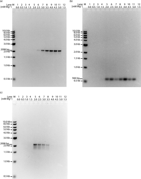
Rycina 3. Przykład miareczkowania Mg2+ w celu optymalizacji eksperymentu PCR z zastosowaniem standardowego 3-stopniowego protokołu PCR. (a) Do amplifikacji genu GAL3 o długości 2098 bp wykorzystano genomowe DNA drożdży S. cerevisiae jako matrycę. W ścieżkach 1–6, gdzie stężenie Mg2+ jest zbyt niskie, nie powstał produkt (ścieżki 1–5) lub powstało go bardzo mało (ścieżka 6). Ścieżki 7–11 reprezentują optymalne stężenia Mg2+ dla tego eksperymentu PCR, na co wskazuje obecność produktu amplikonu o długości 2098 bp. (b) Do amplifikacji amplikonu o długości 566 bp wykorzystano genomowe DNA niecharakteryzowanego bakteriofaga mykobakterii jako matrycę. W ścieżkach 1–4 stężenie Mg2+ jest zbyt niskie, o czym świadczy brak produktu. Ścieżki 5–11 reprezentują optymalne stężenia Mg2+ dla tego PCR, na co wskazuje obecność produktu amplikonu o długości 566 kb. (c) Jako matrycy do amplifikacji genu GAL3 o długości 2098 bp użyto genomowego DNA drożdży S. cerevisiae, tak jak wskazano w panelu a. Jednakże temperaturę przyłączania obniżono z 61 °C do 58 °C, co spowodowało powstanie nieswoistych prążków PCR o zmiennych długościach, tworzących efekt rozmycia (smear) na żelu agarozowym. W ścieżkach 1–4, gdzie stężenie Mg2+ jest zbyt niskie, nie powstał produkt. Ścieżki 5–8 reprezentują optymalne stężenia Mg2+ dla tego PCR, co objawia się obecnością rozmycia i prążka w okolicach wielkości produktu amplikonu 2098 kb. Ścieżki 9–11 wskazują na zbyt rygorystyczne warunki, w których nie powstał produkt. (a-c) Ścieżka 12 stanowi kontrolę negatywną, która nie zawierała DNA matrycowego. W ścieżce M (marker) naniesiono drabinę NEB 1kb Ladder.

Rycina 4. Sterylne probówki stosowane do PCR. (1.) probówka 1,8 ml (2.) pojedyncza cienkościenna probówka do PCR 0,2 ml, (3.) paskowe cienkościenne probówki do PCR 0,2 ml wraz z zakrętkami.

Rysunek 5. Termocykler. Po lewej obraz przedstawiający zamknięty termocykler. Po prawej obraz przedstawiający probówki PCR o cienkich ściankach o pojemności 0,2 ml umieszczone w bloku grzewczym otwartego termocyklera.
Dyskusja
PCR stał się niezbędnym narzędziem w arsenale nauk biologicznych. PCR zmienił bieg nauki, umożliwiając biologom kontrolę nad genomami i tworzenie genów hybrydowych o nowych funkcjach, co pozwala na specyficzne i dokładne testy kliniczne, uzyskiwanie wiedzy na temat genomów i różnorodności, a także po prostu klonowanie genów do dalszych analiz biochemicznych. Zastosowanie PCR jest ograniczone jedynie wyobraźnią naukowca, który z niego korzysta. Istnieje wiele książek i publikacji opisujących nowe, wyspecjalizowane zastosowania PCR, a w następnym pokoleniu nauk biologicznych powstanie ich znacznie więcej. Jednak niezależnie od przewidywanych podejść, podstawowe założenia pozostają niezmienne. PCR, w całym swoim znaczeniu, jest aplikacją in vitro służącą do generowania dużych ilości specyficznego fragmentu DNA.
Projektowanie eksperymentu PCR wymaga przemyślenia i cierpliwości. Wyniki przedstawione na Rysunku 3 obrazują jedno z głównych wyzwań podczas opracowywania strategii optymalizacji PCR. Chodzi o to, że zmiana jednego parametru PCR może wpłynąć na inny. Na przykład, jeśli początkowa reakcja PCR została przeprowadzona w suboptymalnej temperaturze przyłączania (58°C) przy optymalnym stężeniu Mg2+ wynoszącym 2,0 mM, wynikiem będzie rozmazany pas (smear), jak pokazano na Rysunku 3c. Próba wyeliminowania rozmycia mogłaby polegać na ustaleniu warunków PCR z reakcjami zawierającymi 2,0 mM MgCl2 i skorygowaniu temperatury przyłączania do 61°C. Jednak, jak widać na Rysunku 3a, nie doprowadziłoby to do uzyskania produktu. W związku z tym, podczas rozwiązywania problemów z błędnymi wynikami, zaleca się miareczkowanie odczynników, zamiast dodawania jednego stężenia do pojedynczej reakcji. Ponadto najczęstszymi korektami wymaganymi do optymalizacji eksperymentu PCR są zmiana stężenia Mg2+ oraz korekta temperatur przyłączania. Jeśli jednak zmiany te nie zminimalizują lub nie usuną aberracyjnych wyników, miareczkowanie dodatków i/lub zmiana protokołów warunków cykliczności, opisanych w Tabelach 2-6, może pomóc w rozwiązaniu problemu. Jeśli wszystkie powyższe metody zawiodą, należy zaprojektować primery na nowo i próbować ponownie.
Oświadczenia
Nie mam nic do ujawnienia.
Podziękowania
Szczególne podziękowania kieruję do Krisa Reddiego z UCLA za przygotowanie odczynników i wylanie żeli oraz do Erin Sanders z UCLA za inspirację, wskazówki, wsparcie i korektę rękopisu. Chciałbym również podziękować Giancarlo Costagucie i Gregory'emu S. Payne'owi za dostarczenie genomowego DNA drożdży oraz starterów do amplifikacji genu GAL3. Dziękuję również Bhairavowi Shahowi za wykonanie zdjęć sprzętu laboratoryjnego i odczynników wykorzystanych do przygotowania rysunków 2–4. Finansowanie projektu zostało zapewnione przez HHMI (grant HHMI nr 52006944).
Materiały
| Nazwa | Firma | Numer katalogowy | Komentarze |
|---|---|---|---|
| Taq polimeraza | Sigma-Aldrich | D4545-25UN | |
| dNTP | Qiagen | 201225 | |
| Probówki PCR o cienkich ściankach 0,2 ml | Bio-Rad | 223-9469 | |
| Zakrętki do probówek o cienkich ściankach 0,2 ml | Bio-Rad | 223-9472 | |
| EDTA, sól disodowa dwuwodniana | Sigma-Aldrich | E5134 | |
| Trizma-HCl | Sigma-Aldrich | T-3253 | |
| Dedykowany starter fagowy | Invitrogen | 25441F_RL6_Contig2_68k TACTGCAACGCGATGTTGCG | |
| Dedykowany starter fagowy | Invitrogen | 26007R_RL6_ Contig2_68k TCACCATCAATCGTGGCGGT | |
| Dedykowany starter drożdżowy | Integrated DNA Technologies | SacI-Gal3(-256) ACCTGGAGCTCCTATTT TAACATGTGGATTTCTTGAAAGAATGAAATCG | |
| Dedykowany starter drożdżowy | Integrated DNA Technologies | Gal3(1848)-SacI CGGATGGAGCTCGCGT GAAGGTAATTTTTTTTTATGTCTCCG | |
| Termocykler | Bio-Rad | 170-9703 | My Cycler |
| Agaroza | ISC BioExpress | E-3120-500 | |
| Elektroforeza żelowa | Hoeffer Scientific Instruments | Model HE33 | |
| Marker DNA 1 kb | New England Biolabs | N3232S | |
| Zasilacz Bio Rad | Bio-Rad | 164-5050 | PowerPac Basic |
| System Gel Doc | Fotodyne Incorporated | FOTO/Analyst Investigator FX Workstation |
Bibliografia
- Albert, J., Fenyo, E. M. Simple sensitive, and specific detection of human immunodeficiency virus type 1 in clinical specimens by polymerase chain reaction with nested primers. J. Clin. Microbiol. 28, 1560-1564 (1990).
- Baskaran, N., et al. Uniform amplification of a mixture of deoxyribonucleic acids with varying GC content. Genome Res. 6, 633-638 (1996).
- Blanchard, M. M., Taillon-Miller, P., Nowotny, P., Nowotny, V. PCR buffer optimization with uniform temperature regimen to facilitate automation. PCR Methods Appl. 2, 234-240 (1993).
- Borer, P. N., Dengler, B., Tinoco, I. Jr, Uhlenbeck, O. C. Stability of ribonucleic acid double-stranded helices. J. Mol. Biol. 86, 843-853 (1974).
- Breslauer, K. J., Frank, R., Blocker, H., Marky, L. A. Predicting DNA duplex stability from the base sequence. Proc. Natl. Acad. Sci. U.S.A. 83, 3746-3750 (1986).
- Chou, Q., Russell, M., Birch, D. E., Raymond, J., Bloch, W. Prevention of pre-PCR mis-priming and primer dimerization improves low-copy-number amplifications. Nucleic Acids Res. 20, 1717-1723 (1992).
- Coleman, W. B., Tsongalis, G. J. Diagnostics for the clinical laboration. , Humana Press. (2005).
- D'Aquila, R. T., et al. Maximizing sensitivity and specificity of PCR by pre-amplification heating. Nucleic Acids Res. 19, 3749(1991).
- Dieffenbach, C. W., Lowe, T. M., Dveksler, G. S. General concepts for PCR primer design. PCR Methods Appl. 3, S30-S37 (1993).
- Don, R. H., Cox, P. T., Wainwright, B. J., Baker, K., Mattick, J. S. 'Touchdown' PCR to circumvent spurious priming during gene amplification. Nucleic Acids Res. 19, 4008(1991).
- Erlich, H. A., Gelfand, D., Sninsky, J. J. Recent advances in the polymerase chain reaction. Science. 252, 1643-1651 (1991).
- Frey, U. H., Bachmann, H. S., Peters, J., Siffert, W. PCR-amplification of GC-rich regions: 'slowdown PCR. Nat. Protoc. 3, 1312-1317 (2008).
- Haqqi, T. M., Sarkar, G., David, C. S., Sommer, S. S. Specific amplification with PCR of a refractory segment of genomic DNA. Nucleic Acids Res. 16, 11844(1988).
- Henke, W., Herdel, K., Jung, K., Schnorr, D., Loening, S. A. Betaine improves the PCR amplification of GC-rich DNA sequences. Nucleic Acids Res. 25, 3957-3958 (1997).
- Innis, M. A., Gelfand, D. H., Sninsky, J. J., White, T. J. PCR Protocols: A guide to methods and applications. , Academic Press, Inc. San Diego. (1990).
- King, N. Methods in Molecular Biology. 630, Humana Press. (2010).
- Kleppe, K., Ohtsuka, E., Kleppe, R., Molineux, I., Khorana, H. G. Studies on polynucleotides. XCVI. Repair replications of short synthetic DNA's as catalyzed by DNA polymerases. J. Mol. Biol. 56, 341-361 (1971).
- Korbie, D. J., Mattick, J. S. Touchdown PCR for increased specificity and sensitivity in PCR amplification. Nat. Protoc. 3, 1452-1456 (2008).
- Kramer, M. F., Coen, D. M. Enzymatic amplification of DNA by PCR: standard procedures and optimization. Curr. Protoc. Toxicol. Appendix 3, 1-14 (2001).
- Kramer, M. F., Coen, D. M. The polymerase chain reaction. Curr. Protoc. Protein Sci. Appendix 4, Appendix 4J(2002).
- Kreader, C. A. Relief of amplification inhibition in PCR with bovine serum albumin or T4 gene 32 protein. Appl. Environ. Microbiol. 62, 1102-1106 (1996).
- Kwok, S., et al. Identification of human immunodeficiency virus sequences by using in vitro enzymatic amplification and oligomer cleavage detection. J. Virol. 61, 1690-1694 (1987).
- McConlogue, L., Brow, M. A., Innis, M. A. Structure-independent DNA amplification by PCR using 7-deaza-2'-deoxyguanosine. Nucleic Acids Res. 16, 9869(1988).
- Mullis, K. B. The unusual origin of the polymerase chain reaction. Sci. Am. 262, 56-65 (1990).
- Mullis, K. B. Target amplification for DNA analysis by the polymerase chain reaction. Ann. Biol. Clin. (Paris). 48, 579-582 (1990).
- Mullis, K. B. The polymerase chain reaction in an anemic mode: how to avoid cold oligodeoxyribonuclear fusion). PCR Methods Appl. 1, 1-4 (1991).
- Mullis, K. B., Faloona, F. A. Specific synthesis of DNA in vitro via a polymerase-catalyzed chain reaction. Methods Enzymol. 155, 335-350 (1987).
- Paabo, S., Gifford, J. A., Wilson, A. C. Mitochondrial DNA sequences from a 7000-year old brain. Nucleic Acids Res. 16, 9775-9787 (1988).
- Rees, W. A., Yager, T. D., Korte, J., von Hippel, P. H. Betaine can eliminate the base pair composition dependence of DNA melting. Biochemistry. 32, 137-144 (1993).
- Roux, K. H., Hecker, K. H. One-step optimization using touchdown and stepdown PCR. Methods Mol. Biol. 67, 39-45 (1997).
- Rychlik, W., Spencer, W. J., Rhoads, R. E. Optimization of the annealing temperature for DNA amplification in vitro. Nucleic Acids Res. 18, 6409-6412 (1990).
- Saiki, R. K., et al. Primer-directed enzymatic amplification of DNA with a thermostable DNA polymerase. Science. 239, 487-491 (1988).
- Saiki, R. K., et al. Enzymatic amplification of beta-globin genomic sequences and restriction site analysis for diagnosis of sickle cell anemia. Science. 230, 1350-1354 (1985).
- Sambrook, J. A. R., David, W. Molecular Cloning A Laboratory Manual. 2, 3, Cold Spring Harbor Laboratory Press. (2001).
- Sarkar, G., Kapelner, S., Sommer, S. S. Formamide can dramatically improve the specificity of PCR. Nucleic Acids Res. 18, 7465(1990).
- Smit, V. T., et al. KRAS codon 12 mutations occur very frequently in pancreatic adenocarcinomas. Nucleic Acids Res. 16, 7773-7782 (1988).
- Wilson, I. G. Inhibition and facilitation of nucleic acid amplification. Appl. Environ. Microbiol. 63, 3741-3751 (1997).
- Wu, R. Methods in Enzymology. 218, Academic Press, Inc. San Diego. (1993).
Przedruki i uprawnienia
