Opisujemy metody izolowywania komórek somatycznych jajnika mysiego z frakcji stromy wzbogaconej jajnika oraz generowania organoidów somatycznych jajnika mysiego przy użyciu podejścia bez scaffoldu.
Subskrypcja JoVE jest wymagana do oglądania tego materiału. Zaloguj się lub rozpocznij bezpłatny okres próbny.
Artykuł metodologiczny
Badanie mikrośrodowiska jajnika: metody izolacji, hodowli 2D i hodowli organoidalnej komórek somatycznych jajnika mysiego
W tym artykule
Podsumowanie
Streszczenie
Jajnik składa się z heterogenicznych populacji komórek somatycznych, zarówno w folikulu, jak i w otaczającej nim stromie, które są kluczowe dla wsparcia funkcji jajnika i wytwarzania wysokiej jakości gamet. Przedstawiamy metody izolacji komórek somatycznych z mysiego jajnika, w tym komórek endotelialnych, nabłonkowych, steroidogennych, stromalnych i odpornościowych. Kiedy te pierwotne komórki somatyczne jajnika są sadzone i hodowane w tradycyjnym układzie kultury 2D, heterogeniczność komórkowa, organizacja oraz interakcje komórka-komórka i komórka-matryca, zwykle występujące w jajniku, są utracone. Dlatego opisujemy również, jak generować mysie organoidy somatyczne jajnika za pomocą podejścia bez szkieletu. Te organoidy samoodpławiają się, utrzymują różnorodne populacje komórek i wytwarzają matrycę pozakomórkową oraz czynniki wydzielane, w tym cytokiny. Organoidy mogą być wykorzystywane do eksperymentów współhodowlanych i mogą być utrzymywane w kulturze przez co najmniej 3 tygodnie z wysoką żywotnością. Ogólnie rzecz biorąc, modele te umożliwiają badanie fizjologii i patologii jajnika z perspektywy komórek somatycznych.
Wprowadzenie
Chociaż oocyt, czyli komórka płciowa żeńska, jest niezbędny do powstania kolejnego pokolenia, komórki somatyczne w jajniku odgrywają kluczowe role w rozwoju gamet, produkcji hormonów steroidowych i homeostazie jajnika. Oocyty, otoczone komórkami somatycznymi granulozy i teca, tworzą funkcjonalne jednostki jajnika znane jako mieszki. Komórki granulozy są fizycznie połączone z oocytem poprzez projekcje transzonalne i dostarczają oocytowi wsparcia metabolicznego i składników odżywczych, oprócz regulacji zatrzymania mejozy1,2. Ponadto, w odpowiedzi na gonadotropiny, komórki granulozy i teca produkują hormony steroidowe, w tym estrogen, progesteron i androgeny, które mają wpływ poza układem rozrodczym, wpływając na układ krążenia, mięśniowo-szkieletowy i nerwowy, a tym samym wpływając na ogólne zdrowie kobiet3. Mieszki jajnikowe rozwijają się w obrębie niejednorodnego stromy jajnika, złożonej z naczyń krwionośnych, komórek odpornościowych, kilku populacji fibroblastów interstycjalnych i komórek mezenchymalnych, a także wysoce uporządkowanej macierzy zewnątrzkomórkowej (ECM)4. To środowisko stromalne zapewnia fizyczne i chemiczne sygnały, które wpływają na wzrost mieszków, owulację i luteinizację4.
Istnieje kilka metod izolowywania i hodowania gamet i mieszków in vitro, a metody te poszerzyły naszą wiedzę na temat ooogenezy i folikulogeneza5. Jednak kompartyment somatyczny jajnika jest stosunkowo słabo zbadany, częściowo z powodu braku solidnych modeli in vitro. Obecne modele kompartymentu somatycznego jajnika są w dużej mierze ograniczone do pojedynczego typu komórek, takich jak fibroblasty, komórki nabłonkowe powierzchni jajnika, komórki śródbłonka, odpornościowe lub granulozy6,7,8,9,10,11. Hodowla szeroko definiowanych pierwotnych komórek somatycznych jajnika w dwuwymiarowej (2D) hodowli sprzyja populacjom makrofagów z czasem, co prowadzi do utraty różnorodności komórkowej12. Ponadto, tradycyjna dwuwymiarowa (2D) kultura monowarstwowa nie pozwala na zróżnicowane interakcje komórka-komórka i komórka-macierz, które są obecne w jajniku. Eksplanty tkanki jajnikowej zachowują złożoność komórkową i strukturalną natywnej tkanki, ale różnią się składem w zależności od tego, skąd w jajniku zostały pobrane13,14.
Organoidy to trójwymiarowe (3D), wielokomórkowe, miniaturyzowane wersje narządów lub tkanek, które naśladują organizację i funkcje in vivo15,16. Organoidy mogą być generowane z komórek macierzystych lub komórek pierwotnych izolowanych z tkanki normalnej lub chorej15. W kontekście żeńskiego układu rozrodczego, organoidy zostały wygenerowane do modelowania endometrium, jajowodu, szyjki macicy i łożyska17. Jednakże, jeśli chodzi o jajnik, aż do niedawna, modele organoidalne były ograniczone do pojedynczego typu komórek, takich jak nabłonek powierzchni jajnika lub komórki nowotworowe, lub zostały wygenerowane z indukowanych pluripotencjalnych komórek macierzystych, aby przypominać struktury mieszków, a nie stromę jajnika17,18. Ostatnie badania wygenerowały i scharakteryzowały organoidy za pomocą niejednorodnych populacji komórek somatycznych izolowanych z tkanki jajnikowej myszy, makaki rezonansowej i ludzkiej19,20,21. Te modele organoidalne umożliwiły badanie mechanizmów komórkowych leżących u podstaw starzenia się jajników, a także wpływu narażenia na ftalany na skład ECM19,21.
Biorąc pod uwagę krytyczne znaczenie komórek somatycznych, zarówno w mieszku, jak i stromie, dla funkcji jajnika, opisujemy metody izolowania komórek somatycznych z frakcji wzbogaconej w stromę mysich jajników i ich hodowania in vitro przy użyciu tradycyjnej kultury monowarstwowej lub w modelu organoidalnym. Za pomocą bezszachownicowych mikromoldów agarozowych, komórki somatyczne jajnika tworzą organoidy w ciągu 1-3 dni. Organoidy zachowują kluczowe populacje komórek jajnikowych, w tym fibroblasty, makrofagi i komórki steroidogenne, i mogą być kultywowane przez okres do 3 tygodni z wysoką żywotnością. Organoidy somatyczne jajników sprzyjają ocenie transkryptomu i histologicznej, analizie pożywienia uwarunkowanego, współhodowli z innymi typami komórek i metodom przesiewowym związków. Ogólnie rzecz biorąc, te organoidy są solidnym modelem do in vitro badania i modyfikacji kompartymentu somatycznego jajnika i mogą być zastosowane do badań nad rozwojem jajnik
Dostęp ograniczony. Zaloguj się lub rozpocznij wersję próbną, aby wyświetlić tę treść.
Protokół
Wykorzystanie zwierząt do opracowania niniejszego protokołu odbyło się zgodnie z wytycznymi Instytucjonalnego Komitetu ds. Opieki i Wykorzystania Zwierząt na Uniwersytecie Northwestern oraz Wytycznymi Narodowych Instytutów Zdrowia (NIH) dotyczącymi opieki i wykorzystania zwierząt laboratoryjnych (numer zapewnienia dobrostanu zwierząt Uniwersytetu Northwestern: A3283-01; ID protokołu IACUC: IS00013082_IM12). Informacje o producentach i numery katalogowe wszystkich materiałów, w tym silikonowych form do mikrokopalni agarozowych, pożywek i suplementów, podano w Tabeli Materiałów. Przegląd poszczególnych kroków protokołu przedstawiono na Rysunku 1. Wszystkie etapy protokołu należy przeprowadzać przy użyciu odpowiednich środków ochrony osobistej, zgodnie ze wszystkimi instytucjonalnymi przepisami dotyczącymi bezpieczeństwa biologicznego oraz ochrony zdrowia i środowiska, a jeśli nie określono inaczej, w komorze bezpiecznej klasy II.
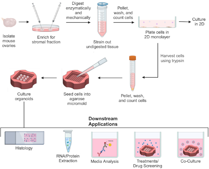
Rysunek 1: Schemat izolacji somatycznych komórek jajnika myszy, generowania organoidów somatycznych jajnika oraz zastosowań końcowych. Jajniki myszy są izolowane, a frakcja zrębu zostaje wzbogacona. Wzbogacona frakcja zrębu jest trawiona enzymatycznie i mechanicznie. Niestrawione tkanki są odfiltrowane. Komórki są wirowane w celu uzyskania osadu, przemywane, przeliczane i wysiewane do hodowli 2D. W celu generowania organoidów, po nocnej hodowli 2D, komórki są zbierane przy użyciu trypsyny, a następnie wirowane, przemywane i przeliczane. Komórki są wysiewane do mikroform mikroagarozowych do hodowli organoidów. Organoidy mogą być wykorzystywane do badań histologicznych, ekstrakcji RNA/białek, analizy pożywki warunkowej, przeprowadzania zabiegów, screeningu leków, a także do kokultur. Kliknij tutaj, aby wyświetlić powiększoną wersję tego rysunku.
1. Przygotowanie pożywek do izolacji i hodowli somatycznych komórek jajnika
UWAGA: Trawienie tkanki jajnika oraz późniejsza izolacja i hodowla pierwotnych komórek somatycznych wymaga czterech rodzajów pożywek: pożywki do sekcji (DM; stosowanej w krokach 2.3–2.4 do sekcji i nakłuwania jajników myszy), pożywki enzymatycznej (EM; stosowanej w krokach 2.5–2.8 do enzymatycznego trawienia jajników myszy wzbogaconych w zrąb), pożywki hamującej (QM; stosowanej w kroku 2.9 w celu spowolnienia trawienia enzymatycznego) oraz pożywki do wysiewu (PM; stosowanej w krokach 2.11–2.14 i kroku 4.6 do płukania, wysiewu i hodowli 2D pierwotnych somatycznych komórek jajnika, a także do inaktywacji trypsyny podczas zbierania komórek z hodowli 2D). Wszystkie pożywki należy przygotować w sterylizowanej komorze bezpieczeństwa biologicznego klasy II lub w dygestorium z przepływem laminarnym. Przepisy na pożywki wymienione poniżej są wystarczające do przetworzenia jajników od 5–6 myszy, lecz należy je przeskalować w zależności od potrzeb. Wszystkie pożywki można przygotować z wyprzedzeniem do tygodnia i przechowywać w 4 °C, jednak enzymy do EM muszą zostać dodane na świeżo w dniu izolacji komórek.
- Przygotować DM (15 mL) zawierające 1% płodowej surowicy bydlęcej (FBS) i 0,5% penicyliny-streptomycyny (PS) w podłożu Leibovitz's L-15. Podłoże należy wysterylizować za pomocą filtra sterylnego.
UWAGA: DM jest buforowane do wymiany z powietrzem otoczenia i wykorzystywane podczas mikrodissekcji jajnika z tkanek otaczających, a także do wzbogacania frakcji zrębowej. - Przygotować EM (10 mL) zawierające 1% FBS i 0,5% PS w αMEM. Dodać 82 jednostki enzymatyczne/mL kolagenazy IV oraz 0,2 mg/mL DNazy I (≥400 jednostek aktywności enzymatycznej/mg). Enzymy należy rozpuścić poprzez odwracanie probówki, a następnie wysterylizować podłoże za pomocą filtra sterylnego.
UWAGA: Podłoże enzymatyczne jest buforowane wodorowęglanem sodu do wymiany gazowej przy 5% CO2 i wykorzystywane do trawienia enzymatycznego fragmentów tkanki jajnika. - Przygotować QM (20 mL) zawierające 10% FBS i 0,5% PS w podłożu αMEM. Podłoże należy wysterylizować za pomocą filtra sterylnego.
UWAGA: QM jest buforowane wodorowęglanem sodu do wymiany gazowej przy 5% CO2 i wykorzystywane do przerwania procesu trawienia enzymatycznego. - Przygotować PM (30 mL) zawierające 10% FBS i 1% PS w RPMI 1640 (zawierającym 25 mM HEPES i 2 mM L-glutaminy). Podłoże należy wysterylizować za pomocą filtra sterylnego.
UWAGA: PM jest buforowane wodorowęglanem sodu do wymiany gazowej przy 5% CO2 i wykorzystywane do wysiewu pierwotnych somatycznych komórek jajnika, a także do hodowli w 2D.
2. Trawienie tkanki jajnika i izolacja pierwotnych komórek somatycznych
UWAGA: Jajniki myszy są izolowane i trawione zarówno enzymatycznie, jak i mechanicznie w celu wyizolowania pierwotnych komórek somatycznych. Jajniki od 5–6 myszy można przetwarzać razem w jednej szalce. Następujące kroki należy przeprowadzić z zachowaniem zasad aseptyki, najlepiej przy użyciu mikroskopu stereoskopowego w komorze z laminarnym przepływem powietrza.
- Do oddzielnych szalek Petri o średnicy 100 mm dodać 10 mL EM, 20 mL QM oraz 20 mL PM. Przed użyciem inkubować szalki w inkubatorze do hodowli tkankowych (środowisko wilgotne, 37 °C i 5% CO2) przez minimum 1 h w celu wyrównania temperatury.
- Przed użyciem ogrzać DM w kąpieli wodnej (37 °C).
- Przeprowadzić sekcję jajników myszy w szalce Petri o średnicy 35 mm zawierającej 3 mL DM. Za pomocą pęsety z cienkim końcem i/lub mikronozyciór oddzielić jajniki od przylegającej tkanki tłuszczowej, jajowodu i kaloty jajnika (Rysunek 2Ai,ii). W razie potrzeby do mikrosekcji użyć mikroskopu stereoskopowego.
- Przenieść wypreparowane jajniki na zegarek o średnicy 45 mm z 2–3 mL DM. Pod mikroskopem stereoskopowym, używając kolejno niedominującej i dominującej ręki, unieruchomić i nakłuć wszystkie obszary jajników igłami insulinowymi 28-G przez około 10 min. Często obracać tkankę jajnika, aby zapewnić przerwanie ciągłości wszystkich obszarów.
UWAGA: Nakłuwanie jajników wzbogaca frakcję stromy poprzez uwalnianie pęcherzyków przedantralnych, a także kompleksów cumulus-oocyta i komórek granulozy z pęcherzyków antralnych (Rysunek 2Aiii–iv). - Używając pęsety z cienkim końcem, przenieść wzbogacone w stromę jajniki myszy do szalki Petri z wyrównanym EM (Rysunek 2Bi).
- Pracując szybko, aby zminimalizować zmiany temperatury i pH mediów, podrzeć wzbogacone w stromę jajniki na kawałki o wielkości około 2–3 mm za pomocą pęsety z cienkim końcem (Rysunek 2Bii).
- Trzymając szalkę Petri pod kątem, wielokrotnie pipetować EM zawierający fragmenty tkanki jajnika za pomocą pipety p1000 ustawionej na 1 mL, 100× razy lub przez 3 min, zależnie od tego, co nastąpi wcześniej (Rysunek 2Biii). W razie potrzeby obracać szalkę Petri, aby upewnić się, że wszystkie fragmenty tkanki zostały poddane pipetowaniu.
UWAGA: Jeśli tkanka jest zbyt duża, aby można ją było pobrać końcówką p1000, końcówkę można przyciąć sterylnym skalpelem, aby poszerzyć otwór. Otwór powinien być wystarczająco szeroki, aby większość (>50%), ale nie wszystkie fragmenty tkanki mogły do niego wejść, co wytworzy efekt ścinania. - Przenieść szalkę Petri zawierającą EM i fragmenty tkanki jajnika do inkubatora do hodowli tkankowych (środowisko wilgotne, 37 °C i 5% CO2) i inkubować łącznie przez 30 min; wyjąć szalkę i wielokrotnie pipetować (100× razy lub przez 3 min) po 15 min, a następnie ponownie po zakończeniu 30-minutowej inkubacji (Rysunek 2Biii).
UWAGA: Po pierwszej 15-minutowej inkubacji fragmenty tkanki powinny być na tyle strawione, aby >80% z nich przechodziło przez końcówkę pipety p1000 bez konieczności jej przycinania. Całkowity czas trawienia i wymagana liczba cykli pipetowania będą zależeć od początkowej wielkości podrzerenych fragmentów tkanki. Ponadto na tempo rozpadu tkanki mogą wpływać wiek i szczep myszy, zmienność między partiami enzymów oraz średnica otworu pipety. Trawienie należy przerwać, gdy fragmenty tkanki jajnika zostaną rozbite na fragmenty o wielkości około 1 mm, które są wciąż widoczne gołym okiem (Rysunek 2Biv). Nadmierne trawienie może prowadzić do utraty komórek i zmniejszenia ich żywotności. - Rozcieńczyć enzymy, aby spowolnić trawienie tkanki, dodając 20 mL uprzednio wyrównanego QM (Rysunek 2Ci).
- Przepuścić zawiesinę komórkową przez sitko komórkowe 40 µm umieszczone nad probówką stożkową 50 mL, aby usunąć niestrawione fragmenty tkanki (Rysunek 2Cii). Odwirować filtrat przy 300 × g przez 5 min w temperaturze pokojowej, aby uzyskać osad komórkowy (Rysunek 2Ciii).
- Przemyć komórki poprzez usunięcie supernatantu aspiracją, resuspensję osadu komórkowego w 1 mL uprzednio wyrównanego PM, przeniesienie zawiesiny komórkowej do probówki do mikrocentryfugi 1,5 mL i ponowne odwirowanie przy 300 × g przez 5 min w temperaturze pokojowej (Rysunek 2Civ).
- Usunąć supernatant, resuspensować osad komórkowy w 1 mL PM, zabarwić alikwot 10 µL komórek błękitem trypanu w celu oceny żywotności i policzyć komórki za pomocą hemocytometru.
UWAGA: Przewidywaną liczbę pierwotnych somatycznych komórek jajnika, w zależności od szczepu i wieku myszy, na tym etapie protokołu izolacji przedstawiono w Tabeli 1. Żywotność komórek na tym etapie powinna wynosić >80%. - Wysiać komórki na naczyniu traktowanym do hodowli tkankowej przy gęstości około 1,25 × 10⁵-1,5 × 10⁵/ cm2 (np. dla liczby komórek około 3 × 10⁶ wysiać komórki do szalki 60 mm traktowanej do hodowli tkankowej, a dla liczby około 1 × 10⁶ wysiać komórki do jednego dołka płytki 6-dołkowej traktowanej do hodowli tkankowej).
- Dodać dodatkowy PM w zależności od całkowitej objętości płytki lub szalki (np. dodać dodatkowe 4 mL PM w przypadku użycia szalki 60 mm lub dodatkowe 2 mL PM w przypadku użycia dołka płytki 6-dołkowej).
UWAGA: Powyższa gęstość wysiewu jest zalecana, jeśli komórki mają być wykorzystane do generowania organoidów po nocnej hodowli 2D. W przypadku dłuższej hodowli 2D komórki można wysiać przy niższej gęstości. Nadmiar medium do wysiewu, który nie został wyrównany, można przechowywać w temperaturze 4 °C do 1 tygodnia. PM jest wymagany zarówno do generowania organoidów, jak i do utrzymania somatycznych komórek jajnika w hodowli 2D. Na tym etapie protokołu somatyczne komórki jajnika można utrzymywać w hodowli monolayerowej 2D (krok 2.14) lub wykorzystać do generowania organoidów (przejść do Sekcji 3). Nocna hodowla 2D przed generowaniem organoidów jest niezbędna do usunięcia oocytów z pęcherzyków we wczesnych stadiach, które mogły przejść przez sitko komórkowe 40 µm.
- Dodać dodatkowy PM w zależności od całkowitej objętości płytki lub szalki (np. dodać dodatkowe 4 mL PM w przypadku użycia szalki 60 mm lub dodatkowe 2 mL PM w przypadku użycia dołka płytki 6-dołkowej).
- Po nocnym wysiewie (minimum 16 h), przemyć komórki solą fizjologiczną buforowaną fosforanem (PBS; bez Mg/Ca, pH 7,0–7,3), aby usunąć martwe komórki i zanieczyszczenia (Rysunek 3A). Po przemyciu dodać świeży PM i wymieniać medium na świeży PM co drugi dzień w trakcie hodowli (Rysunek 3B).
UWAGA: Mimo wysokiej żywotności komórek po izolacji, po nocnym wysiewie zazwyczaj obserwuje się znaczny spadek całkowitej liczby komórek z powodu śmierci komórek lub braku możliwości przytwierdzenia do naczynia do hodowli tkankowej (Rysunek 3A).
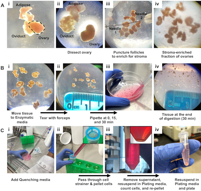
Rycina 2: Schemat izolacji somatycznych komórek jajnika myszy. (A) Reprezentatywne zdjęcia (i, ii) sekcji jajnika myszy oraz (iii, iv) wzbogacania zrębu. Jajnik myszy zaznaczono linią przerywaną w Ai. (B) Reprezentatywne zdjęcia fragmentów tkanki jajnika podczas trawienia mechanicznego i enzymatycznego. Jajniki wzbogacone w zrąb (i) przed i (ii) po rozrywaniu, (iii) fragmenty tkanki jajnika podczas pipetowania oraz (iv) fragmenty tkanki jajnika po trawieniu. (C) Reprezentatywne zdjęcia (i) hamowania trawienia, (ii) filtrowania zawiesiny komórkowej oraz (iii, iv) wirowania somatycznych komórek jajnika myszy w celu uzyskania osadu. Wstawki w Cii i Ciii przedstawiają odpowiednio nietrawione fragmenty tkanki zatrzymane przez sitko komórkowe oraz osad komórkowy po wirowaniu. Kliknij tutaj, aby wyświetlić powiększoną wersję tej ryciny.
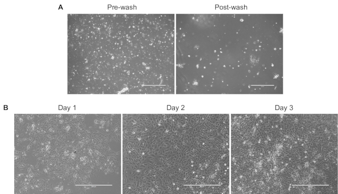
Rysunek 3: Pierwotne somatyczne komórki jajnika mogą być hodowane w 2D. (A) Reprezentatywne obrazy w świetle przechodzącym pierwotnych somatycznych komórek jajnika wysianych w naczyniu 60 mm, 16 h po wysianiu, przed i po płukaniu PBS. (B) Reprezentatywne obrazy w świetle przechodzącym pierwotnych somatycznych komórek jajnika wysianych w płytce 24-dołkowej w ciągu 3 dni hodowli. Paski skali = 400 µm. Kliknij tutaj, aby wyświetlić powiększoną wersję tego rysunku.
3. Przygotowanie mikroform agarose oraz pożywek do generowania i hodowli organoidów
UWAGA: Somatyczne organoidy jajnika są generowane i hodowane w bezrusztowianych mikrowzorcach agarozowych z wykorzystaniem pożywki do organoidów jajnika. Mikrowzorce agarozowe oraz pożywka do organoidów jajnika powinny być przygotowane z zastosowaniem technik aseptycznych w sterylizowanej komorze bezpieczeństwa biologicznego klasy II lub pod nawiewem laminarnym.
- Przygotowanie mikroform z agarozy
- Umieść silikonowe formy do mikrowzorów oraz pęsetę Graefe w torebce do sterylizacji i wysterylizuj w autoklawie w cyklu suchym (Ciśnienie płaszcza: 20 psi, Temperatura komory: 250 °F (121,11 °C), Czas sterylizacji: 15 min).
UWAGA: Kilka form silikonowych można autoklawować i używać jednocześnie, aby szybciej wytwarzać mikroformy z agarozy. - Do każdego dołka 24-dołkowej płytki do hodowli tkanek dodaj 1 mL PBS zawierającego 1% PS w celu przechowywania mikroform z agarozy.
- Rozpuść 1,5% (w/v) sterylną agarozę w PBS, podgrzewając w mikrofalówce, w kąpieli wodnej lub na płycie grzejnej.
UWAGA: Rozpuszczanie agarozy w mikrofalówce w probówce stożkowej o pojemności 50 mL umieszczonej w zlewce 100 mL wypełnionej wodą działa bardzo dobrze (Rycina 4Ai). Oprócz umożliwienia kontrolowanego wrzenia, kąpiel wodna utworzona w zlewce spowalnia ponowne krzepnięcie agarozy. - Pozostaw ciekłą agarozę do ostygnięcia do około 50 °C, aż probówką będzie można wygodnie operować.
- Powoli pipetuj 300–350 µL 1,5% agarozy do formy silikonowej, dbając o to, aby nie przepełnić ani nie niedopełnić formy oraz unikając tworzenia pęcherzyków powietrza (Rycina 4Aii). Upewnij się, że agaroza tworzy płaską warstwę na górze formy, a nie krawędź wklęsłą lub wypukłą (Rycina 4Aiii,iv). Usuwaj pęcherzyki powietrza za pomocą sterylnej końcówki pipety p10, zanim agaroza zacznie krzepnąć (Rycina 4Bi–iii).
- Pozostaw agarozę do stężenia w formie silikonowej przez 2–3 min w temperaturze pokojowej (Rycina 4Biv).
- Po zestaleniu odwróć formę silikonową nad dołkiem 24-dołkowej płytki do hodowli tkanek zawierającej PBS + 1% PS. Ostrożnie wygnij silikon, aby uwolnić mikroformę z agarozy do płytki (Rycina 4Ci).
UWAGA: Mikroformy z agarozy zanurzone w PBS + 1% PS w 24-dołkowej płytce do hodowli tkanek owiniętej parafilmem można przechowywać w 4 °C do 1 miesiąca przed użyciem (Rycina 4Cii). Mikroformy z agarozy należy przygotować co najmniej 16 h przed użyciem, ponieważ mikrodółki często zawierają pęcherzyki powietrza bezpośrednio po odformowaniu i zanurzeniu w PBS + 1% PS (Rycina 4Ciii). Jednakże pęcherzyki te znikają podczas przechowywania (Rycina 4Civ). Przed użyciem do hodowli organoidów wkładki z mikrodółkami z agarozy należy dokładnie zbadać pod mikroskopem, aby potwierdzić, że mikrodółki nie zlały się ani nie zostały uszkodzone podczas odlewania (Rycina 4Di–iv). Wadliwe mikroformy nie są zalecane do generowania i hodowli organoidów. Zaleca się przygotowanie nadmiarowej liczby mikroform z agarozy, aby uwzględnić ok. 25% wadliwych egzemplarzy.
- Umieść silikonowe formy do mikrowzorów oraz pęsetę Graefe w torebce do sterylizacji i wysterylizuj w autoklawie w cyklu suchym (Ciśnienie płaszcza: 20 psi, Temperatura komory: 250 °F (121,11 °C), Czas sterylizacji: 15 min).
- Przygotowanie pożywki dla organoidów jajnika
- W komorze z laminarnym przepływem powietrza dodaj suplementy 1 i 2 do pożywki wzrostowej dla organoidów (mysz) oraz 1% PS do pożywki podstawowej zgodnie z instrukcjami producenta. Przygotowaną pożywkę dla organoidów jajnika przechowuj w 4 °C do 2 tygodni przed użyciem.
UWAGA: Suplementy 1 i 2 można rozdzielić na alikwoty i przechowywać w -20 °C.
- W komorze z laminarnym przepływem powietrza dodaj suplementy 1 i 2 do pożywki wzrostowej dla organoidów (mysz) oraz 1% PS do pożywki podstawowej zgodnie z instrukcjami producenta. Przygotowaną pożywkę dla organoidów jajnika przechowuj w 4 °C do 2 tygodni przed użyciem.
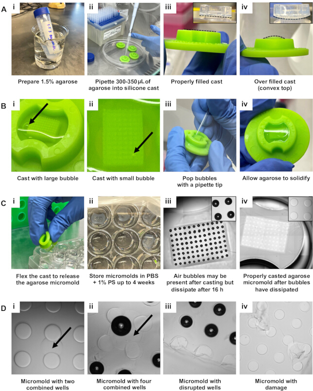
Rycina 4: Schemat przedstawiający przygotowanie mikrootworów agarozowych. (A) Reprezentatywne zdjęcia (i) preparatu 1,5% agarozy, (ii) pipetowania do form silikonowych, (iii) prawidłowo wypełnionych oraz (iv) przepełnionych form silikonowych. Linie przerywane w Aiii i Aiv pokazują odpowiednio płaskie i wypukłe dna mikrootworów agarozowych. (B) Reprezentatywne zdjęcia (i,ii) wypełnionych form silikonowych z pęcherzykami powietrza w agarozie, (iii) usuwania pęcherzyków końcówką pipety przed zestaleniem agarozy oraz (iv) zestalonego mikrootworu agarozowego w formie silikonowej. (C) Reprezentatywne zdjęcia (i) usuwania zestalonych mikrootworów agarozowych z form silikonowych, (ii) przechowywania mikrootworów agarozowych w płytce 24-dołkowej zawierającej PBS + 1% PS, (iii) pęcherzyków obecnych w mikrootworach bezpośrednio po odlewaniu oraz (iv) wolnego od pęcherzyków, prawidłowo odlanego mikrootworu agarozowego po 16 h przechowywania. (D) Reprezentatywne zdjęcia mikrootworów z (i,ii) połączonymi dołkami, (iii) uszkodzonymi dołkami lub (iv) defektami. Skróty: PBS = roztwór soli fizjologicznej buforowany fosforanem; PS = penicylina-streptomycyna. Kliknij tutaj, aby wyświetlić powiększoną wersję tej ryciny.
4. Generowanie i hodowla organoidów
UWAGA: Po całonocnej hodowli 2D pierwotne somatyczne komórki jajnika są zbierane z hodowli 2D i wysiewane do mikiforem Agarozy w celu wytworzenia organoidów. Następujące kroki należy przeprowadzić z zachowaniem zasad aseptyki w sterylnej komorze bezpieczeństwa biologicznego klasy II.
- Podgrzać PM oraz pożywkę do organoidów do 37 °C w kąpieli wodnej, a 0,05% trypsynę doprowadzić do temperatury pokojowej.
- Używając pęsety Graefe, ostrożnie przenieść mikiformy do dołków nowej płytki 24-dołkowej (Rycyna 5Ai).
- Do każdego dołka płytki 24-dołkowej zawierającego agarową mikiformę dodać 500 µL pożywki do organoidów jajnika (Rycyna 5Aii). Upewnić się, że mikiformy są całkowicie zanurzone w pożywce, a następnie inkubować mikiformy i pożywkę przez co najmniej 30 min w inkubatorze (środowisko wilgotne, 37 °C, 5% CO2).
- Usunąć PM z adherentnych somatycznych komórek jajnika poprzez aspirację, a następnie przemyć komórki buforem PBS pozbawionym Mg/Ca.
- Dodać 0,05% trypsynę (1–2 mL trypsyny dla płytki 6-dołkowej lub szalki 60 mm), aby zebrać komórki z hodowli 2D. Inkubować komórki z trypsyną przez 2–3 min w temperaturze 37 °C w inkubatorze.
- Potwierdzić pod mikroskopem, że komórki oddzieliły się od podłoża, a następnie inaktywować trypsynę, dodając dwie objętości podgrzanego PM na każdą jedną objętość użytej trypsyny.
- Przenieść zawiesinę komórkową do probówki stożkowej 15 mL i odwirować komórki przy 300 × g przez 5 min w temperaturze pokojowej.
- Resuspendować komórki w 1 mL podgrzanego PM, zabarwić alikwot komórek błękitem trypanowym w celu oceny żywotności i policzyć komórki za pomocą hemocytometru.
UWAGA: Przewidywana liczba pierwotnych somatycznych komórek jajnika, w zależności od szczepu i wieku myszy, po całonocnej hodowli 2D i następującej po niej trypsynizacji, znajduje się w Tabeli 1. Żywotność komórek po trypsynizacji zazwyczaj przekracza 90%. - Odwirować komórki (300 × g przez 5 min w temperaturze pokojowej) i resuspendować osad komórkowy w pożywce do organoidów jajnika do gęstości 3,33 × 10⁶ żywych komórek na 1 mL pożywki, tj. 2,5 × 10⁵ żywych komórek na 75 µL pożywki.
UWAGA: W tym przypadku na jedną agarową mikiformę wysiewa się 2,5 × 10⁵ komórek. Zatem całkowita liczba mikiform, które można przygotować, zależy od całkowitej liczby komórek. Organoidy można wytworzyć przy użyciu mniejszej liczby niż 2,5 × 10⁵ komórek na agarową mikiformę, jednak może to wpłynąć na rozmiar organoidów, kinetykę agregacji, względny skład komórkowy oraz funkcje, w tym produkcję hormonów. - Wyjąć zinkubowane agarowe mikiformy z inkubatora, upewniając się, że są one zorientowane tak, aby komora wysiewu komórek była skierowana do góry, a następnie usunąć pożywkę do organoidów jajnika z komór wysiewowych (Rycyna 5Aiii).
- Usunąć pożywkę z komór wysiewowych mikiform, używając pęsety Graefe do uniesienia i przechylenia mikiform lub poprzez pipetowanie. Aby usunąć pożywkę z komór wysiewowych za pomocą pipety, należy delikatnie trzymać końcówkę pipety nad narożnikiem komory wysiewowej podczas zasysania pożywki. Nie należy dotykać bezpośrednio agarowej mikiformy, aby nie uszkodzić mikrowyżłobień.
UWAGA: Komory wysiewu komórek powinny być jak najsuchsze, tak aby mikrowyżłobienia były widoczne gołym okiem, lecz zewnętrzna część mikiform powinna nadal być otoczona pożywką do organoidów jajnika (Rycyna 5Aiii).
- Usunąć pożywkę z komór wysiewowych mikiform, używając pęsety Graefe do uniesienia i przechylenia mikiform lub poprzez pipetowanie. Aby usunąć pożywkę z komór wysiewowych za pomocą pipety, należy delikatnie trzymać końcówkę pipety nad narożnikiem komory wysiewowej podczas zasysania pożywki. Nie należy dotykać bezpośrednio agarowej mikiformy, aby nie uszkodzić mikrowyżłobień.
- Dodać 75 µL zawiesiny komórkowej (tj. 2,5 × 10⁵ komórek) bezpośrednio do komory wysiewu każdej agarowej mikiformy, unikając tworzenia pęcherzyków powietrza (Rycyna 5Bi). Włożyć płytkę 24-dołkową z powrotem do inkubatora.
- Od jednej do dwóch godzin po wysianiu komórek dodać dodatkowe 250 µL podgrzanej pożywki do organoidów jajnika wokół agarowych mikiform, uważając, aby nie zakłócić treści komór wysiewowych (Rycyna 5Bii).
UWAGA: Pierwotne somatyczne komórki jajnika powinny zacząć agregować w ciągu 1–24 h po wysianiu w agarowych mikiformach (Rycyna 5Biii). - Wymienić pożywkę otaczającą agarowe mikiformy na 750 µL świeżej pożywki do organoidów jajnika w dniu następującym po wysianiu komórek, a następnie co drugi dzień przez cały okres hodowli.
- Aby zmienić pożywkę, należy trzymać płytkę pod niewielkim kątem, usuwając zużytą pożywkę otaczającą agarowe mikiformy. Nie należy usuwać pożywki z komór wysiewowych. Świeżą pożywkę należy delikatnie pipetować w stronę ścianki dołka, aby nie zakłócić komór wysiewowych.
UWAGA: Kondycjonowana pożywka z wymian może być pobierana i przechowywana w temperaturze -80 °C do analizy czynników wydzielanych przez organoidy. Po uformowaniu się organoidów (1–3 dni po wysianiu komórek) pożywkę można zastąpić pożywką zawierającą białka rekombinowane, inhibitory itp., aby zbadać wpływ różnych zabiegów na funkcję organoidów lub do screeningu związków.
- Aby zmienić pożywkę, należy trzymać płytkę pod niewielkim kątem, usuwając zużytą pożywkę otaczającą agarowe mikiformy. Nie należy usuwać pożywki z komór wysiewowych. Świeżą pożywkę należy delikatnie pipetować w stronę ścianki dołka, aby nie zakłócić komór wysiewowych.
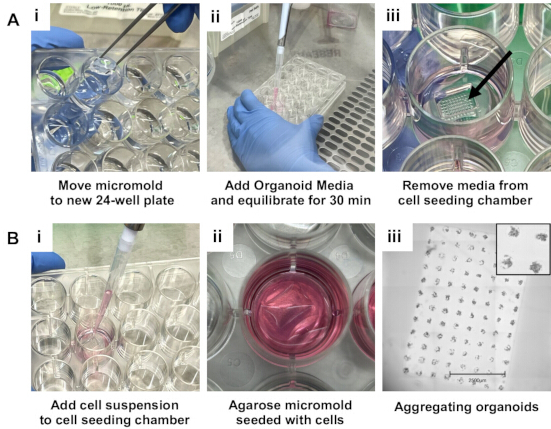
Rycina 5: Schemat generowania somatycznych organoidów jajnika. (A) Reprezentatywne obrazy (i) przenoszenia agarowych mikrootformowań do płytki hodowlanej za pomocą pęsety Graefe, (ii) równoważenia mikrootformowań agarowych w pożywce do organoidów jajnika (Ovarian Organoid Media) oraz (iii) usuwania pożywki z komór wysiewowych. Strzałka w Aiii wskazuje pustą komorę wysiewową. (B) Reprezentatywne obrazy (i,ii) wysiewu komórek do agarowych mikrootformowań oraz (iii) skanu w świetle przechodzącym mikrootformowania agarowego zawierającego agregujące somatyczne komórki jajnika. Pasek skali (Biii): 2500 µm. Kliknij tutaj, aby wyświetlić powiększoną wersję tej ryciny.
5. Wykorzystanie organoidów w zastosowaniach następczych
UWAGA: Organoidy można wykorzystać w wielu zastosowaniach następczych, w tym w histologii, badaniach biologii molekularnej (np. analizach RNA/białek), analizie pożywek warunkowanych oraz w kokulturach.
- Histologia
UWAGA: Organoidy można poddać obróbce w celu oceny punktów końcowych histologicznych bez usuwania ich z agarozowych mikform. W rzeczywistości organoidy w mikformach agarozowych działają podobnie do mikrotablic tkankowych, co umożliwia wysokoprzepustową ocenę histologiczną kilku, rozmieszczonych w stałych odstępach organoidów jednocześnie.- Rozpuścić 1,5% (w/v) sterylną agarozę w PBS poprzez gotowanie.
- Po hodowli somatycznych organoidów jajnika trzymać płytkę pod kątem i usunąć medium otaczające mikformy agarozowe.
- Pod mikroskopem stereotypowym usunąć medium z komór wysiewowych komórek, delikatnie zbliżając końcówkę pipety do narożnika każdej komory wysiewowej i ostrożnie odsysając medium, aby nie zaburzyć organoidów ani nie uszkodzić samej mikformy.
- Ostrożnie pipetować 100–125 µL ciekłej 1,5% agarozy schłodzonej do około 50 °C do narożnika każdej komory wysiewowej komórek, aby wypełnić i uszczelnić komory wysiewowe, dbając o to, by nie zaburzyć organoidów (Rysunek 6Ai).
- Pozostawić agarozę do stężenia przez 2–3 min w temperaturze pokojowej.
- Dodać 750 µL zmodyfikowanego utrwalacza Davidsona lub 4% paraformaldehydu do każdej studni i utrwalać organoidy w zamkniętych mikformach agarozowych przez noc w temperaturze 4 °C.
- Po utrwaleniu usunąć utrwalacz i przemyć mikformy agarozowe 70% etanolem.
UWAGA: Mikformy można przechowywać w 70% etanolu w temperaturze 4 °C do czasu obróbki tkanek (odwodnienia, przejaśniania i infiltracji woskiem). Podczas obróbki tkanek należy kontrolować orientację mikform agarozowych. - Po obróbce tkanek zatopić mikformy agarozowe w parafinie w taki sposób, aby mikformy były płasko ułożone w blokach parafiny i aby dno mikform było pierwszym elementem poddanym cięciu (Rysunek 6Aii).
- Wyciąć bloki parafiny w plastry o grubości 5 µm i umieścić preparaty tkankowe na naładowanych szkiełkach mikroskopowych.
UWAGA: Obecność mikrowyrobisk można zweryfikować wzrokowo podczas cięcia zarówno w plastrach tkankowych, jak i w blokach parafiny (Rysunek 6Aiii,iv). Obecność organoidów w przekrojach mikform można potwierdzić, badając preparaty pod mikroskopem (Rysunek 6Av). Zamiast umieszczać 2–3 kolejne przekroje na jednym szkiełku, umieszczenie jednego szkiełka 2–3 przekrojów oddalonych od siebie o 50 µm (tj. co 10ty przekrój) zwiększa prawdopodobieństwo, że większość organoidów zostanie odwzorowana w przynajmniej jednym przekroju na każdym szkiełku. Organoidy są rozmieszczone w stałych odstępach w przekrojach tkankowych, co ułatwia obrazowanie po barwieniu histologicznym (Rysunek 6Avi). - Pozostawić szkiełka do wyschnięcia w inkubatorze w temperaturze 37 °C przez noc.
- Przeprowadzić barwienie histologiczne lub immunohistochemiczne lub przechowywać szkiełka w temperaturze pokojowej19.
- Pozyskiwanie organoidów do zastosowań molekularnych
UWAGA: Po hodowli organoidy można usunąć z mikform agarozowych i poddać różnym zastosowaniom z zakresu biologii molekularnej, w tym ekstrakcji RNA lub białek w celu oceny ekspresji genów lub białek.- Trzymać płytkę zawierającą mikformy agarozowe pod lekkim kątem i usunąć medium otaczające mikformy agarozowe za pomocą pipety.
- Dodać 750 µL PBS w temperaturze pokojowej do studni zawierających mikformy agarozowe.
- Użyć pęsety Graefe, aby odwrócić i potrząsnąć mikformami agarozowymi w celu uwolnienia organoidów do otaczającego PBS. Potwierdzić uwolnienie organoidów z mikform agarozowych pod mikroskopem.
- Usunąć puste mikformy agarozowe za pomocą pęsety Graefe i przenieść PBS zawierający organoidy do probówek do mikrowirówki za pomocą pipety.
- U pelletować organoidy poprzez wirowanie przy 10 000 × g przez 5 min w temperaturze pokojowej.
- Usunąć nadmiar cieczy poprzez aspirację i przystąpić do ekstrakcji RNA lub białek. Alternatywnie, zamrozić gwałtownie pellet komórkowy i przechowywać w temperaturze -80 °C19.
- Zatrzymywanie warunkowanego medium z organoidów do analizy
UWAGA: Warunkowane medium z organoidów można zbierać w trakcie całej hodowli w celu analizy czynników wydzielanych przez organoidy, w tym hormonów, cytokin i czynników wzrostu. Reprezentatywne wyniki analizy cytokin w warunkowanym medium z organoidów przedstawiono na Rysunku 7A.- Trzymać płytkę zawierającą mikformy agarozowe pod lekkim kątem i usunąć warunkowane medium otaczające mikformy za pomocą pipety. Nie usuwać medium z komór wysiewowych komórek, aby uniknąć zaburzenia organoidów.
- Przenieść warunkowane medium do probówek do mikrowirówki i przystąpić do analizy medium lub zamrozić gwałtownie i przechowywać w temperaturze -80 °C19,22.
UWAGA: Opcjonalnie, wirować warunkowane medium przy 10 000 × g przez 5 min, aby usunąć martwe komórki lub resztki komórkowe. Przenieść nadfiltr do nowych probówek do mikrowirówki przed analizą lub przechowywaniem medium.
- Współhodowla organoidów
UWAGA: Somatyczne organoidy jajnika można współhodować z innymi komórkami i tkankami za pomocą kilku metod. Reprezentatywne wyniki współhodowli organoidów z komórkami raka jajnika i pęcherzykami, zgodnie z metodami opisanymi odpowiednio w krokach 5.4.1 i 5.4.3, przedstawiono na Rysunku 7B,C.- Aby wytworzyć organoidy przy użyciu kombinacji pierwotnych somatycznych komórek jajnika myszy i innych typów komórek w celu zbadania efektów bezpośrednich interakcji komórka-komórka, należy postępować zgodnie z krokami 5.4.1.1–5.4.1.4.
- Postępować zgodnie z krokami 4.1–4.9, aby przemyć pierwotne somatyczne komórki jajnika po nocnej hodowli 2D, pozyskać komórki z hodowli 2D przy użyciu 0,05% trypsyny, u pelletować, policzyć i resuspendować komórki w medium do organoidów jajnika w gęstości 2,5 × 10⁵ żywych komórek na 75 µL medium.
- Jeśli inny interesujący typ komórek jest adherentny, pozyskać komórki z hodowli 2D. Policzyć komórki, u pelletować je przez wirowanie i resuspendować w medium do organoidów jajnika w gęstości 2,5 × 10⁵ żywych komórek na 75 µL medium.
- Połączyć zawiesinę pierwotnych somatycznych komórek jajnika z zawiesiną innego interesującego typu komórek w znanym stosunku.
- Postępować zgodnie z krokami 4.10–4.13, aby wysiać powstałą zawiesinę komórkową do mikform agarozowych i hodować organoidy.
- Aby przenieść mikformy agarozowe zawierające uformowane organoidy do płytek 24-dołkowych zawierających inne komórki lub organoidy, należy postępować zgodnie z krokami 5.4.2.1–5.4.2.4.
UWAGA: Aby ocenić wpływ czynników wydzielanych przez organoidy na inne typy komórek i odwrotnie, uformowane organoidy mogą pozostać w mikformach agarozowych, a całe mikformy można przenieść do płytki 24-dołkowej zawierającej inne komórki lub organoidy. Te metody współhodowli można stosować, gdy medium do organoidów jajnika może być użyte do hodowli obu typów komórek/tkanek.- Podgrzać medium do organoidów jajnika do 37 °C w kąpieli wodnej.
- Trzymać płytkę zawierającą mikformy agarozowe pod kątem i usunąć medium otaczające mikformy agarozowe za pomocą pipety.
- Używając pęsety Graefe, ostrożnie przenieść mikformy do płytek 24-dołkowych zawierających inne komórki lub organoidy.
- Dodać 750 µL świeżego medium do organoidów jajnika, aby otoczyło mikformy agarozowe.
- Aby usunąć uformowane organoidy z mikform agarozowych i przenieść je do nowych mikform agarozowych, należy postępować zgodnie z krokami 5.4.3.1–5.4.3.13.
UWAGA: Jeśli medium do organoidów jajnika nie jest pożądanym medium do eksperymentów ze współhodowlą, model opisany w kroku 5.4.2 stwarza problem znacznego przeniesienia medium, ponieważ mikformy agarozowe są nasycone medium do organoidów jajnika. Uformowane organoidy (1–3 dni po początkowym wysiewie komórek) można przenieść z ich mikform agarozowych do nowych mikform agarozowych w celu dalszej hodowli lub współhodowli, aby zapobiec przeniesieniu medium.- Podgrzać pożądane medium do współhodowli do 37 °C w kąpieli wodnej.
UWAGA: Media oparte na DMEM, αMEM i F12 zostały z powodzeniem zastosowane w eksperymentach współhodowli z somatycznymi organoidami jajnika. - Używając pęsety Graefe, ostrożnie przenieść nowe mikformy agarozowe z PBS +1% PS do studni płytki 24-dołkowej zawierającej inne komórki lub organoidy do współhodowli.
- Upewnić się, że mikformy są całkowicie zanurzone w medium, dodać w razie potrzeby dodatkowe pożądane medium do współhodowli i równoważyć mikformy oraz medium przez co najmniej 30 min w inkubatorze (środowisko nawilżone, 37 °C z 5% CO2).
- Trzymać płytkę zawierającą mikformy agarozowe z uformowanymi organoidami pod lekkim kątem i usunąć medium otaczające mikformy agarozowe za pomocą pipety.
- Dodać pożądane medium do współhodowli wokół mikform agarozowych z uformowanymi organoidami.
- Użyć pęsety Graefe, aby odwrócić i potrząsnąć mikformami agarozowymi w celu uwolnienia organoidów do otaczającego medium. Potwierdzić uwolnienie organoidów z mikform agarozowych pod mikroskopem.
UWAGA: Jeśli pierwotne somatyczne komórki jajnika nie zagregowały się odpowiednio, organoidy mogą nie pozostać nienaruszone po usunięciu z mikform agarozowych. Składanie się organoidów może trwać 1–3 dni, w zależności od szczepu i wieku myszy, z których izolowano somatyczne komórki jajnika. - Usunąć puste mikformy agarozowe za pomocą pęsety Graefe i przenieść medium zawierające organoidy do probótek do mikrowirówki za pomocą pipety.
- Wirować organoidy przy 300 × g przez 5 min w temperaturze pokojowej.
- Pod mikroskopem stereotypowym usunąć nadfiltr za pomocą pipety, pozostawiając 50–75 µL medium zawierającego organoidy.
- Wyjąć zrównoważone mikformy z inkubatora i upewnić się, że mikformy są zorientowane tak, aby komora wysiewowa komórek była skierowana do góry.
- Usunąć medium z komór wysiewowych komórek za pomocą pęsety Graefe lub pipety.
UWAGA: Studnie nie powinny zawierać więcej niż 500 µL medium, aby można było opróżnić komory wysiewowe komórek mikform. - Przenieść organoidy bezpośrednio do komory wysiewowej każdej nowej mikformy agarozowej za pomocą pipety. Wrócić z płytką 24-dołkową do inkubatora.
- Po 1–2 h dodać dodatkowe 250 µL pożądanego medium wokół zewnętrznej strony mikform agarozowych, uważając, aby nie zaburzyć komór wysiewowych komórek.
- Podgrzać pożądane medium do współhodowli do 37 °C w kąpieli wodnej.
- Aby wytworzyć organoidy przy użyciu kombinacji pierwotnych somatycznych komórek jajnika myszy i innych typów komórek w celu zbadania efektów bezpośrednich interakcji komórka-komórka, należy postępować zgodnie z krokami 5.4.1.1–5.4.1.4.
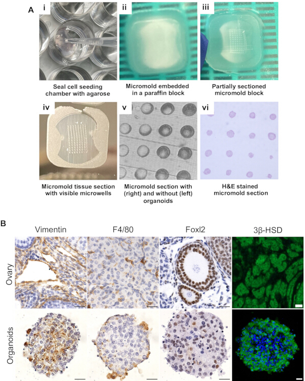
Rycina 6: Kluczowe typy komórek jajnika zostają zachowane w somatycznych organoidach jajnika. (A) Reprezentatywne obrazy (i) uszczelniania mikroformy agarozą po zakończeniu hodowli, (ii) nieprzeciętej mikroformy oraz (iii) sekcjonowanej mikroformy zatopionej w bloku parafiny. Mikrootwory można zwizualizować na (iv) zamontowanych skrawkach, a obecność organoidów w sekcjonowanych mikrootworach można (v) potwierdzić pod mikroskopem przed (vi) barwieniem histologicznym. (B) Reprezentatywne obrazy skrawków tkanki jajnika myszy oraz skrawków somatycznych organoidów jajnika po barwieniu immunohistochemicznym z użyciem przeciwciał przeciwko Vimentin, F4/80, Foxl2 oraz 3β-HSD. Paski skali = 20 µm. Skróty: H&E = hematoksylina i eozyna. Rycina została opracowana za zgodą autorów Dipali et al.19. Aby zobaczyć powiększoną wersję tej ryciny, kliknij tutaj.
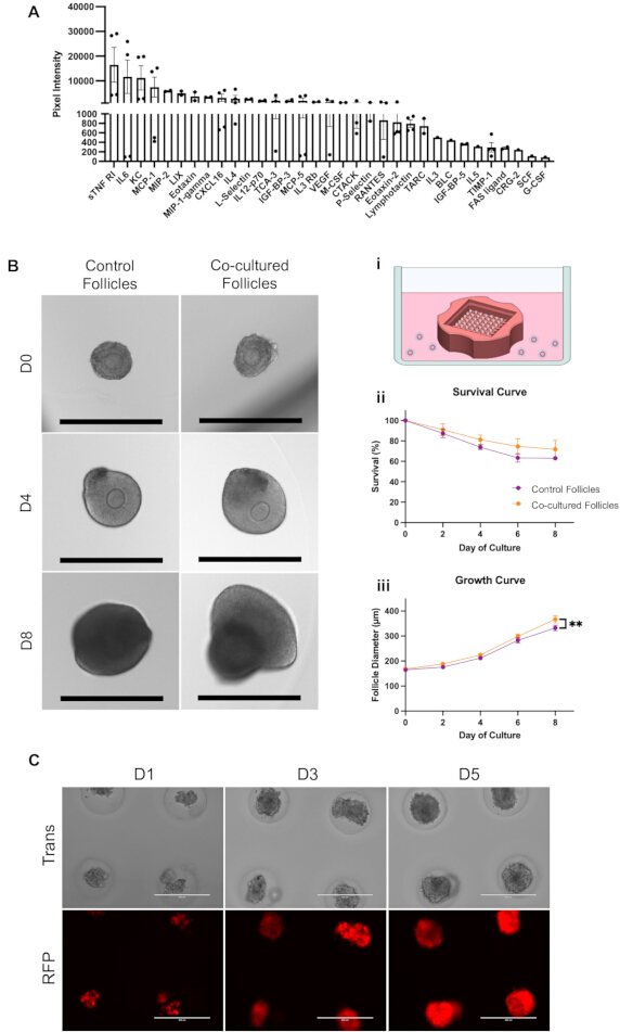
Rycina 7: Kokultura somatycznych organoidów jajnika z pęcherzykami i komórkami raka jajnika. (A) Wydzielanie cytokin przez organoidy mierzono w każdym mikformie agarozowej w 5. dniu hodowli i znormalizowano względem pożywki warunkowej z mikform agarozowych bez organoidów. N = 4 mikformy agarozowe. (B) Reprezentatywne obrazy w świetle przechodzącym pęcherzyków jajnikowych w dniach 0, 4 i 8 hodowli z organoidami lub bez nich (kontrola) oraz (i) schemat modelu kokultury. Paski skali = 400 µm. Kwantyfikacja (ii) przeżywalności pęcherzyków oraz (iii) wzrostu w ciągu 8 dni hodowli z organoidami lub bez nich (kontrola). Dane przedstawiono jako średnia ± SD. Niektóre słupki błędów są zbyt małe, aby były widoczne. N = 56 pęcherzyków na grupę. **P < 0.01. (C) Reprezentatywne obrazy w świetle przechodzącym (góra) i fluorescencyjne (dół) pierwotnych somatycznych komórek jajnika hodowanych w stosunku 10:1 z komórkami raka jajnika znakowanymi czerwonym białkiem fluorescencyjnym (OVCAR8-RFP) w organoidach w dniach 1, 3 i 5 hodowli. Paski skali = 400 µm. Skrót: RFP = czerwone białko fluorescencyjne. Kliknij tutaj, aby wyświetlić powiększoną wersję tej ryciny.
6. Wskazówki dotyczące rozwiązywania problemów
- Problem 1: Liczba wyizolowanych pierwotnych somatycznych komórek jajnika jest niższa niż przewidywano.
UWAGA: Jeśli fragmenty tkanki jajnika są stale mniejsze niż 1 mm lub nie są widoczne gołym okiem po trawieniu enzymatycznym, liczba wyizolowanych komórek może być niska z powodu obniżonej żywotności wynikającej z nadmiernego trawienia. Jeśli fragmenty tkanki jajnika są większe niż 1 mm po trawieniu enzymatycznym, liczba wyizolowanych komórek może być niska z powodu niewystarczającego trawienia tkanki jajnika.- Jeśli fragmenty tkanki jajnika są większe niż 1 mm po trawieniu enzymatycznym, należy wykonać kroki 6.1.1.1–6.1.1.2.
- Należy potwierdzić, że Collagenase IV oraz DNase I znajdują się w zalecanym terminie przydatności do użycia i nie utraciły aktywności enzymatycznej z powodu niewłaściwego przechowywania lub, w przypadku DNase I, wielokrotnych cykli zamrażania i rozmrażania.
- Należy zwiększyć liczbę powtórzeń pipetowania tkanki jajnika, zmniejszyć średnicę otworu końcówki p1000, jeśli została ona odcięta w celu poszerzenia otworu, i/lub wydłużyć czas inkubacji w EM.
- Jeśli fragmenty tkanki jajnika są stale znacznie mniejsze niż 1 mm po trawieniu enzymatycznym, należy zmniejszyć liczbę powtórzeń pipetowania tkanki jajnika, zwiększyć średnicę otworu końcówki p1000 poprzez odcięcie jej w celu poszerzenia otworu i/lub skrócić całkowity czas inkubacji w EM.
- Jeśli fragmenty tkanki jajnika są większe niż 1 mm po trawieniu enzymatycznym, należy wykonać kroki 6.1.1.1–6.1.1.2.
- Problem 2: Więcej niż ~25% odlewanych agarozy mikform form posiada defekty
- Należy potwierdzić, że forma silikonowa jest wolna od pozostałości agarozy i zanieczyszczeń. W razie potrzeby należy przemyć formy silikonowe w PBS i wysterylizować ponownie w autoklawie. W razie potrzeby należy ponownie podgrzać 1% agarozę używaną do odlewania, ponieważ agaroza, która zaczyna krzepnąć, może nie wypełnić równomiernie formy silikonowej i/lub doprowadzić do powstania pęcherzyków powietrza.
- Po naniesieniu agarozy do formy silikonowej należy pozwolić jej na całkowite stężenie, ponieważ przedwczesne usunięcie mikroformy agarozowej z formy może spowodować uszkodzenie mikrowyżłobień.
- Należy potwierdzić, że formy silikonowe nie są niedopełnione, ponieważ może to zmniejszyć integralność strukturalną mikroformy agarozowej, prowadząc do jej uszkodzenia podczas wyginania silikonu w celu usunięcia mikroformy agarozowej.
- Dodatkowo producent form silikonowych zaleca, aby nie były one autoklawowane więcej niż 12 razy. Jeśli forma silikonowa była poddawana nadmiernej sterylizacji, należy ją wymienić.
- Problem 3: Organoidy nie powstają w więcej niż 10% mikrowyżłobień
- Należy potwierdzić, że formy silikonowe nie są przepełnione podczas odlewania mikroform agarozowych, ponieważ może to prowadzić do nierównomiernego rozkładu komórek w mikrowyżłobieniach, faworyzując mikrowyżłobienia centralne w mikroformie agarozowej nad mikrowyżłobieniami zewnętrznymi.
- Należy unikać tworzenia pęcherzyków powietrza podczas dozowania zawiesiny komórkowej do komory wysiewu komórek w mikroformach agarozowych, ponieważ pęcherzyki mogą zaburzyć rozkład komórek w mikrowyżłobieniach.
- W razie potrzeby należy rozpocząć od ilości zawiesiny komórkowej większej niż 75 µL (przy gęstości 3.33 × 10⁶ żywych komórek na 1 mL) na mikroformę, aby upewnić się, że pęcherzyki nie powstają z powodu niewystarczającej objętości w końcówce pipety.
- Należy wydłużyć odstęp czasu między wysiewem komórek a dodaniem pożywki do organoidów jajnika na zewnątrz mikroformy agarozowej, aby zapewnić komórkom wystarczający czas na osiadanie w mikrowyżłobieniach.
Dostęp ograniczony. Zaloguj się lub rozpocznij wersję próbną, aby wyświetlić tę treść.
Wyniki
Aby określić skład komórkowy somatycznych organoidów jajnika, przeprowadziliśmy immunohistochemię przekrojów tkanek organoidów po 6 dniach hodowli, wykorzystując przeciwciała przeciwko kluczowym typom komórek jajnika. Organoidy zawierały fibroblasty wykazujące pozytywną reakcję z wimentyną, makrofagi pozytywne dla F4/80, a także populacje komórek steroidogennych (Rysunek 6B)19. Wykorzystaliśmy przeciwciało przeciwko Foxl2 do znakowania komórek ziarnistych i luteinizow...
Dostęp ograniczony. Zaloguj się lub rozpocznij wersję próbną, aby wyświetlić tę treść.
Dyskusja
W niniejszym artykule opisano metody izolacji pierwotnych komórek somatycznych jajnika mysiego z frakcji wzbogaconej w stromę jajnika, w sposób zachowujący heterogeniczność komponentu somatycznego jajnika. Pierwotne komórki somatyczne jajnika mysiego mogą być hodowane w 2D monowarstwie lub wykorzystywane do generowania organoidów somatycznych jajnika za pomocą bezoporowych form mikromatryc mikromolowych z agarozy. Mikroformy z agarozy zawierają 96 mikrodołków do tworzenia organoidów i pasują do pojedynczej komórki płytki...
Dostęp ograniczony. Zaloguj się lub rozpocznij wersję próbną, aby wyświetlić tę treść.
Oświadczenia
Autorzy nie mają żadnych sporów interesów do ujawnienia.
Podziękowania
To dzieło zostało sfinansowane przez Narodowy Instytut Zdrowia Dziecka i Rozwoju Człowieka (R01HD093726 dla F.E.D. i T32HD094699 dla S.S.D. i E.J.Z.), Narodowy Instytut Raka (F31CA257300 dla S.S.D.), oraz fundusze start-upowe z Departamentu Położnictwa i Ginekologii (dla F.E.D.). Chcemy wyrazić wdzięczność dr Candace Tingen oraz laboratorium dr Teresy Woodruff, a także dr Jennifer Rowley, za ich podstawowe badania i optymalizację metod izolowania pierwotnych mysich komórek somatycznych jądra i charakterystykę tych komórek.
Dostęp ograniczony. Zaloguj się lub rozpocznij wersję próbną, aby wyświetlić tę treść.
Materiały
| Nazwa | Firma | Numer katalogowy | Komentarze |
|---|---|---|---|
| 0.2 μm, Polyethersulfone syringe filter | Corning | 431229 | |
| 1.5 mL microcentrifuge tubes | MIDSCI | PR-MCT15 | |
| 100 mL glass beaker | Fisherbrand | FB100100 | |
| 100 mm Petri dishes | Corning | 351029 | |
| 15 mL conical tubes | ThermoFisher Scientific | 339651 | |
| 24-well plates (TC-treated) | Corning | 353047 | |
| 28-gauge insulin needles | Exel International | 26027 | |
| 35 mm Petri dishes | Corning | 351008 | |
| 3β-HSD antibody | Cosmo Bio | K0607 | 1:1000 |
| 4% Paraformaldehyde | Electron Microscopy Sciences | 1574100 | |
| 40 µm cell strainer | Corning | 431750 | |
| 45 mm watch glass | Electron Microscopy Sciences | 7054345 | |
| 50 mL concial tubes | ThermoFisher Scientific | 339652 | |
| 60 mL sterile syringe | Air-Tite | ML60 | |
| 60 mm TC-treated plates | Corning | 353037 | |
| 6-well plates (TC-treated) | Corning | 353046 | |
| Agarose | Hoefer | GR140-500 | Gel strength >1200 g/cm2; Gel Temperature 36 °C |
| Autoclave sterilization pouch | Fisherbrand | 01-812-55 | |
| Biotinylated Goat Anti-Mouse IgG | Vector Laboratories | BA-9200-1.5 | 1:200 |
| Biotinylated Goat Anti-Rabbit IgG | Vector Laboratories | PK-6101 | 1:200 |
| Biotinylated Goat Anti-Rat IgG | Vector Laboratories | BA-9400-1.5 | 1:100 |
| Bluing reagent | Fisherbrand | 23-245681 | |
| Centrifuge with swing bucket rotor for 50 mL conical tubes | ThermoFisher Scientific | Sorvall X1R Pro-MD | |
| Class II biosafety cabinet | The Baker Company | SG403A | |
| Cleaved Caspase-3 (CC3) antibody | Cell Signaling Technology | 9579T | 1:250 |
| CO2 incubator | ThermoFisher Scientific | Heracell VIOS 160i CO2 Incubator | |
| Collagenase, Type IV, powder | Gibco, Fisher Scientific | 17-104-019 | |
| C-Series Mouse Cytokine Antibody Array 3 Kit | RayBiotech | AAM-CYT-3-8 | |
| Cytoseal XYL | Epredia | 83124 | |
| Deoxyribonuclease I from bovine pancreas | Millipore Sigma | DN25-100MG | Dnase I |
| Dissecting microscissors | World Precision Instruments | WPI-14003 | |
| Dissecting scissors | World Precision Instruments | WPI-15922 | |
| Dissection microscope | Leica | MZ9.5, S9 series | |
| DPBS, no calcium, no magnesium | Gibco, Fisher Scientific | 14-190-250 | pH 7.0–7.3 |
| Eosin | Fisherbrand | 23-314631 | |
| F4/80 antibody | Bio-Rad | MCA497G | 1:50 |
| Fetal Bovine Serum, certified, heat inactivated, United States | Gibco, ThermoFisher Scientific | 10082147 | |
| Fine-tipped dissecting forceps | World Precision Instruments | WPI-14098 | |
| FOXL2 antibody | Abcam | ab246511 | 1:200 |
| Graefe Forceps | Fine Science Tools | 1105010 | |
| Hematoxylin | EK Industries | EKI 47971GL | |
| Hemocytometer | MedOne | DHCN015 | |
| Imaging System | ThermoFisher Scientific | EVOS FL Auto | |
| IntestiCult Organoid Growth Medium (Mouse) | STEMCELL Technologies | 06005 | Basal medium, Supplements 1 and 2 |
| Laminar flow hood | IVFtech | IVFtech sterile workstation | |
| Leibovitz's L-15 Medium | Gibco, Fisher Scientific | 11415114 | |
| Microscope slides | Mercedes Scientific | MER 7255/90/WH/CC | |
| MicroTissues 3D Petri Dish micro-mold spheroids | Millipore Sigma | Z764043-6EA | Silicone casts for agarose micromolds |
| Modified Davidson's Fixative | Electron Microscopy Sciences | 6413350 | |
| OVCAR8-RFP cells | Gift from Joanna Burdette PhD, University of Illinois Chicago | ||
| Penicillin-Streptomycin | Millipore Sigma | P4333-100ML | |
| Picrosirius Red staining solution | StatLab | STPSRPT | |
| RPMI 1640 Medium (ATCC modification) | Gibco, Fisher Scientific | A1049101 | |
| Tabletop centrifuge | Eppendorf | Centrifuge 5430 R | |
| Trypan blue | ThermoFisher Scientific | T10282 | |
| Trypsin-EDTA (0.05%), phenol red | Gibco, ThermoFisher Scientific | 25300054 | |
| Vimentin antibody | Cell Signaling Technology | 5741S | 1:100 |
| αMEM, GlutaMAX Supplement, no nucleosides | Gibco, Fisher Scientific | 32561102 |
Bibliografia
- Thomas, F. H., Vanderhyden, B. C. Oocyte-granulosa cell interactions during mouse follicular development: regulation of kit ligand expression and its role in oocyte growth. Reprod Biol Endocrinol. 4 (1), 19(2006).
- Robker, R. L., Hennebold, J. D., Russell, D. L. Coordination of ovulation and oocyte maturation: a good egg at the right time. Endocrinology. 159 (9), 3209-3218 (2018).
- Casey, G. Sex hormones and health. Nurs N Z. 23 (1), 24-28 (2017).
- Kinnear, H. M., et al. The ovarian stroma as a new frontier. Reproduction. 160 (3), R25-R39 (2020).
- Simon, L. E., Kumar, T. R., Duncan, F. E. In vitro ovarian follicle growth: a comprehensive analysis of key protocol variables. Biol Reprod. 103 (3), 455-470 (2020).
- Spector, I., Derech-Haim, S., Boustanai, I., Safrai, M., Meirow, D. Anti-Müllerian hormone signaling in the ovary involves stromal fibroblasts: a study in humans and mice provides novel insights into the role of ovarian stroma. Hum Reprod. 39 (11), 2551-2564 (2024).
- Kedem, A., et al. Activated ovarian endothelial cells promote early follicular development and survival. J Ovarian Res. 10 (1), 64(2017).
- Converse, A., et al. Multinucleated giant cells are hallmarks of ovarian aging with unique immune and degradation-associated molecular signatures. PLoS Biol. 23 (6), e3003204(2025).
- Bafor, E. E., et al. Isolation of single cells from individual mouse ovaries for flow cytometry and functional analysis. STAR Protoc. 4 (4), 102710(2023).
- Shepherd, T. G., Thériault, B. L., Campbell, E. J., Nachtigal, M. W. Primary culture of ovarian surface epithelial cells and ascites-derived ovarian cancer cells from patients. Nat Protoc. 1 (6), 2643-2649 (2006).
- Beschta, S., et al. A rapid and robust method for the cryopreservation of human granulosa cells. Histochem Cell Biol. 156 (5), 509-517 (2021).
- Tingen, C. M., et al. A macrophage and theca cell-enriched stromal cell population influences growth and survival of immature murine follicles in vitro. Reproduction. 141 (6), 809-820 (2011).
- O'Mara, A. E., et al. Chronic mirabegron treatment increases human brown fat, HDL cholesterol, and insulin sensitivity. J Clin Invest. 130 (5), 2209-2219 (2020).
- Zink, N., Stock, A. -K., Vahid, A., Beste, C. On the neurophysiological mechanisms underlying the adaptability to varying cognitive control demands. Front Hum Neurosci. 12, 411(2018).
- Zhao, Z., et al. Organoids. Nat Rev Methods Primers. 2 (1), 94(2022).
- Yang, S., et al. Organoids: the current status and biomedical applications. MedComm. 4 (3), e274(2023).
- Alzamil, L., Nikolakopoulou, K., Turco, M. Y. Organoid systems to study the human female reproductive tract and pregnancy. Cell Death Differ. 28 (1), 35-51 (2021).
- Pierson Smela, M. D., et al. Directed differentiation of human iPSCs to functional ovarian granulosa-like cells via transcription factor overexpression. eLife. 12, e83291(2023).
- Dipali, S. S., et al. Self-organizing ovarian somatic organoids preserve cellular heterogeneity and reveal cellular contributions to ovarian aging. Aging Cell. 25 (1), e70333(2026).
- Di Nisio, V., et al. Silk-ovarioids: establishment and characterization of a human ovarian primary cell 3D-model system. Hum Reprod Open. 2025 (3), hoaf042(2025).
- Nikanfar, S., Leonel, E. C. R., Damdimopoulou, P., Flaws, J. A., Amorim, C. A. Effects of phthalate exposure on human ovarian extracellular matrix composition: insights from a 3D spheroid model. Environ Res. 279, 121797(2025).
- Briley, S. M., et al. Reproductive age-associated fibrosis in the stroma of the mammalian ovary. Reproduction. 152 (3), 245-260 (2016).
- Converse, A., Zaniker, E. J., Amargant, F., Duncan, F. E. Recapitulating folliculogenesis and oogenesis outside the body: encapsulated in vitro follicle growth. Biol Reprod. 108 (1), 5-22 (2023).
- Dean, M., Jin, V., Russo, A., Lantvit, D. D., Burdette, J. E. Exposure of the extracellular matrix and colonization of the ovary in metastasis of fallopian-tube-derived cancer. Carcinogenesis. 40 (1), 41-51 (2019).
- Charalambous, C., Webster, A., Schuh, M. Aneuploidy in mammalian oocytes and the impact of maternal ageing. Nat Rev Mol Cell Biol. 24 (1), 27-44 (2023).
- Rowley, J. E., et al. Low molecular weight hyaluronan induces an inflammatory response in ovarian stromal cells and impairs gamete development in vitro. Int J Mol Sci. 21 (3), 1036(2020).
- Amargant, F., Vieira, C., Pritchard, M. T., Duncan, F. E. Systemic low-dose anti-fibrotic treatment attenuates ovarian aging in the mouse. Geroscience. 47 (3), 3475-3495 (2024).
Dostęp ograniczony. Zaloguj się lub rozpocznij wersję próbną, aby wyświetlić tę treść.
Przedruki i uprawnienia
Przeglądaj więcej artykułów
