A 66-year-old man with obesity and locally advanced prostate cancer developed severe rhabdomyolysis (peak creatine kinase [CK] >122,000 U/L) and acute kidney injury shortly after undergoing robot-assisted radical prostatectomy, which lasted 250 minutes and was performed in the steep Trendelenburg lithotomy position.
Opis przypadku
Rhabdomyolysis Following Robot-Assisted Radical Prostatectomy
W tym artykule
Podsumowanie
Streszczenie
Robot-assisted radical prostatectomy remains a favorable treatment option for locally advanced prostate cancer in patients with obstructive lower urinary tract symptoms. However, the steep Trendelenburg lithotomy position required for this procedure can result in serious complications, particularly in patients with obesity. Here, the case of a 66-year-old man with obesity (BMI, 35 kg/m2), insulin-dependent diabetes mellitus, and multiple comorbidities is reported, who underwent robot-assisted radical prostatectomy (console time, approximately 4 h) for clinically staged cT3a cN1 prostate cancer. Immediately after surgery, the patient developed severe gluteal pain, sensorimotor deficits, and massive rhabdomyolysis, resulting in anuric acute kidney injury and respiratory insufficiency. The clinical course was further complicated by heparin-induced thrombocytopenia type II and a grade IV gluteal pressure ulcer. A 22-day intensive care stay was required, including continuous hemodiafiltration and transfusion of 12 units of erythrocytes. With multidisciplinary management, renal function partially recovered, and the patient was discharged with minimal residual neuropathic deficits. This case highlights the importance of comprehensive preoperative risk assessment and provides clinically relevant insights into the recognition and multidisciplinary management of severe complications associated with prolonged positioning during robot-assisted radical prostatectomy.
Wprowadzenie
Robot-assisted radical prostatectomy (RARP) offers excellent oncologic and functional outcomes for patients with prostate cancer1. However, the procedure requires prolonged steep Trendelenburg positioning, which may compress muscle compartments and impair tissue perfusion, particularly in patients with obesity2. Reperfusion after repositioning can subsequently result in extensive muscle breakdown (rhabdomyolysis), with the release of myoglobin potentially leading to acute kidney injury and other systemic complications3.
Obesity and prolonged operative time may increase complications and are considered major risk factors for positioning-related rhabdomyolysis1,2,4,5. Previous studies have shown that an open-legs position can markedly reduce postoperative creatine kinase levels and potentially prevent clinically significant rhabdomyolysis without compromising surgical access6. This relatively simple modification, combined with careful patient selection and vigilant postoperative monitoring, may offer advantages over conventional lithotomy positioning6.
Positioning-related rhabdomyolysis following RARP has been reported in 1–6% of cases, with the risk increasing substantially in patients with a BMI >30 kg/m2 and operative or console time exceeding 4 h7. Well-leg compartment syndrome represents a similarly serious positioning-related complication involving an overlapping pathophysiologic mechanism of prolonged tissue compression, ischemia, and subsequent reperfusion injury8,9,10. This case report aims to facilitate the early identification of patients at increased risk for positioning-related rhabdomyolysis and to highlight preventive measures that may improve patient safety during robotic pelvic surgery.
Case Presentation
The patient was a 66-year-old man with a BMI of 35 kg/m2 and grade II obesity. The medical history included insulin-dependent type 2 diabetes mellitus (HbA1c, 8.8%), diabetic polyneuropathy, lumbar spinal canal stenosis at L3–S1 with disc herniation, arterial hypertension, coronary artery disease with a previous myocardial infarction, sick sinus syndrome requiring pacemaker implantation, chronic pancreatitis, and bladder outlet obstruction that had been managed with a suprapubic catheter for several months.
Before surgery, the patient underwent cardiologic evaluation. The cardiologist identified stable coronary artery disease with preserved left ventricular function and no evidence of acute ischemia. The patient was considered fit for the planned procedure, with continuation of his usual cardiac medications and perioperative pacemaker monitoring. The patient was not receiving statin therapy at the time of surgery. Preoperative thyroid function testing showed normal TSH.
Preoperative staging demonstrated locally advanced prostate cancer, clinically staged as cT3a cN1 cM0, with an initial prostate-specific antigen level of 14 ng/mL and a Gleason score of 4 + 5 = 9 in 12/12 biopsy cores. Digital rectal examination revealed a large, firm prostate with findings suspicious for bilateral extracapsular extension. Transrectal ultrasonography demonstrated a prostate volume >120 mL with capsular irregularity. Intraoperatively, the patient was positioned in the Trendelenburg position at 30° for approximately 4 h; gel padding was applied to the gluteal region, sacrum, and heels. No prolonged intraoperative hypotension was documented. Although preoperative staging indicated cT3a disease, intraoperative inspection revealed infiltration of the levator ani and perivesical fat not identified on preoperative imaging, and T4 disease was entirely unsuspected. The unexpectedly extensive local invasion substantially increased the technical difficulty of dissection
Diagnosis, Assessment, and Plan
Following multidisciplinary discussion, the patient was scheduled for robot-assisted radical prostatectomy with bilateral pelvic lymphadenectomy. The decision to proceed with radical prostatectomy rather than definitive radiotherapy plus long-term androgen deprivation therapy was made after a multidisciplinary tumor board discussion.
Four key factors were considered in the decision to proceed with radical prostatectomy. First, the patient had a very-high-risk disease, characterized by a Gleason score of 4 + 5 = 9 in all 12 biopsy cores, cT3a cN1 staging, and a prostate volume exceeding 120 mL, for which radical prostatectomy with extended pelvic lymph node dissection offered local disease control and accurate pathological staging. Second, the patient had chronic bladder outlet obstruction requiring long-term suprapubic catheterization, which was considered likely to be aggravated by radiotherapy. Third, a dedicated cardiologic evaluation documented adequate cardiac fitness for the planned surgical procedure. Finally, after comprehensive counseling regarding the risks and potential benefits of the available treatment strategies, the patient expressed an informed preference for a single-stage surgical approach.
The procedure was performed with the patient in the conventional steep Trendelenburg lithotomy position. Total operative time was 250 minutes, including approximately 240 minutes of console time. Intraoperatively, the tumor was locally advanced, with infiltration of the levator ani and perivesical fat, resulting in an apical R2 resection margin.
Immediately after extubation, the patient reported severe gluteal pain radiating to the thighs and developed incomplete sensorimotor deficits suggestive of a transverse neurologic syndrome at the T12/L1 level. Anuria subsequently developed. Laboratory investigations demonstrated a marked increase in creatine kinase (>122,000 U/L), a peak serum creatinine of 2.98 mg/dL, and metabolic acidosis, consistent with rhabdomyolysis-associated acute kidney injury11.
Contrast-enhanced computed tomography and magnetic resonance imaging of the pelvis and spine excluded acute spinal cord compression and hematoma. Repeated neurologic evaluations indicated axonal polyneuropathy secondary to rhabdomyolysis. Based on the clinical presentation, laboratory findings, and imaging results, the working diagnosis was severe positioning-related rhabdomyolysis with crush syndrome physiology. The initial 'T12/L1 transverse syndrome' reflected the sensory-motor level of the presenting deficits and did not indicate spinal cord injury, which imaging and the subsequent course excluded. Primary compartment syndrome was also considered in the differential diagnosis; however, conservative management was pursued as the patient's clinical and laboratory findings improved with supportive treatment12.
Protokół
The patient consented in writing to all treatment steps and anonymous publication. The study complies with the Declaration of Helsinki and was approved by the ethics committees of the Westfalen-Lippe Medical Association and the University of Muenster (2023–500-f-S) for retrospective data management.
1. Preoperative risk stratification
- The patient underwent a thorough history and physical examination focusing on metabolic syndrome, BMI, diabetes mellitus, arterial hypertension, cardiac history, spinal pathology, and preexisting bladder dysfunction.
- A cardiological consultation, including echocardiography and assessment for myocardial ischemia, was obtained because of the patient's history of coronary artery disease.
- Baseline laboratory tests, including serum creatine kinase, renal function parameters, and a coagulation profile, were obtained.
- The patient was counseled regarding positioning-related complications because of the high-risk profile, including a BMI ≥35 kg/m2 and the anticipated prolonged operative time.
2. Intraoperative positioning and monitoring
- The patient was placed in the steep Trendelenburg lithotomy position. The open-legs position, with the thighs abducted and minimal pressure on the calves, was considered an alternative to reduce positioning-related complications.
- Gel padding was applied to pressure-sensitive areas, including the gluteal region, sacrum, and heels.
- Pneumoperitoneum pressure was maintained at 8–15 mmHg, and invasive arterial blood pressure was monitored continuously.
- The exact console time and total operative time were recorded.
3. Surgical technique
- A transperitoneal robot-assisted radical prostatectomy with bilateral pelvic lymphadenectomy was performed.
- Wide excision was performed according to the intraoperative findings, with the aim of achieving optimal oncologic clearance.
- A suprapubic catheter was placed at the end of the procedure.
4. Immediate postoperative surveillance
- The patient was transferred for close postoperative monitoring because of his high-risk profile and postoperative clinical condition.
- Serum creatine kinase, myoglobin, electrolytes, and renal function parameters were monitored postoperatively only in anticipated high-risk patients (BMI ≥ 30 kg/m2, anticipated operative time ≥ 4 h, or prolonged Trendelenburg positioning, diabetes mellitus, peripheral vascular disease, pre-existing neuropathy, or significant cardiovascular comorbidity) at 6–12, 24, and 48 hours after surgery. This schedule is not recommended for routine patients undergoing robot-assisted radical prostatectomy.
- Fluid therapy and measures to promote diuresis were initiated after the marked elevation in creatine kinase was identified.
5. Intensive Care Unit management of established rhabdomyolysis
- Invasive hemodynamic monitoring and aggressive fluid resuscitation were provided.
- Continuous hemodiafiltration was initiated after the patient developed anuric acute kidney injury.
- Packed red blood cells were transfused as required to maintain an adequate hemoglobin level.
- Anticoagulation was adjusted following confirmation of heparin-induced thrombocytopenia, with argatroban used as an alternative anticoagulant.
- Respiratory support, including high-flow nasal oxygen and noninvasive ventilation, was provided for respiratory insufficiency as clinically required.
- Multidisciplinary management involved urology, nephrology, neurology, cardiology, general surgery, orthopedic surgery, and wound-care specialists. Frequent repositioning, wound management, and physiotherapy were implemented during the patient's intensive care course.
Wyniki
Despite immediate admission to the intensive care unit and protocol-driven management, the patient required 22 days of advanced supportive care. Figure 1 shows the intraoperative Trendelenburg-lithotomy positioning used for the procedure. Creatine kinase levels peaked at >122,000 U/L on the first postoperative day and gradually declined with continuous renal replacement therapy and supportive measures. Peak serum creatinine reached 2.98 mg/dL, accompanied by metabolic acidosis and anuria consistent with rhabdomyolysis-associated acute kidney injury. Continuous hemofiltration was initiated and later discontinued as renal function progressively recovered. Detailed serial trends in estimated glomerular filtration rate, myoglobin, and serum potassium were not available for retrospective analysis and are therefore not reported.
The patient received 12 units of packed red blood cells. Renal function gradually improved, allowing discontinuation of continuous hemodiafiltration. Neurological examination and imaging confirmed axonal polyneuropathy with right-predominant paraparesis, which progressively improved during rehabilitation. Following consultation with the orthopedic surgery team, compartment fasciotomy was not required. Abdominal compartment syndrome was excluded from the outset because the clinical findings were localized predominantly to the gluteal region.
A grade IV gluteal pressure ulcer subsequently developed and was successfully managed with surgical debridement under general anesthesia and vacuum-assisted closure therapy (Clavien–Dindo grade IIIb). The patient was transferred from the intensive care unit to the peripheral ward on postoperative day 22 and was discharged several weeks later in stable condition, with continued outpatient wound care and physiotherapy.
Final histopathological examination confirmed pT4 pN1 (6/28), Gleason score 4 + 5 = 9, R2 prostate adenocarcinoma with a tumor volume of 19.95 cm3. During the subsequent months of follow-up, the patient's neurological and systemic symptoms resolved, and prostate-specific antigen levels remained undetectable under androgen deprivation therapy.
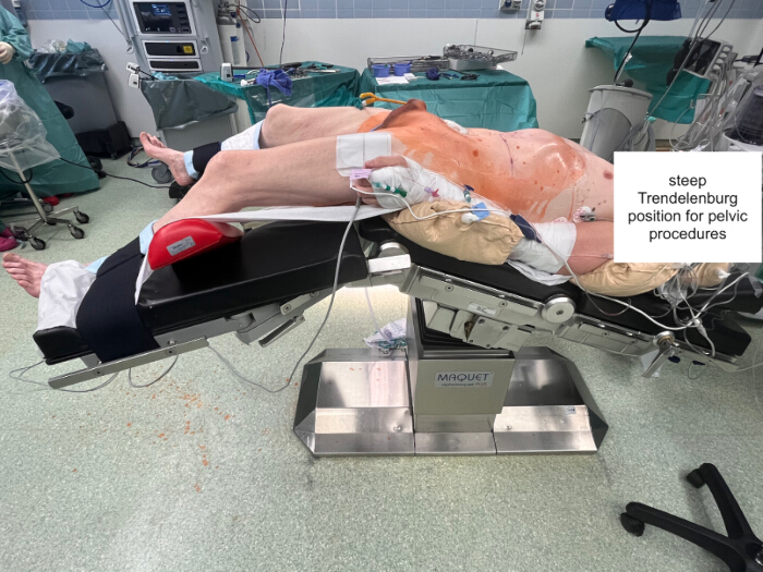
Figure 1: Steep Trendelenburg-lithotomy positioning used during robot-assisted pelvic surgery. Please click here to view a larger version of this figure.
Dyskusja
This case illustrates how robot-assisted radical prostatectomy can lead to serious postoperative complications. The patient had grade II obesity (BMI 35 kg/m2), poorly controlled insulin-dependent diabetes mellitus (HbA1c 8.8%), diabetic polyneuropathy, lumbar spinal stenosis, coronary artery disease, and chronic bladder outlet obstruction requiring a long-term suprapubic catheter. These factors increased the perioperative risk and raised important questions regarding the selection of radical surgery compared with radiation therapy combined with androgen deprivation therapy. Surgery was selected as the first step in a multimodal treatment strategy, which might include adjuvant radiation with or without hormonal therapy to improve local tumor control, given the very-high-grade locally advanced disease, pre-existing catheter dependence, documented cardiac fitness, and the patient's informed preference. Nevertheless, the postoperative course demonstrates that substantial complications can occur in high-risk patients despite careful preoperative assessment.
Despite the use of gel padding and a relatively low pneumoperitoneum pressure of 8 mmHg, prolonged conventional steep Trendelenburg lithotomy positioning may have contributed to mechanical compression of the dependent gluteal and lower-extremity muscle compartments, resulting in extensive muscle injury, massive myoglobin release, acute kidney injury, and secondary axonal polyneuropathy.
Pneumoperitoneum was maintained at 8 mmHg. Although no evidence directly links pneumoperitoneum pressure to rhabdomyolysis, increased intra-abdominal pressure can impair tissue perfusion, and prolonged pneumoperitoneum has been associated with acute renal impairment following robotic prostatectomy. Comparative evidence regarding padding systems specifically for robotic pelvic surgery remains limited, and randomized data demonstrating the superiority of a specific padding system for preventing rhabdomyolysis after robot-assisted prostatectomy are lacking.
Open-leg lithotomy positioning primarily reduces compression of the calves and lower extremities and, consequently, the risk of well-leg compartment syndrome; however, it does not eliminate prolonged pressure on the gluteal regions and posterior thighs, which remain dependent and compressed against the operating table during steep Trendelenburg positioning. In the present case, the predominantly gluteal injury was consistent with this mechanism. Alternative positioning strategies, including the supine split-leg position, have been adopted at several institutions and may reduce lower-limb and positional pressure while preserving surgical access, although comparative evidence regarding gluteal pressure injury specifically remains limited.
The temporal sequence and distribution of the clinical findings strongly supported a diagnosis of positioning-related rhabdomyolysis. Immediately after surgery, severe gluteal pain radiating to the thighs developed, accompanied by an incomplete sensorimotor transverse syndrome at the T12/L1 level. This was followed by a marked increase in creatine kinase levels to >122,000 U/L. The localization of pain and subsequent development of a grade IV gluteal pressure ulcer corresponded to the dependent areas subjected to prolonged compression against the operating table during steep Trendelenburg positioning. This pattern was consistent with ischemia-reperfusion injury. In obesity, prolonged compression of the gluteal compartments, potentially exacerbated by inadequate pressure distribution and the head-down position, may reduce tissue perfusion. Subsequent reperfusion upon returning the patient to the supine position can contribute to extensive muscle injury and necrosis11.
Other potential causes were systematically evaluated. Urgent computed tomography and magnetic resonance imaging excluded spinal cord compression and hematoma, while the immediate postoperative onset made drug-induced, infectious, or exertional rhabdomyolysis less likely. The high BMI of 35 kg/m2 and prolonged operative duration of 250 min were also consistent with reported risk factors for positioning-related rhabdomyolysis13. Longer console times and elevated BMI have additionally been investigated as factors associated with perioperative outcomes following robot-assisted radical prostatectomy14,15. Together, the timing of symptom onset, the anatomical distribution of muscle injury, a marked elevation in creatine kinase, and the exclusion of alternative diagnoses supported positioning-related rhabdomyolysis as the most likely diagnosis.
Prompt recognition of the complication and transfer to the intensive care unit enabled early initiation of continuous renal replacement therapy, hemodynamic support, and multidisciplinary management. Renal function gradually recovered, and the neurological deficits improved during rehabilitation. However, the development of a grade IV gluteal pressure ulcer demonstrated the additional risk of severe pressure-related tissue injury in this setting and the complexity of managing critically ill patients with prolonged immobility.
The present case occurred following prolonged conventional steep Trendelenburg lithotomy positioning. The open-leg position has been proposed as an alternative approach for reducing positioning-related complications. In a propensity score-matched analysis, this modification substantially reduced postoperative creatine kinase levels and eliminated clinically apparent rhabdomyolysis in the study group6. The proposed mechanism involves reducing external pressure on the calves to <20 mmHg while maintaining adequate surgical exposure. However, because the open-leg position primarily reduces lower-extremity compression, it may not eliminate prolonged pressure on the dependent gluteal regions during steep Trendelenburg positioning.
These findings emphasize the importance of preoperative risk stratification, consideration of alternative positioning strategies, appropriate pressure-relieving measures, and structured early postoperative monitoring in patients undergoing prolonged robotic pelvic surgery. Particular attention should be given to patients with obesity, diabetes, pre-existing neuropathy, spinal disease, or an anticipated prolonged operative time. When positioning-related rhabdomyolysis occurs, early recognition and close interdisciplinary collaboration among urology, intensive care, nephrology, neurology, orthopedic surgery, and wound-care teams may help limit systemic complications and long-term functional impairment16.
Universal baseline or serial creatine kinase (CK) measurements are not recommended for all patients undergoing robot-assisted radical prostatectomy. Instead, baseline CK assessment is recommended for high-risk patients, including those with a BMI ≥30 kg/m2, an anticipated console or operative time ≥4 h, diabetes mellitus, peripheral vascular disease, pre-existing neuropathy, or known cardiac compromise7,11,14,15. In this high-risk group, serial monitoring of CK, myoglobin, electrolytes, and renal function at 6–12, 24, and 48 h postoperatively is recommended, as CK elevation typically occurs during the first 24–72 h after muscle injury13. Routine admission to an intensive care unit or intermediate care setting for all high-risk patients may not be feasible due to resource limitations; therefore, transfer to a monitored setting is recommended when postoperative clinical signs or relevant laboratory abnormalities, such as marked CK elevation, myoglobinuria, or rising serum creatinine, develop. An operative duration of ≥4 h is suggested as a trigger for postoperative laboratory monitoring based on reported associations between prolonged operative or console duration and perioperative complications7,13,14. Continuous invasive arterial blood pressure monitoring may also be considered in high-risk patients to facilitate early detection of hypotension and to maintain adequate perfusion of dependent muscle compartments.
This report has several limitations. First, as a single-patient retrospective case report, it cannot establish causality or provide generalizable risk estimates. Second, comprehensive serial laboratory data, including serum creatinine, estimated glomerular filtration rate (eGFR), myoglobin, and potassium levels during the intensive care unit stay, were unavailable. Availability of these measurements would have provided a more complete characterization of the clinical trajectory. Anti-HMGCR immune-mediated necrotizing myopathy and inherited metabolic myopathies were considered in the differential diagnosis, but additional testing was not performed. The acute perioperative onset, localized muscle injury involving dependent pressure areas, absence of prior statin use, and absence of a personal or family history of myopathy or exertional rhabdomyolysis made these alternative diagnoses less likely.
Oświadczenia
The authors have nothing to disclose. The authors used a language model (Gemini) exclusively for language polishing and grammatical refinement. The authors controlled, verified, and approved all scientific content and assume full responsibility for the final manuscript.
Podziękowania
The authors have no acknowledgments.
Materiały
| Nazwa | Firma | Numer katalogowy | Komentarze |
|---|---|---|---|
| Argatroban | Bayer | — | Alternative anticoagulation for confirmed HIT type II |
| Continuous veno-venous hemodiafiltration (CVVHDF) system | Prismaflex | — | Renal replacement therapy for anuric AKI and hyperkalemia |
| da Vinci X Surgical System | Intuitive Surgical | da Vinci X | Robot-assisted radical prostatectomy with bilateral pelvic lymphadenectomy |
| Gel padding and pressure-relief materials | Allen Medical | — | Intraoperative protection of the gluteal region, sacrum, and heels |
| High-flow nasal oxygen device | Optiflow | — | Respiratory support for partial respiratory insufficiency |
| Human albumin 20% | Various | — | Volume expansion and oncotic support during ICU stay |
| Packed red blood cells | Blood bank | — | Transfusion for severe anemia (12 units total) |
| Piperacillin-tazobactam | Not provided | — | Broad-spectrum antibiotic used for treatment of respiratory tract colonization/infection |
| Suprapubic catheter | Uromed | — | Bladder drainage before and after surgery |
| Vacuum-assisted closure (VAC) system | 3M Acelity | — | Management of grade IV gluteal pressure ulcer |
Bibliografia
- Novara G, et al. Systematic review and meta-analysis of perioperative outcomes and complications after robot-assisted radical prostatectomy. Eur Urol. 2012;62(3):431-452.
- Karaoren G, Bakan N, Kucuk EV, Gumus E. Is rhabdomyolysis an anaesthetic complication in patients undergoing robot-assisted radical prostatectomy? J Minim Access Surg. 2017;13(1):29-36.
- Galyon SW, Richards KA, Pettus JA, Bodin SG. Three-limb compartment syndrome and rhabdomyolysis after robotic cystoprostatectomy. J Clin Anesth. 2011;23(1):75-78.
- Onagi A, et al. Transient renal dysfunction due to rhabdomyolysis after robot-assisted radical prostatectomy. Int Urol Nephrol. 2020;52(10):1877-1884.
- Yang RH, Chu YK, Huang CW. Compartment syndrome following robotic-assisted prostatectomy: rhabdomyolysis in bone scintigraphy. Clin Nucl Med. 2013;38(5):365-366.
- Tsubouchi K, et al. Efficacy of the opened legs position for protecting against postoperative rhabdomyolysis after robot-assisted radical prostatectomy: A propensity score-matched analysis of perioperative outcomes. Int J Urol. 2022;29(10):1132-1138.
- Maerz DA, Beck LN, Sim AJ, Gainsburg DM. Complications of robotic-assisted laparoscopic surgery distant from the surgical site. Br J Anaesth. 2017;118(4):492-503.
- Raman SR, Jamil Z. Well leg compartment syndrome after robotic prostatectomy: a word of caution. J Robot Surg. 2009;3(2):105-107.
- Endo Y, et al. A case of well leg compartment syndrome after robot-assisted laparoscopic prostatectomy: with review. J Med Invest. 2022;69(1.2):145-147.
- Fukuda M, et al. Well leg compartment syndrome following robot-assisted radical cystectomy in the lithotomy position: a case report. JA Clin Rep. 2021;7(1):13.
- Pariser JJ, et al. Rhabdomyolysis after major urologic surgery: epidemiology, risk factors, and outcomes. Urology. 2015;85(6):1328-1332.
- Wen T, Deibert CM, Siringo FS, Spencer BA. Positioning-related complications of minimally invasive radical prostatectomies. J Endourol. 2014;28(6):660-667.
- Mattei A, et al. Positioning injury, rhabdomyolysis, and serum creatine kinase-concentration course in patients undergoing robot-assisted radical prostatectomy and extended pelvic lymph node dissection. J Endourol. 2013;27(1):45-51.
- Farzat M, Elsherif M, Wagenlehner FM. How May Longer Console Times Influence Outcomes after Robot-Assisted Radical Prostatectomy (RARP)? J Clin Med. 2023;12(12). doi:10.3390/ jcm12124022.
- Farzat M, Sharabaty I, Tanislav C, Alsaid Y, Wagenlehner FM. BMI Impact on Readmissions for Patients Undergoing Robot-Assisted Radical Prostatectomy: A Monocentric, Single-Surgeon Serial Analysis of 500 Cases. J Clin Med. 2023;12(12). doi:10.3390/ jcm12123908
- Gezginci E, et al. Postoperative pain and neuromuscular complications associated with patient positioning after robotic assisted laparoscopic radical prostatectomy: a retrospective non-placebo and non-randomized study. Int Urol Nephrol. 2015;47(10):1635-1641.