This study compared 3-month cognitive outcomes after acute ischemic stroke in the anterior and posterior circulations using routine MRI and the Montreal Cognitive Assessment.
Artykuł badawczy
Anterior Versus Posterior Circulation Acute Ischemic Stroke and 3-Month Cognitive Profiles: A Single-Center Cohort Study
W tym artykule
Podsumowanie
Streszczenie
Post-stroke cognitive impairment (PSCI) is common, but its pattern may differ between vascular territories. This single-center cohort included adults with first-ever, magnetic resonance imaging (MRI)-confirmed acute ischemic stroke admitted between May 2022 and June 2024. Infarcts were classified as anterior or posterior circulation, and cognition was assessed in person at 3 months using the Beijing version of the Montreal Cognitive Assessment (MoCA). The primary PSCI definition was an education-adjusted MoCA score <26, with a sensitivity analysis using <22. Of 173 enrolled patients, 167 completed follow-up (91 anterior and 76 posterior circulation infarctions). PSCI occurred in 50/91 (54.9%) and 27/76 (35.5%) patients, respectively (p = 0.012). The unadjusted odds ratio was 2.21 (95% confidence interval, 1.18–4.14), and the adjusted odds ratio was 2.18 (95% confidence interval, 1.06–4.49; p = 0.035). With the <22 cutoff, the corresponding prevalences were 31.9% and 15.8%, and the adjusted odds ratio was 2.43 (95% confidence interval, 1.06–5.54; p = 0.035). After false-discovery-rate correction, differences in visuospatial/executive, naming, delayed-recall, language, abstraction, and orientation performance remained, whereas attention did not differ. Cognitive outcomes differed between vascular-territory groups despite similar global disability scores; these group-level associations do not establish a causal effect of territory or a domain-specific diagnosis.
Wprowadzenie
Stroke is a major cause of death and long-term disability worldwide, and the number of people living with its consequences remains high1,2. Follow-up after a stroke often focuses on motor recovery, recurrent vascular events, and the ability to perform basic activities. Cognitive problems may be less apparent in patients who regain physical independence, yet they can still interfere with rehabilitation and everyday functioning. Even patients with mild neurological deficits may have difficulty with planning, remembering instructions, or managing multiple tasks. Because these deficits may not be captured by neurological or global disability scales, cognition should be assessed directly during follow-up.
PSCI ranges from mild deficits in one or more cognitive functions to major neurocognitive disorder and often has multiple contributing causes. The index infarct, previous silent vascular injury, cerebral small-vessel disease, neurodegeneration, mood symptoms, delirium, and recurrent events may all contribute3,4,5. Reported frequencies differ widely across studies because the populations, exclusion criteria, test instruments, diagnostic thresholds, and timing of assessment are not the same. Reviews nevertheless show that cognitive impairment without dementia is common during the first year after stroke and that post-stroke dementia remains an important long-term problem6,7,8,9. Age, education, stroke severity, recurrent stroke, vascular disease, and imaging abnormalities have also been associated with poorer cognitive outcome10. Cognitive testing during the acute admission may be affected by fatigue, sleep disruption, metabolic disturbance, emotional stress, or fluctuating neurological symptoms. Assessment at 3 months allows some of these acute influences to subside while remaining relevant to rehabilitation planning.
The Montreal Cognitive Assessment (MoCA) is widely used in stroke clinics because it can be completed in a short visit and covers visuospatial/executive function, naming, delayed recall, attention, language, abstraction, and orientation11. It is generally more sensitive than the Mini-Mental State Examination for mild deficits, although its performance varies with language, education, test timing, and the cutoff used12. Current European guidance supports structured screening after stroke but also notes that a screening score is not a substitute for a full neuropsychological diagnosis13. In patients with stroke, test performance may be affected by aphasia, visual loss, motor impairment, and educational level. In cohort studies, MoCA subtest scores can describe between-group patterns, but they should not be interpreted as diagnoses of isolated cognitive-domain impairment.
Infarct location is one factor that may shape the cognitive profile. Cognitive impairment can remain after otherwise good neurological recovery14, and lesion-symptom studies have identified both strategic regions and distributed networks linked to poor cognitive outcome15,16. Cognitive effects may vary with lesion size, laterality, tract disconnection, cortical-subcortical network involvement, and pre-existing brain disease17. Anterior circulation infarcts may involve frontal, lateral temporal, parietal, basal ganglia, or internal-capsule networks, whereas posterior circulation infarcts may involve the occipital lobes, medial temporal structures, thalamus, brainstem, or cerebellum. Each vascular territory contains heterogeneous lesion locations. Although anterior-versus-posterior classification is less anatomically precise than detailed lesion mapping, it can be applied to routine MRI cohorts.
This study compared PSCI at 3 months after the first-ever acute ischemic stroke in the anterior and posterior circulations. The primary outcome was the proportion of patients with an education-adjusted MoCA score <26. Secondary and exploratory outcomes included PSCI severity, total MoCA score, individual subtest scores, non-full subtest frequencies, and the 3-month modified Rankin Scale (mRS). A sensitivity analysis used a lower MoCA threshold (<22), supported by stroke-specific diagnostic-accuracy evidence. The objective was to determine whether vascular-territory groups differed in the frequency and pattern of MoCA-defined impairment, while accounting for the anatomical heterogeneity within each territory.
Protokół
All procedures involving human participants were reviewed and approved by the Medical Ethics Committee of Hebei General Hospital (NO.2025-LW-0317). The committee approved the retrospective use of clinical cases and waived the requirement for individual informed consent for this analysis. Data were deidentified before analysis and sharing. All the materials are used in the Table of Materials.
Study design and ethical oversight
A single-center observational cohort study was conducted in the Department of Neurology of Hebei General Hospital. Eligible patients admitted between May 2022 and June 2024 were enrolled and reassessed 3 months after stroke onset. Vascular territory, categorized as anterior or posterior circulation, was the exposure, and MoCA-defined PSCI at 3 months was the primary outcome. Reporting followed the STROBE recommendations for observational studies18. Each participant was assigned a study code, and direct identifiers were removed from the analysis file. Of 173 enrolled patients, six lacked the 3-month cognitive assessment and were excluded from the analytic cohort; contemporaneous screening counts and individual reasons for noncompletion were not available and therefore were not reconstructed.
Participant screening and eligibility
Consecutive adults aged 18–79 years who presented within 14 days of symptom onset with a first-ever acute ischemic stroke were screened. Only patients who were clinically stable, conscious, and able to participate in the planned assessments were included. The dates of symptom onset, admission, diagnostic MRI, and enrollment were recorded to confirm eligibility.
Acute ischemic stroke was confirmed according to the applicable Chinese guideline and required an acute lesion on diffusion-weighted MRI. Patients with transient ischemic attack without infarction or intracranial hemorrhage were excluded. Medical records were reviewed, and the patient or family was interviewed regarding pre-stroke cognition. They were asked whether memory, language, judgment, or independence in daily activities had declined before the index stroke. Patients with documented cognitive impairment, dementia, or a major psychiatric or behavioral disorder that would prevent a valid post-stroke assessment were excluded. Patients with traumatic brain injury, hemorrhagic cerebrovascular disease, severe intracranial complications, active malignancy, or clinically important cardiac, hepatic, renal, or gastrointestinal disease that would interfere with assessment or follow-up were excluded. Patients who could not complete the full MoCA because of severe aphasia, apraxia, uncorrected visual or hearing impairment, or another major communication barrier were also excluded. The findings may not apply to patients with severe stroke who could not complete the MoCA. Structural MRI was reviewed for marked background brain disease. Patients with moderate or severe atrophy, defined as a maximum transverse third-ventricle diameter ≥10 mm together with other imaging signs of atrophy, or with confluent leukoaraiosis, were excluded. Patients with an MRI-incompatible implanted device, an unsafe metallic foreign body, or claustrophobia that prevented completion of the required sequences were also excluded.
Baseline clinical data collection
During the index admission, age, sex, years of education, smoking, heavy alcohol use, hypertension, diabetes mellitus, coronary heart disease, hyperlipidemia, and the admission NIHSS score were recorded on the case-report form. Smoking was defined as continuous or cumulative smoking for more than 6 months before stroke, at an average of at least one cigarette per day. Heavy alcohol use was defined as a history of more than 1 year with an average intake of at least one bottle of beer or approximately 50 g of spirits per day. Hypertension was defined as systolic blood pressure ≥140 mmHg and/or diastolic blood pressure ≥90 mmHg on two consecutive measurements, or a previous diagnosis treated with antihypertensive medication. Diabetes was defined by typical symptoms and at least one of the following: random glucose ≥11.1 mmol/L, fasting glucose ≥7.0 mmol/L, a 2-h oral glucose tolerance test value ≥11.1 mmol/L, or a previous diagnosis managed with diet or medication. Coronary heart disease was defined from a documented previous diagnosis, specialist assessment, coronary angiography, or coronary computed tomography angiography. Hyperlipidemia was defined from a previous diagnosis with lipid-lowering treatment or fasting total cholesterol ≥5.2 mmol/L, low-density lipoprotein cholesterol ≥3.4 mmol/L, and/or triglycerides ≥1.82 mmol/L.
A trained clinician administered the National Institutes of Health Stroke Scale (NIHSS) after admission. The total score was entered into the analysis file, and the item-level form was retained in the source record when available. Because the NIHSS weights some anterior circulation findings more heavily than posterior circulation symptoms, it was treated as a measure of observed neurological deficit rather than infarct volume. Patients received routine acute and secondary-prevention care according to clinical indications. None of the 173 enrolled participants received intravenous thrombolysis, endovascular thrombectomy, or bridging therapy; therefore, within-cohort confounding by reperfusion treatment could not be evaluated, and generalizability to reperfusion-treated populations is limited.
MRI acquisition and vascular-territory classification
Brain MRI was performed using a 3.0 T system. The imaging protocol included axial T1-weighted, axial T2-weighted, axial fluid-attenuated inversion recovery (FLAIR), sagittal T1-weighted, diffusion-weighted imaging (DWI), and time-of-flight magnetic resonance angiography (TOF-MRA). The scanner used in the study is listed in the Table of Materials. TOF-MRA was acquired with a 240 × 210 mm field of view, 1.4 mm section thickness, −0.7 mm interslice spacing, repetition time (TR) of 24 ms, echo time (TE) of 3.2 ms, a 384 × 160 matrix, one excitation, and 140 sections. Axial T1-FLAIR was acquired with a 240 × 192 mm field of view, 5 mm section thickness, 1.5 mm interslice spacing, TR of 1,750 ms, TE of 25.0 ms, a 320 × 224 matrix, one excitation, and 20 sections. Axial T2-weighted imaging was acquired with a 240 × 240 mm field of view, 5 mm section thickness, 1.5 mm spacing, TR of 4,841 ms, TE of 103.8 ms, a 416 × 416 matrix, one excitation, and 20 sections.
Axial T2-FLAIR was acquired with a 240 × 240 mm field of view, 5 mm section thickness, 1.5 mm spacing, TR of 7,000 ms, TE of 137.2 ms, a 256 × 224 matrix, one excitation, and 20 sections. Sagittal T1-weighted imaging was acquired with a 240 × 240 mm field of view, 5 mm section thickness, 1.5 mm spacing, TR of 2,250 ms, TE of 27.7 ms, a 288 × 224 matrix, one excitation, and 18 sections. Axial DWI was acquired with a 240 × 240 mm field of view, 5 mm section thickness, 1.5 mm spacing, TR of 4,880 ms, TE of 77.4 ms, a 130 × 160 matrix, one excitation, and 40 sections. The DWI lesion was assessed for consistency with the acute clinical syndrome, and the remaining sequences were reviewed for hemorrhage, atrophy, and confluent white matter change.
An experienced radiologist and a clinician, masked to the detailed clinical history, independently reviewed lesion location using DWI, the remaining MRI sequences, and vascular imaging. Disagreements were adjudicated by a senior radiologist. MRI location was combined with the Oxfordshire Community Stroke Project classification19. Lesions supplied mainly by the internal carotid, anterior cerebral, or middle cerebral arteries were classified as anterior circulation infarctions; lesions supplied mainly by the vertebral, basilar, or posterior cerebral arteries were classified as posterior circulation infarctions. Laterality (left, right, or bilateral), cortical involvement (cortical, subcortical, or both), the principal anatomical site, and lesion multiplicity (single or multiple acute lesions) were recorded. The prespecified territory category was used for the primary analysis and was not changed after cognitive outcomes were known. Infarct volume and automated lesion segmentation were not available.
Three-month functional and cognitive assessment
At approximately 3 months after stroke onset, participants were invited to an in-person follow-up visit. Telephone contact was used only to arrange the visit; the full MoCA was administered face-to-face. Six participants did not complete the 3-month assessment. Baseline characteristics of these participants are provided in Supplementary Table 1; specific reasons for noncompletion were unavailable. At the visit, disability was assessed using the mRS, which was treated as a functional outcome and was not included in the PSCI definition. Cognition was assessed with the authorized Beijing version of the full 30-point MoCA, a version previously evaluated in Chinese populations20,21. Two trained clinicians used the same instructions and scoring form and remained masked to detailed medical histories. One point was added for participants with ≤12 years of formal education, with the adjusted total capped at 30. The primary screening definition classified education-adjusted MoCA scores ≥26 as normal and scores <26 as PSCI. Because stroke-specific diagnostic accuracy varies by setting and threshold, a sensitivity analysis used <22, consistent with the 21/22 threshold supported by a recent meta-analysis22. Descriptive severity categories were 18–25 (mild), 10–17 (moderate), and 0–9 (severe); these study categories were not formal neurocognitive diagnoses. The seven MoCA subtest scores were analyzed in their original ranges. A subtest score below its maximum was labeled non-full only as an exploratory descriptive dichotomy; continuous score distributions remained the primary subtest presentation.
Data quality and statistical analysis
A deidentified dataset with one row per participant was constructed. Permitted ranges, category codes, duplicate nonmissing study identifiers, and agreement between the raw MoCA total and component scores were checked before analysis. The final dataset contained 173 rows; 167 participants had complete 3-month outcomes and six were lost to follow-up. Normally distributed continuous variables were summarized as mean ± standard deviation and compared using an independent-samples t-test. Skewed or ordinal variables were summarized as median (interquartile range) and compared using the Mann–Whitney U test. Categorical variables were reported as n (%) and compared using Pearson chi-square or an exact method when cells were sparse. Ordered PSCI severity categories were compared using a rank-based test. Percentages used the relevant vascular-territory group as the denominator unless otherwise stated.
The unadjusted odds ratio for PSCI in the anterior versus posterior circulation group was calculated with a 95% confidence interval. The prespecified multivariable logistic model included vascular territory, age, sex, education ≤12 years, admission NIHSS score, hypertension, diabetes, and hyperlipidemia. The same model was repeated using adjusted MoCA <22 as a sensitivity outcome. Total MoCA and subtest scores were compared using rank-based tests. Non-full subtest frequencies were compared using chi-square or exact tests. To address multiplicity, Benjamini–Hochberg false-discovery-rate correction was applied separately to the seven subtest-score comparisons and the seven non-full-score comparisons; raw p values and adjusted q values are reported. All tests were two-sided. Participants with complete 3-month outcomes and complete model covariates were included, and missing cognitive outcomes were not imputed. Enrollment comprised all eligible consecutive cases during the fixed study period; no prospective sample-size calculation was performed. A descriptive post hoc calculation based on the observed primary proportions and an equal-group approximation yielded approximately 71.6% power, which was not treated as a substitute for prospective planning.
Reproducibility checkpoints and troubleshooting
Before table and figure generation, the final locked dataset was checked for permitted ranges, duplicated nonmissing identifiers, MoCA component-sum mismatches, and consistency of education adjustment. No range violations, component-sum mismatches, or duplicated nonmissing identifiers were found. The six cases without 3-month outcomes were included in the deidentified supplementary dataset but excluded from outcome analyses. All tables and figures were regenerated from the corrected locked dataset using the same analysis script, and percentages were checked against their vascular-territory denominators.
Wyniki
Participant flow and baseline characteristics
The cohort included 173 patients with first-ever MRI-confirmed acute ischemic stroke. Six did not complete the 3-month assessment, leaving 167 patients for analysis: 91 with anterior and 76 with posterior circulation infarction (Figure 1). Mean age was 62.02 ± 10.06 years in the anterior group and 62.74 ± 8.53 years in the posterior group. Men accounted for 67.0% and 71.1% of the groups, respectively. Age, sex, hypertension, diabetes, coronary heart disease, hyperlipidemia, smoking, heavy alcohol use, and education ≤12 years did not differ significantly (Table 1). Admission NIHSS score was lower in the anterior group [median, 2 (interquartile range, 1–3)] than in the posterior group [3 (2–4); p = 0.015]. The six participants lost to follow-up comprised three anterior and three posterior circulation cases; their available baseline characteristics are listed in Supplementary Table 1. Lesion laterality was left-sided in 46/91 anterior and 38/76 posterior cases, right-sided in 44/91 and 38/76, and bilateral in 1/91 and 0/76, respectively. Cortical or combined cortical-subcortical involvement occurred in 44/91 anterior and 19/76 posterior cases; subcortical-only lesions occurred in 47/91 and 57/76. Multiple acute lesions were recorded in 20/91 and 11/76. Detailed principal anatomical sites are reported in Supplementary Table 2.
Primary cognitive outcome
Overall, 77/167 patients (46.1%) met the primary MoCA-based PSCI criterion. PSCI occurred in 50/91 patients in the anterior circulation group and 27/76 in the posterior circulation group (54.9% versus 35.5%, p = 0.012; Table 2 and Figure 2A). The unadjusted odds ratio was 2.21 (95% confidence interval, 1.18–4.14). In the adjusted model, the odds ratio for anterior versus posterior circulation infarction was 2.18 (95% confidence interval, 1.06–4.49; p = 0.035; Supplementary Table 3). Using adjusted MoCA <22, PSCI occurred in 29/91 (31.9%) and 12/76 (15.8%) patients, respectively; the adjusted odds ratio was 2.43 (95% confidence interval, 1.06–5.54; p = 0.035; Supplementary Table 4). Individual total-score distributions and the full adjusted-model estimates are shown in Supplementary Figures 1 and 2, respectively.
PSCI severity and global scores
In the anterior circulation group, 41 patients had no PSCI, 30 had mild PSCI, 15 had moderate PSCI, and 5 had severe PSCI. The corresponding numbers in the posterior group were 49, 20, 7, and 0. Severity distributions differed (p = 0.005; Figure 2B). Median education-adjusted MoCA score was 25 (19–27) in the anterior group and 26 (25–28) in the posterior group (p = 0.004). Median mRS scores were 3 (0–3) and 2 (1–3), respectively, and did not differ (p = 0.996; Table 2).
MoCA subtest scores
Compared with the posterior group, the anterior group had lower median scores for visuospatial/executive function [4 (2–4) versus 5 (4–5)], naming [3 (2–3) versus 3 (3–3)], delayed recall [4 (2–4) versus 4 (4–5)], language [3 (2–3) versus 3 (3–3)], and abstraction [2 (1–2) versus 2 (2–2)]. Orientation was higher in the anterior group [6 (5–6) versus 5 (4–6)]. Each of these comparisons had a false-discovery-rate q < 0.001. Attention scores were 4 (3–5) and 5 (4–5), respectively (raw p = 0.149; q = 0.149). Several subtests had the same median but different score distributions (Table 3).
Non-full subtest scores
A non-full score was more common in the anterior group for visuospatial/executive function (86.8% versus 46.1%; q < 0.001), naming (41.8% versus 3.9%; q < 0.001), delayed recall (89.0% versus 71.1%; q = 0.004), language (44.0% versus 6.6%; q < 0.001), and abstraction (47.3% versus 7.9%; q < 0.001). Attention did not differ (84.6% versus 81.6%; q = 0.601). A non-full orientation score was more common in the posterior group (75.0% versus 48.4%; q < 0.001) (Figure 3). This dichotomy is descriptive, collapses different degrees of score loss, and does not constitute a validated diagnosis of impairment in a specific cognitive domain.
DATA AVAILABILITY:
Deidentified participant-level data and the accompanying data dictionary are provided in Supplementary Table 5, an Excel workbook containing separate Data and Data Dictionary worksheets. The R analysis code used to reproduce the reported tables and figures is provided in Supplementary File 1. Direct identifiers have been removed.
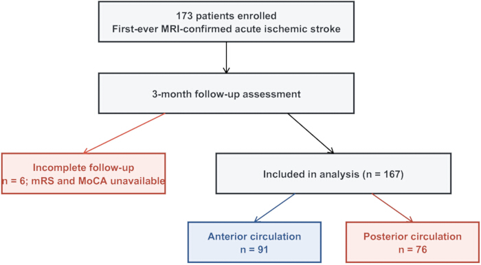
Figure 1: Study flow. Among 173 enrolled patients with first-ever MRI-confirmed acute ischemic stroke, six did not complete the 3-month outcome assessment. The analysis included 167 patients: 91 with anterior and 76 with posterior circulation infarction. MRI, magnetic resonance imaging. Please click here to view a larger version of this figure.
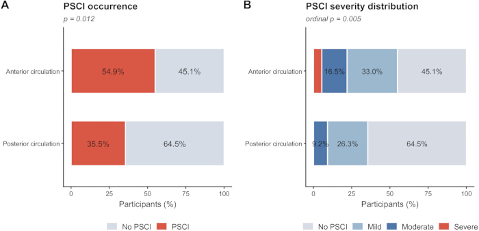
Figure 2: Post-stroke cognitive impairment at 3 months. (A) Patients with and without MoCA-defined PSCI in the anterior and posterior circulation groups (Pearson chi-square p = 0.012). (B) Distribution of no, mild, moderate, and severe PSCI according to the study categories (ordinal comparison p = 0.005). Abbreviations: MoCA = Montreal Cognitive Assessment; PSCI = post-stroke cognitive impairment. Please click here to view a larger version of this figure.
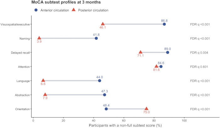
Figure 3: MoCA subtest profiles at 3 months. Points show the percentage of patients with a score below the maximum for each subtest in the anterior and posterior circulation groups. Labels report Benjamini–Hochberg false discovery rate q-values for the non-full-score comparisons. The non-full-score measure is descriptive and does not constitute a validated domain-specific diagnosis. Please click here to view a larger version of this figure.
| Characteristic | Anterior | Posterior | p value | Test |
| Age, years | 62.02 ± 10.06 | 62.74 ± 8.53 | 0.625 | Independent-samples t-test |
| Admission NIHSS score | 2 (1-3) | 3 (2-4) | 0.015 | Mann-Whitney U test |
| Male sex | 61 (67.03) | 54 (71.05) | 0.576 | Pearson chi-square test |
| Hypertension | 66 (72.53) | 56 (73.68) | 0.867 | Pearson chi-square test |
| Diabetes mellitus | 34 (37.36) | 31 (40.79) | 0.651 | Pearson chi-square test |
| Coronary heart disease | 7 (7.69) | 7 (9.21) | 0.724 | Pearson chi-square test |
| Hyperlipidemia | 31 (34.07) | 16 (21.05) | 0.063 | Pearson chi-square test |
| Smoking | 30 (32.97) | 29 (38.16) | 0.485 | Pearson chi-square test |
| Heavy alcohol use | 24 (26.37) | 23 (30.26) | 0.578 | Pearson chi-square test |
| Education ≤12 years | 51 (56.04) | 36 (47.37) | 0.264 | Pearson chi-square test |
Table 1: Baseline characteristics of patients with anterior and posterior circulation infarction. Data are mean ± standard deviation, median (interquartile range), or n (%). Abbreviations: NIHSS = National Institutes of Health Stroke Scale.
| Outcome | Anterior | Posterior | Effect / test | p value |
| PSCI, n (%) | 50 (54.95) | 27 (35.53) | Unadjusted OR 2.21 (95% CI 1.18-4.14) | 0.012 |
| No PSCI, n (%) | 41 (45.05) | 49 (64.47) | ||
| Mild PSCI, n (%) | 30 (32.97) | 20 (26.32) | Ordinal comparison | 0.005 |
| Moderate PSCI, n (%) | 15 (16.48) | 7 (9.21) | ||
| Severe PSCI, n (%) | 5 (5.49) | 0 (0.00) | ||
| Education-adjusted MoCA score | 25 (19-27) | 26 (25-28) | Mann-Whitney U test | 0.004 |
| mRS score | 3 (0-3) | 2 (1-3) | Mann-Whitney U test | 0.996 |
| Anterior vs posterior circulation | Exposure | Reference | Adjusted OR 2.18 (95% CI 1.06-4.49) | 0.035 |
Table 2: Cognitive and functional outcomes at 3 months. The adjusted odds ratio was obtained from a logistic regression model containing vascular territory, age, sex, education ≤12 years, admission NIHSS score, hypertension, diabetes, and hyperlipidemia. Abbreviations: CI = confidence interval; MoCA = Montreal Cognitive Assessment; mRS = modified Rankin Scale; PSCI = post-stroke cognitive impairment.
| MoCA subtest | Anterior score, median (IQR) | Posterior score, median (IQR) | Score raw p | Score FDR q | Anterior non-full, n (%) | Posterior non-full, n (%) | Non-full raw p | Non-full FDR q |
| Visuospatial/executive | 4 (2-4) | 5 (4-5) | <0.001 | <0.001 | 79 (86.81) | 35 (46.05) | <0.001 | <0.001 |
| Naming | 3 (2-3) | 3 (3-3) | <0.001 | <0.001 | 38 (41.76) | 3 (3.95) | <0.001 | <0.001 |
| Delayed recall | 4 (2-4) | 4 (4-5) | <0.001 | <0.001 | 81 (89.01) | 54 (71.05) | 0.003 | 0.004 |
| Attention | 4 (3-5) | 5 (4-5) | 0.149 | 0.149 | 77 (84.62) | 62 (81.58) | 0.601 | 0.601 |
| Language | 3 (2-3) | 3 (3-3) | <0.001 | <0.001 | 40 (43.96) | 5 (6.58) | <0.001 | <0.001 |
| Abstraction | 2 (1-2) | 2 (2-2) | <0.001 | <0.001 | 43 (47.25) | 6 (7.89) | <0.001 | <0.001 |
| Orientation | 6 (5-6) | 5 (4-6) | <0.001 | <0.001 | 44 (48.35) | 57 (75.00) | <0.001 | <0.001 |
Table 3: MoCA subtest results by vascular territory. Data are median (interquartile range) or n (%). Raw p values and Benjamini–Hochberg false-discovery-rate q values are reported. A non-full subtest score is any score below the maximum and is a descriptive exploratory measure. MoCA, Montreal Cognitive Assessment.
SupplementaryFigure 1: Distribution of education-adjusted Montreal Cognitive Assessment (MoCA) scores by vascular territory. Boxes show the interquartile range, center lines show medians, and points show individual observations.Please click here to download this file.
Supplementary Figure 2: Multivariable associations with post-stroke cognitive impairment (PSCI). Points show adjusted odds ratios, and horizontal lines show 95% confidence intervals from the prespecified logistic model. Abbreviations: NIHSS = National Institutes of Health Stroke Scale.Please click here to download this file.
Supplementary Table 1: Available baseline characteristics of the six participants who did not complete the 3-month follow-up. This table summarizes the available demographic and clinical characteristics of participants lost to follow-up. These data provide context for assessing potential attrition bias in the study.Please click here to download this file.
Supplementary Table 2: Lesion laterality, cortical involvement, multiplicity, and principal anatomical site by vascular territory. This table presents the distribution of lesion characteristics across different vascular territories. It provides a detailed overview of the anatomical patterns and distribution of ischemic lesions among participants.Please click here to download this file.
Supplementary Table 3: Full multivariable logistic regression model for the primary PSCI outcome. This table presents the complete regression model, including the covariates evaluated for their association with post-stroke cognitive impairment (PSCI). Adjusted odds ratios, confidence intervals, and p values are reported to facilitate interpretation of the associations.Please click here to download this file.
Supplementary Table 4: Sensitivity analysis using education-adjusted MoCA <22. This table presents the sensitivity analysis performed using an education-adjusted Montreal Cognitive Assessment (MoCA) score below 22 as the threshold for cognitive impairment. The same covariates as in the primary model were included to assess the robustness of the findings.Please click here to download this file.
Supplementary Table 5: Deidentified participant-level dataset and data dictionary. The Excel workbook contains separate Data and Data Dictionary worksheets. The Data worksheet provides deidentified participant-level information, while the Data Dictionary defines the variables and their coding to facilitate interpretation and reproducibility.Please click here to download this file.
Supplementary File 1: R analysis code used to reproduce the reported tables, statistical estimates, and figures.Please click here to download this file.
Dyskusja
In this cohort, 46.1% of patients met the primary education-adjusted MoCA definition of PSCI 3 months after a first-ever ischemic stroke. PSCI was more frequent, and the severity distribution was less favorable, in the anterior circulation group. The association persisted after adjustment and in the <22 sensitivity analysis, but the wide confidence intervals and residual confounding preclude causal interpretation. False-discovery-rate-adjusted analyses supported group differences in several MoCA subtests, whereas attention did not differ. These screening results describe group-level score distributions and not isolated cognitive-domain diagnoses. The overall PSCI frequency is within the range reported in previous reviews, although selection, timing, and diagnostic definitions limit direct comparison3,7,8,9,22.
The higher PSCI frequency in the anterior circulation group may relate to differences in affected networks, but the broad territory classification contains substantial anatomical heterogeneity. In this cohort, anterior lesions were most often centered in the basal ganglia, frontal lobe, or temporal lobe, whereas posterior lesions were most often in the brainstem, cerebellum, medial temporal lobe, thalamus, or occipital lobe (Supplementary Table 2). Left-right distributions were similar, but cortical involvement was more frequent in the anterior group and subcortical involvement in the posterior group. These descriptive differences could contribute to the observed cognitive pattern23,24,25. Strategic location and network disconnection may matter as much as lesion volume15,16,17; however, infarct volume, automated segmentation, and lesion-network mapping were unavailable. Territory, therefore, cannot be interpreted as the sole anatomical explanation or used for individual prediction.
The admission NIHSS score was one point higher in the posterior circulation group despite a lower PSCI rate. This apparent divergence may reflect the scale's unequal sensitivity to anterior and posterior circulation signs and differences between acute focal deficits and later cognitive network effects26,27. NIHSS is not a surrogate for lesion volume, especially in posterior circulation stroke. Adjustment for admission NIHSS did not remove the association, but unmeasured lesion burden, stroke subtype, and other clinical factors could still explain part of the difference. Similar 3-month mRS scores further indicate that a global disability scale does not capture all aspects of cognitive recovery.
The study used routine clinical assessments, 3.0 T MRI, prespecified vascular-territory classification, and an in-person Beijing MoCA at 3 months. The conventional <26 threshold was used for comparability with the original MoCA framework. Results were similar when PSCI was defined using the lower, stroke-specific cutoff of <22 supported by diagnostic-accuracy evidence20,21,22. Nevertheless, cutoff performance depends on language, education, case mix, and reference diagnosis. A low MoCA score should prompt contextual clinical assessment rather than serve as a stand-alone diagnosis. Rehabilitation planning should remain individualized according to symptoms, lesion features, mood, sensory function, education, and daily-life impact28.
This study has several limitations. First, it was conducted at one center with a modest sample, including only 27 primary-outcome cases in the posterior circulation group. The adjusted estimate was imprecise, and the approximate observed-effect post hoc power was 71.6%; subgroup analyses were likely underpowered. Second, exclusion of patients unable to complete the MoCA and those with marked atrophy or confluent leukoaraiosis probably selected milder survivors and may have underestimated PSCI prevalence, limiting generalizability. Third, pre-stroke cognition was based on history rather than a validated informant instrument such as the IQCODE or AD8, so pre-existing decline may have been misclassified as post-stroke impairment. Fourth, the model did not include depression, anxiety, sleep, fatigue, medication effects, recurrent events, stroke subtype, white-matter-disease burden, lesion volume, or network disconnection. No participant received reperfusion therapy, preventing evaluation of that factor and limiting applicability to treated populations. Fifth, six participants lacked follow-up outcomes, and reasons for noncompletion were unavailable. Finally, the MoCA is a screening instrument with subtest ceiling effects; non-full scores collapse different degrees of deficit, and even false-discovery-rate-adjusted subtest findings cannot establish domain-specific diagnoses.
In summary, anterior circulation infarction was associated with a higher frequency and less favorable severity distribution of MoCA-defined PSCI at 3 months in this cohort. The association was directionally consistent under <26 and <22 thresholds, and mRS scores were similar between groups. The findings support direct cognitive screening during follow-up but remain group-level associations based on broad vascular-territory classification. They do not establish a causal or independently predictive role for territory and should not be used to infer an individual's cognitive profile. Multicenter studies incorporating prospective power calculation, validated pre-stroke cognitive assessment, detailed volumetric lesion mapping, formal neuropsychological testing, and longitudinal follow-up are required.
Oświadczenia
The authors declare that they have no competing interests.
AUTHORS’ CONTRIBUTIONS:
Yuan Zhang contributed to study design, patient assessment, data management, statistical analysis, and drafting of the manuscript. Yang Zhang, Jianfeng Li, and Yuxuan Gu contributed to patient assessment, follow-up, and data management. Shibo Dong reviewed the imaging and contributed to the imaging methods. Hongshan Chu supervised clinical quality control and reviewed the manuscript. Hebo Wang conceived and supervised the study, obtained funding, and reviewed the manuscript. All authors approved the final version.
Podziękowania
This study was supported by the Medical Science Research Project of Hebei (Grant No. 20230342). The authors thank the neurology and imaging staff of Hebei General Hospital for assistance with patient assessment and data checking. During revision, the authors used OpenAI Codex for language editing. The authors reviewed and verified all resulting content and take full responsibility for the manuscript.
Materiały
| Nazwa | Firma | Numer katalogowy | Komentarze |
|---|---|---|---|
| 3.0 T magnetic resonance imaging system | Philips Medical Systems | Achieva 3.0 T | Scanner used for T1WI, T2WI, FLAIR, DWI, and TOF-MRA |
| IBM SPSS Statistics | IBM Corp. | Version 27.0 | Statistical analysis |
| Modified Rankin Scale | Not applicable | 0–6 scale | Three-month functional outcome assessment |
| Montreal Cognitive Assessment | MoCA Clinic and Institute | Authorized Beijing version | Standard full 30-point cognitive screening instrument |
| National Institutes of Health Stroke Scale | NINDS | NIHSS | Admission neurological deficit assessment |
Bibliografia
- GBD 2019 Stroke Collaborators. Global, regional, and national burden of stroke and its risk factors, 1990-2019: a systematic analysis for the Global Burden of Disease Study 2019. Lancet Neurol. 2021;20(10):795-820.
- Feigin VL, et al. World Stroke Organization: Global Stroke Fact Sheet 2025. Int J Stroke. 2025;20(2):132-144.
- El Husseini N, et al. Cognitive impairment after ischemic and hemorrhagic stroke: a scientific statement from the American Heart Association/American Stroke Association. Stroke. 2023;54(6):e272-e291.
- Gorelick PB, et al. Vascular contributions to cognitive impairment and dementia: a statement for healthcare professionals from the American Heart Association/American Stroke Association. Stroke. 2011;42(9):2672-2713.
- Sachdev P, et al. Diagnostic criteria for vascular cognitive disorders: a VASCOG statement. Alzheimer Dis Assoc Disord. 2014;28(3):206-218.
- Pendlebury ST, Rothwell PM. Prevalence, incidence, and factors associated with pre-stroke and post-stroke dementia: a systematic review and meta-analysis. Lancet Neurol. 2009;8(11):1006-1018.
- Sexton E, et al. Systematic review and meta-analysis of the prevalence of cognitive impairment no dementia in the first year post-stroke. Eur Stroke J. 2019;4(2):160-171.
- Barbay M, et al. Systematic review and meta-analysis of prevalence in post-stroke neurocognitive disorders in hospital-based studies. Dement Geriatr Cogn Disord. 2018;46(5-6):322-334.
- Mijajlović MD, et al. Post-stroke dementia - a comprehensive review. BMC Med. 2017;15(1):11.
- Filler J, et al. Risk factors for cognitive impairment and dementia after stroke: a systematic review and meta-analysis. Lancet Healthy Longev. 2024;5(1):e31-e44.
- Nasreddine ZS, et al. The Montreal Cognitive Assessment, MoCA: a brief screening tool for mild cognitive impairment. J Am Geriatr Soc. 2005;53(4):695-699.
- Shi D, Chen X, Li Z. Diagnostic test accuracy of the Montreal Cognitive Assessment in the detection of post-stroke cognitive impairment under different stages and cutoffs: a systematic review and meta-analysis. Neurol Sci. 2018;39(4):705-716.
- Quinn TJ, et al. European Stroke Organization and European Academy of Neurology joint guidelines on post-stroke cognitive impairment. Eur J Neurol. 2021;28(12):3883-3920.
- Jokinen H, et al. Post-stroke cognitive impairment is common even after successful clinical recovery. Eur J Neurol. 2015;22(9):1288-1294.
- Weaver NA, et al. Strategic infarct locations for post-stroke cognitive impairment: a pooled analysis of individual patient data from 12 acute ischaemic stroke cohorts. Lancet Neurol. 2021;20(6):448-459.
- Zhao L, et al. Strategic infarct location for post-stroke cognitive impairment: a multivariate lesion-symptom mapping study. J Cereb Blood Flow Metab. 2018;38(8):1299-1311.
- Biesbroek JM, Weaver NA, Biessels GJ. Lesion location and cognitive impact of cerebral small vessel disease. Clin Sci (Lond). 2017;131(8):715-728.
- von Elm E, et al. The Strengthening the Reporting of Observational Studies in Epidemiology (STROBE) statement: guidelines for reporting observational studies. PLoS Med. 2007;4(10):e296.
- Bamford J, et al. Classification and natural history of clinically identifiable subtypes of cerebral infarction. Lancet. 1991;337(8756):1521-1526.
- Yu J, Li J, Huang X. The Beijing version of the Montreal Cognitive Assessment as a brief screening tool for mild cognitive impairment: a community-based study. BMC Psychiatry. 2012;12:156.
- Zuo L, et al. Screening for cognitive impairment with the Montreal Cognitive Assessment in Chinese patients with acute mild stroke and transient ischaemic attack: a validation study. BMJ Open. 2016;6(7):e011310.
- Wei X, et al. Which cutoff value of the Montreal Cognitive Assessment should be used for post-stroke cognitive impairment? A systematic review and meta-analysis on diagnostic test accuracy. Int J Stroke. 2023;18(8):908-916.
- Cummings JL. Frontal-subcortical circuits and human behavior. Arch Neurol. 1993;50(8):873-880.
- Aggleton JP, Brown MW. Episodic memory, amnesia, and the hippocampal-anterior thalamic axis. Behav Brain Sci. 1999;22(3):425-444; discussion 444-489.
- Kraft A, et al. Neurological and neuropsychological characteristics of occipital, occipito-temporal and occipito-parietal infarction. Cortex. 2014;56:38-50.
- Sato S, et al. Baseline NIH Stroke Scale score predicting outcome in anterior and posterior circulation strokes. Neurology. 2008;70(24 Pt 2):2371-2377.
- Kazi SA, Siddiqui M, Majid S. Stroke outcome prediction using admission NIHSS in anterior and posterior circulation stroke. J Ayub Med Coll Abbottabad. 2021;33(2):274-278.
- Winstein CJ, et al. Guidelines for adult stroke rehabilitation and recovery: a guideline for healthcare professionals from the American Heart Association/American Stroke Association. Stroke. 2016;47(6):e98-e169.