This narrative review evaluates shared injury mechanisms across regulated cell-death pathways in ischemic stroke. It distinguishes causal evidence from pathway-associated markers and integrates cell type, injury phase, and neurovascular context to identify testable therapeutic priorities.
Artykuł przeglądowy
Molecular Networks of Cell Death in Ischemic Stroke: From Mechanisms to Interventions
W tym artykule
Podsumowanie
Streszczenie
Ischemic stroke involves a complex network of interconnected processes, including bioenergetic failure, excitotoxicity, ionic dyshomeostasis involving Na+, K+, Ca2+, and Cl−, oxidative stress, DNA damage, and neurovascular dysfunction. Apoptosis, necroptosis, pyroptosis, ferroptosis, and parthanatos contribute to experimental ischemic injury with different levels of mechanistic support. Autophagy primarily maintains cellular homeostasis and modulates these pathways. Autophagy-dependent cell death requires separate evidence that the autophagic machinery causes death. These processes operate across the ischemic core and penumbra and evolve during ischemia, reperfusion, and recovery. Their effects extend beyond neurons to microglia, astrocytes, brain microvascular endothelial cells, pericytes, and other neurovascular components. This narrative review evaluates pathway execution, shared molecular nodes, and the spatial and temporal limits of the available evidence. It emphasizes receptor-complex regulation, mitochondrial and lysosomal function, phospholipid peroxidation, DNA damage, and inflammatory signaling as points of convergence. Preclinical interventions are assessed according to their models, molecular endpoints, and functional outcomes. A cell- and stage-specific network perspective supports the development of targeted therapies, while the efficacy and safety of coordinated pathway intervention in patients remain to be established.
Wprowadzenie
Stroke remains a leading cause of death and long-term disability worldwide. The Global Burden of Disease 2021 analysis describes a substantial burden across 204 countries and territories, including ischemic and hemorrhagic subtypes1. Intravenous thrombolysis and mechanical thrombectomy restore perfusion in eligible patients, but treatment windows and eligibility restrict access. Reperfusion injury, hemorrhagic transformation, and persistent neurological injury are additional challenges after treatment, rather than equivalent restrictions on its application2,3,4. Recanalization therefore does not resolve every component of tissue injury. Bioenergetic failure, glutamate excitotoxicity, disturbed Na+, K+, Ca2+, and Cl− homeostasis, excessive reactive oxygen species (ROS) generation, endoplasmic reticulum (ER) stress, and deoxyribonucleic acid (DNA) damage affect the neurovascular unit (NVU) and its surrounding microenvironment. Neurons, astrocytes, microglia, oligodendrocytes, pericytes, and cerebrovascular endothelial cells can be contributors to, and targets of, this injury5,6,7.
The Nomenclature Committee on Cell Death (NCCD) defines regulated cell death (RCD) by molecular mechanisms that can be modified genetically or pharmacologically8. This framework moves beyond a morphological division into necrosis and apoptosis. Nevertheless, the involvement of a protein associated with an RCD pathway does not establish execution of that pathway. Experimental support for apoptosis, necroptosis, pyroptosis, ferroptosis, and parthanatos differs by cell type, model, and endpoint. Their respective contributions in patients are less clearly resolved2,7,9,10. Autophagy requires particular care: it is principally a lysosomal degradation and adaptation process, whereas autophagy-dependent cell death (ADCD) requires a causal dependence of death on autophagy machinery8,11. This review therefore examines autophagy as a regulator of injury and survival, without treating every autophagy-associated phenotype as a sixth established death modality in stroke.
Broad reviews by Tuo et al., Mao et al., and Ren et al. already describe neuronal death mechanisms and therapeutic targets2,3,9. The contribution of this review is an evidence-qualified synthesis of their intersections across neurovascular cell types and injury phases. This review connects pathway-specific execution criteria to a spatial and temporal map, then evaluates whether shared nodes support causal coupling, parallel responses, or only association. Recent ferroptosis and autophagy-ferroptosis reviews inform this network perspective12,13,14. Cuproptosis is an emerging, putative additional mechanism in ischemic stroke, but its evidence base does not justify assigning it equal status here15. The therapeutic sections use this synthesis to identify testable combinations and timing constraints, rather than infer clinical efficacy from molecular overlap.
Przegląd i perspektywa
1. Scope and literature-search approach
This narrative review began with the existing reference collection and reviewer-recommended literature; the initial collection had no prospectively recorded search or screening protocol. A citation-led update on October 6, 2026, used Europe PMC to access MEDLINE/PubMed and PubMed Central records. Six searches combined “ischemic stroke” OR “cerebral ischemia” with apoptosis, necroptosis, autophagy, pyroptosis, ferroptosis, or parthanatos. Publication-date filters extended through the search date without a restrictive lower limit. DOI and bibliographic checks and consultation of reference lists, publisher records, and repositories clarified mechanisms and experimental models.
Selection was purposive, prioritizing primary focal-ischemia, ischemia/reperfusion, and oxygen-glucose deprivation studies with pathway perturbation, execution endpoints, or functional outcomes. Foundational nonstroke studies defined mechanisms; reviews provided context. Intracerebral-hemorrhage-only studies did not establish ischemic-stroke mechanisms, and expression-only findings were treated as associations. No effect-size pooling or formal risk-of-bias assessment was performed. Selection and publication bias, experimental-model concentration, and limited human evidence constrain this synthesis; it does not claim systematic coverage.
2. Core mechanisms and standards of evidence
- Apoptosis
Apoptosis involves characteristic cell shrinkage, chromatin condensation, nuclear fragmentation, membrane blebbing, apoptotic-body formation, and phagocytic clearance16. Death receptors recruit Fas-associated death domain protein (FADD) and death-inducing signaling complex (DISC) machinery to activate caspase-8/1017,18. Caspase-8 activates caspase-3/7 or cleaves BH3-interacting domain death agonist (BID), connecting receptor signals to mitochondria19. Tumor necrosis factor receptor 1 (TNFR1) complex I supports survival/inflammation; cytosolic complexes can favor apoptosis or necroptosis20,21. B-cell lymphoma 2 (BCL-2)-associated X protein (BAX) and BCL-2 antagonist/killer 1 (BAK) promote mitochondrial outer membrane permeabilization (MOMP), cytochrome c release, and apoptotic protease-activating factor 1 (Apaf-1)/caspase-9 activation22,23. Limited MOMP may be sublethal, so mitochondrial injury alone is insufficient evidence of irreversible apoptosis24 (Figure 1).
Mitochondria also regulate ROS and metabolism and participate in other death-associated stress responses. Their dysfunction is therefore a convergence point, whereas cytochrome c-dependent caspase activation supplies more specific evidence of mitochondrial apoptosis. Long non-coding ribonucleic acids regulate apoptotic networks in experimental stroke25. Distinguishing initiation, execution, and sublethal stress prevents assignment of all mitochondrial injury to one program24. - Necroptosis
Canonical necroptotic execution requires receptor-interacting protein kinase 3 (RIPK3) and mixed lineage kinase domain-like protein (MLKL). Receptor-interacting protein kinase 1 (RIPK1)-dependent complex assembly leads to MLKL phosphorylation, oligomerization, and membrane injury26,27,28,29. RIPK1 is not universally required: Toll-like receptor signaling through Toll/interleukin-1 receptor-domain-containing adaptor-inducing interferon-β (TRIF), or Z-DNA-binding protein 1 (ZBP1), can recruit RIPK3 independently8,30. Endosomal sorting complexes required for transport III (ESCRT-III) can repair MLKL-associated membrane damage and delay lysis31. Protein abundance and inflammatory changes therefore cannot replace execution measurements (Figure 2). - Autophagy
Macroautophagy delivers cytoplasmic cargo to lysosomes11. Beclin 1 (BECN1)/class III phosphatidylinositol 3-kinase (PI3KC3) supports nucleation, while autophagy-related (ATG) proteins form ATG12-ATG5-ATG16L1 conjugation machinery supporting microtubule-associated protein 1 light chain 3 (LC3) lipidation11,32. Mechanistic target of rapamycin complex 1 (mTORC1) restrains unc-51-like kinase 1 (ULK1); AMP-activated protein kinase (AMPK) inhibits mTORC1 and can directly phosphorylate ULK14,33,34. Lysosomal calcium channels supply additional nutrient-response regulation35. LC3-II accumulation can reflect increased formation or impaired clearance. Dynamic flux measurements, lysosomal controls, and interpretation of sequestosome 1 (p62) are necessary11. Autophagy-dependent cell death (ADCD) requires death to depend causally on autophagic machinery (Figure 3).
BECN1 and ATG proteins also affect development, adaptation, and cellular clearance. Protection or injury after their manipulation is not automatically an autophagy-specific death phenotype11,32. Effective lysosomal degradation must be distinguished from accumulation of autophagic intermediates. These controls are particularly important when an intervention changes several stress responses simultaneously11. - Pyroptosis
Inflammasomes activate caspase-1, which matures interleukin (IL)-1β/IL-18 and cleaves gasdermin D (GSDMD); its N-terminal fragment forms pores36,37,38. Cytosolic lipopolysaccharide (LPS) activates human caspase-4/5 or murine caspase-11, which also cleave GSDMD39,40. Caspase-3-mediated gasdermin E (GSDME) cleavage is a distinct route41. Inflammasome assembly, cytokine release, pore formation, and terminal lysis are distinguishable. Nicorandil-associated suppression of cleaved caspase-1/GSDMD supplies intervention evidence, whereas GSDMD deletion, transplantation, and neutrophil depletion support causal peripheral-immune contribution42,43 (Figure 4).
The noncanonical LPS-sensing mechanism defines a general gasdermin route; its description does not establish an LPS-driven sequence in ischemic brain. Similarly, GSDME cleavage by caspase-3 should not be depicted as direct cleavage by caspase-4/5/11. Cell identity and localization of cleaved execution factors determine the interpretation of tissue-level signals8,41. - Ferroptosis
Ferroptosis combines iron-dependent phospholipid peroxidation with insufficient antioxidant defense44,45,46,47. Nuclear receptor coactivator 4 (NCOA4)-dependent ferritinophagy releases stored iron48,49. Acyl-coenzyme A synthetase long-chain family member 4 (ACSL4) supplies oxidation-sensitive lipid substrates50. System xc− comprises solute carrier family 7 member 11 (SLC7A11) and solute carrier family 3 member 2 (SLC3A2), supporting cystine uptake and glutathione (GSH) synthesis. GSH-dependent glutathione peroxidase 4 (GPX4) reduces phospholipid hydroperoxides44,51. Ferroptosis suppressor protein 1 (FSP1)/coenzyme Q (CoQ) and mitochondrial dihydroorotate dehydrogenase (DHODH) provide parallel defenses52,53. GTP cyclohydrolase 1 (GCH1)/tetrahydrobiopterin (BH4) adds another defense system46. Lipid oxidation should be paired with complementary endpoints and pharmacological, preferably genetic, rescue (Figure 5). Broader neurological iron-homeostasis frameworks provide mechanistic context without establishing stroke-specific causality54. - Parthanatos
Excessive poly(ADP-ribose) polymerase 1 (PARP1) activity produces poly(ADP-ribose) (PAR), promotes apoptosis-inducing factor (AIF) redistribution, and connects DNA damage to caspase-independent chromatin degradation55,56,57. Depletion of oxidized nicotinamide adenine dinucleotide (NAD+) contributes to injury but is not diagnostic56. AIF recruits macrophage migration inhibitory factor (MIF), whose nuclease activity executes DNA cleavage. Disrupting AIF-MIF interaction or MIF nuclease activity protected the experimental ischemic brain58. This intracellular role differs from MIF cytokine signaling (Figure 6). - Interpreting execution and causal evidence
Assignment requires molecular execution, death measurements, and causal perturbation8. Terminal deoxynucleotidyl transferase dUTP nick end labeling (TUNEL), lactate dehydrogenase (LDH), reactive oxygen species (ROS), cytokines, and total protein abundance are nonspecific. Infarct or behavioral improvement establishes relevance, but can reflect vascular, immune, or metabolic effects. Genetic rescue, target engagement, and concordant interventions strengthen attribution. Table 1 summarizes evidence combinations11,47,59; Figure 7 maps regulatory intersections whose causal strength is considered below.
Pharmacological rescue is more informative when dose, timing, off-target effects, and tissue target engagement are measured. Genetic loss should ideally be complemented by re-expression or a separation-of-function mutant. Concordant interventions at different pathway levels help distinguish execution-specific protection from broad cytoprotection. These standards guide the appraisal; they do not imply that every cited experiment satisfies all criteria8.
3. Regulated cell death and autophagy in ischemic stroke
- Apoptosis
Apoptotic signaling is prominent in peri-infarct tissue but is not exclusively penumbral60. Mild transient ischemia studies showed delayed DNA fragmentation, caspase activation, and caspase-inhibitor protection61,62. A window extending to 9 h in a 30-min mouse middle cerebral artery occlusion (MCAO) model cannot define a clinical window. Fas ligand (FasL) mutation supports receptor involvement, while FasL-associated microglial responses complicate exclusively neuronal attribution63,64. IL-17A and endoplasmic reticulum (ER)-stress interventions connect inflammation with apoptotic signaling65,66,67. BCL-2 overexpression reduced neuronal loss and cytochrome c/caspase activation68. X-linked inhibitor of apoptosis protein (XIAP) and second mitochondria-derived activator of caspases (SMAC), also termed direct inhibitor-of-apoptosis-binding protein with low isoelectric point (DIABLO), further regulate execution69. - Necroptosis
RIPK1 phosphorylation, RIPK3/MLKL activation, and necrostatin-1 protection support necroptotic signaling in rat ischemia70,71. Complementary RIPK1 kinase-inactive, RIPK3-deficient, and MLKL-deficient mouse experiments reduced infarction, neurological injury, and inflammation72. Neuronal signaling modifies microglial/macrophage responses, while microglial tumor necrosis factor-α (TNF-α) promotes endothelial injury and blood-brain barrier (BBB) disruption73,74. N-myc downstream-regulated gene 2 (NDRG2)/RIPK1-associated astrocyte findings and voltage-dependent anion channel 1 (VDAC1) oligomerization extend the cellular and mitochondrial context75,76,77, without substituting for MLKL execution evidence.
TUNEL colocalization in the pharmacological experiments identifies DNA injury, not a necroptosis-specific endpoint70. Genetic protection provides complementary support, but reduced inflammation can accompany altered death signaling and must be assessed independently72. The neuronal, astrocytic, and vascular results together argue for cell-resolved analysis rather than attribution of an entire infarct to a single executor. - Autophagy
Mitochondrial quality control, energy stress, and lysosomal capacity shape the autophagic response78. Kaempferol and epigallocatechin-3-gallate studies reported protection alongside different autophagy-marker changes79,80; they cannot establish a universal protective-early/lethal-late rule. Histone H4 lysine 16 acetylation research linked protection to restored lysosomal function and bafilomycin-sensitive effects81. Endothelial autophagy-associated occludin degradation can instead impair the BBB82. Piezo-type mechanosensitive ion channel component 1 (PIEZO1), hypoxia-inducible factor 1α (HIF-1α)/BCL-2-interacting protein 3 (BNIP3), and ER stress provide further regulatory intersections83,84,85,86. These findings concern flux, substrates, and adaptation; ADCD still requires causal demonstration.
Persistent stress during reperfusion can impair lysosomal clearance and convert an initially adaptive response into ineffective cargo processing. Neuronal organelle maintenance and endothelial degradation of a junctional substrate have different consequences81,82. Cell type, stress intensity, treatment timing, and substrate therefore explain more than a simple increase or decrease in autophagosome abundance. - Pyroptosis
Neuron-localized cleaved caspase-1 with cortical GSDMD cleavage supports inflammatory signaling, but does not establish neuron-specific execution87. GSDMD-ablation experiments primarily support a microglial contribution88. Stimulator of interferon genes (STING) and NLR family pyrin domain-containing 3 (NLRP3) perturbation support microglial regulatory coupling89; C-X3-C motif chemokine ligand 1 (CX3CL1) and the CHRNA7-FAM7A fusion gene (CHRFAM7A) act as modulators90,91. Astrocytic lipocalin-2 (LCN2)/24p3 receptor (24p3R) signaling and endothelial NLRP3-associated injury identify additional contexts92,93,94. Table 2 separates cell localization, inflammasome activation, gasdermin execution, and evidential limits.
Astrocytic LCN2 loss/re-expression and receptor interference strengthen the regulatory connection to NLRP3-associated injury92. Endothelial inhibitor-associated protection should remain distinct from endothelial execution-factor genetic rescue93. Whole-brain cytokines can originate from resident or recruited immune cells, and gasdermin pores can precede terminal lysis. Neither cytokine reduction nor cortical cleavage alone resolves the affected cell population. - Ferroptosis
Mitochondrial ferritin deficiency worsened iron/lipid oxidation and injury, while NCOA4-mediated ferritin degradation supplies another susceptibility mechanism95,96. Thrombin-linked ACSL4 activation and ACSL4 manipulation connect reperfusion/coagulation with lipid remodeling97,98. Astragaloside IV-associated ACSL4 RNA modification suggests upstream regulation99. Nuclear factor erythroid 2-related factor 2 (NRF2) deficiency and pharmacological modulation support antioxidant protection100. SLC7A11 modification and GPX4 regulation add molecular control points101,102. These results do not make any single iron or antioxidant marker diagnostic of ferroptosis.
NRF2 participates in antioxidant, inflammatory, and metabolic responses, so its benefit cannot be reduced to an exclusive GPX4 switch. Parallel FSP1/CoQ and DHODH defenses also affect susceptibility52,53. Their relative stroke-specific contributions across cells and injury stages remain incompletely mapped103. Total iron, lipid-damage products, or altered GPX4 abundance must be interpreted alongside rescue and execution measurements47. - Parthanatos
During ischemia/reperfusion, superoxide reacts with nitric oxide to generate peroxynitrite, connecting excitotoxic and inflammatory signals with oxidative/nitrosative DNA damage and PARP1 hyperactivation104. PARP1 inhibition/deficiency reduces experimental injury105, but also affects repair and inflammation. PAR accumulation, nuclear AIF redistribution, chromatin degradation, and AIF-MIF perturbation provide stronger attribution58. Caspase-independent death alone is insufficient. - Spatial, temporal, and cellular organization
Core and penumbra evolve with perfusion and metabolic capacity. Reperfusion changes oxidative load, immune access, and vascular permeability2,4. Transient MCAO research linked reperfusion-associated necroptosis to vascular injury and subsequent transforming growth factor-β-activated kinase 1 (TAK1) loss to apoptotic signaling106. This is a model-specific sequence, not a universal pathway transition. Endothelial ferroportin 1 (FPN1) deletion reduced acute iron-related injury but impaired recovery when deficiency persisted107. Neutrophil injury and endothelial occludin loss further demonstrate that cell identity and substrate matter43,82. Single-cell analysis of 11-keto-β-boswellic acid (KBA) plus Z-guggulsterone suggested distinct microglial and astrocytic responses108. Table 3 and Figure 8 organize these observations. Pericytes, oligodendrocytes, later repair phases, and human cell-resolved execution remain insufficiently mapped. Single-cell/spatial signatures generate hypotheses and require genetic and serial execution measurements.
Binary proinflammatory/anti-inflammatory microglial labels compress more diverse states. Serial sampling and perfusion measurements are needed to determine whether programs occur sequentially within one cell, concurrently in different cells, or through intercellular amplification. Age, sex, diabetes, hypertension, and other clinically relevant conditions should be incorporated when testing whether an experimental network persists across stroke settings.
4. Crosstalk among cell death pathways
- Receptor complexes and RIPK1 regulation
Caspase-8 availability, RIPK1 activity, ubiquitin scaffolds, and cellular stress determine receptor-associated branching. Disruptor of telomeric silencing 1-like (DOT1L) and protein arginine methyltransferase 1 (PRMT1) studies identify epigenetic regulation of RIPK1-associated apoptosis/necroptosis109,110. Active MLKL can stimulate NLRP3, providing a connection to inflammatory amplification111. These findings support shared regulation, not an obligatory three-pathway sequence.
DOT1L modifies the signaling environment and should not be represented as a structural component of the TNFR1 complex109. RIPK1-independent recruitment of RIPK3 also limits a universal TNFR1/RIPK1 cascade. The strongest available interpretation is a regulated branch point and inflammatory amplification, with the resulting execution route dependent on the cellular context. - Mitochondria and lysosomes
BCL-2 phosphorylation and BECN1 connect apoptotic restraint with autophagy in remote-conditioning experiments112. Myeloid cell leukemia 1 (MCL1)/BECN1 colocalization is spatial association, not proof that MCL1 maintains protective flux113. RIPK1 inhibition accompanies altered LC3/cathepsin B and heat shock protein 70.1B-related lysosomal stabilization114,115. Oridonin connects RIPK3/mitophagy-associated changes with caspase-9 injury116. Organelle preservation may lower several death signals without directly switching execution pathways.
Resolving the MCL1/BECN1 hypothesis requires perturbing their interaction while measuring flux and apoptotic execution in the same cells113. Likewise, static LC3 changes or inhibitor protection cannot determine the causal direction of RIPK1-associated lysosomal injury. Preserving organelle function may suppress several downstream signals without proving direct coupling between their execution mechanisms. - Oxidative stress and lipid peroxidation
Ferritin-related and YTH N6-methyladenosine RNA-binding protein 1 (YTHDF1)-dependent tumor protein p53 studies connect genotoxic stress with SLC7A11 and lipid injury117,118. Because p53 also regulates apoptosis, separation-of-function experiments are needed to establish direct coupling. High mobility group box 1 (HMGB1)/alpha kinase 1 (ALPK1) perturbation alters microglial NLRP3/caspase-1/GSDMD and ferroptosis-associated readouts119. A common regulator and overlapping oxidative markers do not prove that either execution mechanism obligatorily activates the other.
ACSL4 provides a complementary metabolic intersection between vascular/coagulation signals and membrane susceptibility97,98. Changes in lipid substrates and inflammatory markers do not establish a direct ACSL4-to-gasdermin execution pathway. Lipidomics, temporal sampling, and cell-specific perturbation should distinguish shared substrate regulation from a sequence in which one death mechanism triggers another. - DNA damage and inflammasomes
Myeloid-derived MIF-associated RIPK1 vascular injury differs from intracellular MIF nuclease-mediated parthanatos120. RIPK3-AIF interaction in global ischemia suggests another intersection whose relation to MLKL and MIF requires clarification121. Spautin-1-associated suppression of inflammasome/gasdermin signals cannot establish that excessive autophagy caused pyroptosis, because the drug also affects ubiquitin-specific peptidases 10 and 13 (USP10/USP13)122. Taurine-upregulated gene 1 (TUG1) knockdown-associated phosphatase and tensin homolog-induced kinase 1 (PINK1)/Parkin mitophagy illustrates quality-control restraint of inflammatory injury123. Subcellular location and dynamic flux distinguish these possibilities. - PANoptosis
PANoptosis integrates pyroptotic, apoptotic, and necroptotic machinery through coordinated complexes124,125,126. Mixed-tissue marker overlap is insufficient. Ischemia experiments reported apoptosis-associated speck-like protein containing a caspase recruitment domain (ASC) co-immunoprecipitation with ZBP1, NLRP3, caspase-8, and phosphorylated MLKL, with S100 calcium-binding protein A10 (S100A10)/sonic hedgehog-associated modulation127. This supports complex interactions, while component necessity remains incompletely established. Vagus nerve stimulation supplied preclinical sirtuin 1 (SIRT1)-associated intervention evidence128. NAD+-sensing NLR family caspase recruitment domain-containing 5 (NLRC5) PANoptosomes outside stroke generate hypotheses rather than demonstrate a parthanatos-to-PANoptosis bridge in ischemic brain129.
S100A10 knockdown exacerbated experimental injury, while recombinant sonic hedgehog modified the response127. Complex interaction evidence advances the framework beyond coincident markers, but does not establish that every associated component is necessary for neuronal death or rescue. Circulating SIRT1 associations likewise cannot demonstrate clinical efficacy of PANoptosis-directed treatment128. These distinctions keep coordinated execution separate from upstream regulatory association.
5. Therapeutic targeting and translation
- Antiapoptotic strategies
Intranasal Fas-blocking peptide and systemic conjugate studies support receptor-targeted delivery, with possible inflammatory contributions130,131. Artemisinin-associated mitochondrial/caspase protection differs from postischemic caspase rescue132. Pretreatment, genetic susceptibility, and treatment after onset must be distinguished; reduced injury alone does not establish selective antiapoptotic action (Table 4).
Artemisinin experiments included pretreatment of neuronal models before oxygen-glucose deprivation/reoxygenation; inhibition or silencing of extracellular signal-regulated kinase (ERK) weakened protection132. This supports a regulatory link but not a treatment window after clinical onset. Mitochondrial membrane potential, ROS, and caspase measurements should be interpreted together, and broad mitochondrial preservation should not be described as selective inhibition of one apoptotic step. - Antinecroptotic strategies
MLKL-directed and necrostatin-related interventions remain preclinical and require species-specific target validation30,133. EX-527, a SIRT1 inhibitor, reduced infarction and improved survival without improving neurological deficits in one focal-ischemia study134. Histological protection, functional recovery, and inhibition of MLKL-dependent lysis are separate outcomes. - Autophagy and lysosomal function
Astrocyte exosomal nicotinamide phosphoribosyltransferase (NAMPT) and ginsenoside Rg1 studies support different AMPK/mTOR-associated interventions135,136. Hypoxic preconditioning establishes adaptation, not treatment efficacy after onset137. Therapy should preserve effective cargo clearance; changes in LC3 or p62 require dynamic controls and cannot establish selective ADCD modulation11,81.
Increased LC3-II does not establish enhanced flux, and reduced LC3-II can reflect either less formation or better clearance. Interpretation of p62 also requires consideration of transcription11. The lysosomal study is more informative because clearance and bafilomycin-sensitive benefit were examined81. Treatment should be chosen according to the affected substrate and cell population rather than the direction of a static marker change. - Antipyroptotic strategies
MCC950-associated NLRP3 inhibition, including treatment beginning 24 h after mouse stroke, and VX-765-associated caspase-1 inhibition support intervention at different signaling levels138,139. Cytokine reduction alone does not prove prevention of gasdermin-mediated death. Cellular target engagement and functional outcomes should accompany execution measurements.
The delayed MCC950 experiment evaluated subacute injury after treatment initiated at 24 h, illustrating a model-specific opportunity beyond the earliest ischemic phase138. It does not establish a universal window or demonstrate that all anti-inflammatory benefit represents prevented pyroptotic death. Resident microglia, recruited leukocytes, and endothelial cells require separate exposure and execution assessment. - Antiferroptotic strategies
Iron availability, lipid remodeling, and antioxidant capacity provide distinct targets. Hydroxysafflor yellow A and rosmarinic-acid liposomes support ferritinophagy/iron-homeostasis hypotheses140,141. Baicalein and calycosin studies implicate lipid oxidation and ACSL4-related susceptibility142,143. NRF2-linked barrier protection supplies another context144. Persistent FPN1 suppression may impair recovery107; genetic knockout is not itself treatment. Antioxidant and immune effects require comparison with selective ferroptosis perturbations.
Calycosin-associated benefit was weakened by ACSL4 overexpression, strengthening the substrate-susceptibility connection while leaving broader drug effects possible143. Iron-transport interventions should also distinguish acute protection from repair. Comparisons with genetic perturbation and lipid-oxidation rescue are needed before assigning the complete effect of an antioxidant or delivery formulation to ferroptosis inhibition. - Parthanatos-related interventions
PJ34 and olaparib supply PARP-related cytoprotection without establishing exclusive parthanatos inhibition145,146. Crocetin studies measured PAR and nuclear AIF alongside oxidant/mitochondrial changes147, strengthening mechanistic attribution. AIF-MIF perturbation offers greater execution specificity. Dose, delayed administration, physiological DNA repair, and permanent-versus-reperfused ischemia require explicit consideration.
Olaparib showed protection within a limited tested dose range rather than a monotonic response146. Crocetin's permanent-occlusion findings cannot be transferred automatically to reperfused stroke147. PAR accumulation and nuclear AIF strengthen pathway attribution, but preservation of repair, metabolism, and inflammation remains relevant when assessing PARP-directed benefit and adverse effects. - Cell-specific combinations and translational priorities
NRF2-related antioxidant capacity is a shared regulatory node. Acetyl-11-keto-β-boswellic acid (AKBA) was associated with NRF2/heme oxygenase 1 (HO-1)-related protection in experimental cerebral ischemia and oxygen-glucose deprivation; the original report has a published figure correction148,149. KBA plus Z-guggulsterone is a different combination that produced distinct microglial and astrocytic transcriptomic responses108. Such findings support cell-specific hypotheses, not proof of pathway elimination or clinical superiority. A salvianolic-acid review connects redox, inflammatory, and organelle mechanisms with therapeutic development and clinical formulations150. Formulation-level findings cannot establish pathway-specific efficacy in patients. Combinations need component-versus-combination controls, tissue exposure, target engagement, and serial execution measurements. Spatial mapping should be paired with cell-specific perturbation. Testing alongside reperfusion in aged/comorbid models should assess hemorrhage, sustained neurological function, and repair. Network-directed therapy remains a research strategy requiring defined cellular targets and treatment phases.
Wnioski
Ischemic brain injury involves interconnected stress responses and regulated death mechanisms whose contributions vary across cells, regions, and time. Apoptosis has extensive mechanistic support, while necroptosis, pyroptosis, ferroptosis, and parthanatos have strengthened the experimental understanding of vascular, inflammatory, lipid, and genotoxic injury. Their relative execution in human stroke remains less certain. Autophagy principally regulates homeostasis and other death pathways. ADCD requires a separate causal demonstration. Shared molecular nodes and emerging PANoptosome evidence provide a basis for testing coordinated intervention, while marker overlap alone does not prove pathway switching or combination efficacy. Cell-specific perturbation, temporal sampling, and clinically relevant functional outcomes are needed to turn this network framework into targeted therapeutic hypotheses.
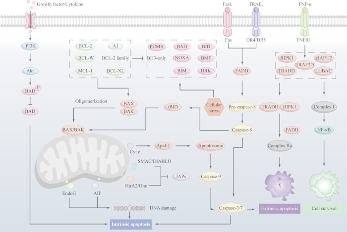
Figure 1: Apoptotic signaling pathways. Death receptors recruit adaptor and initiator-caspase complexes. Caspase-8 links receptor signaling to mitochondria through BID cleavage. BAX/BAK-mediated mitochondrial outer membrane permeabilization (MOMP) permits cytochrome c release, apoptotic protease-activating factor 1 (Apaf-1)-dependent apoptosome formation, and caspase-9/3/7 activation. B-cell lymphoma 2 (BCL-2)-family proteins and inhibitor-of-apoptosis proteins regulate these steps. Tumor necrosis factor receptor 1 (TNFR1) complex I supports survival and inflammatory signaling, whereas cytosolic death complexes can promote apoptosis or necroptosis. Extensive MOMP commonly supports death commitment, whereas limited MOMP is not necessarily immediately lethal. Apoptosis-inducing factor (AIF)/endonuclease G (EndoG)-associated caspase-independent effects are distinguished from classical caspase-dependent apoptosis. The schematic summarizes general molecular relationships rather than a proven sequence in every ischemic cell. Original schematic created by the authors. Please click here to view a larger version of this figure.
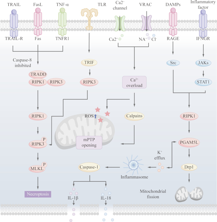
Figure 2: Necroptotic signaling. TNFR1-associated receptor-interacting protein kinase 1 (RIPK1) signaling can recruit receptor-interacting protein kinase 3 (RIPK3) and mixed lineage kinase domain-like protein (MLKL) when the regulatory context permits necroptosis. Toll/interleukin-1 receptor-domain-containing adaptor-inducing interferon-β (TRIF)-dependent Toll-like receptor signaling provides a RIPK1-independent route to RIPK3. MLKL phosphorylation and membrane injury represent central execution events. Mitochondrial, calcium, and inflammatory signals contribute to contextual amplification rather than universal pathway requirements. Endosomal sorting complexes required for transport III (ESCRT-III)-mediated membrane repair can oppose terminal lysis. Changes in protein expression or cytokine release alone do not establish necroptotic execution. Original schematic created by the authors. Please click here to view a larger version of this figure.
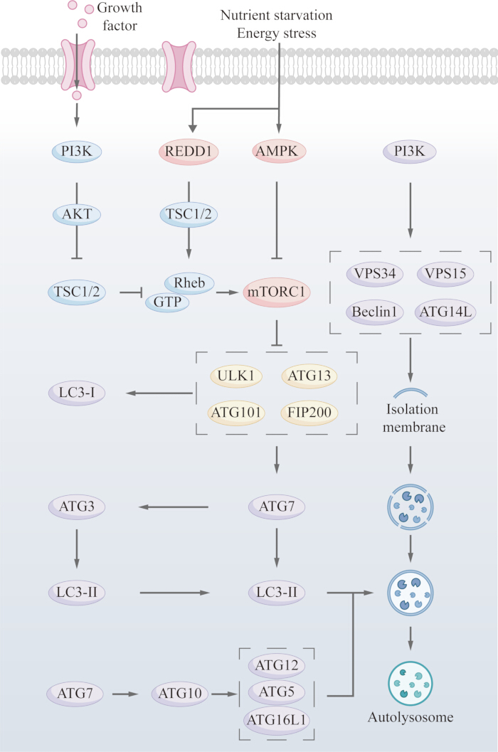
Figure 3: Regulation of autophagy. Growth-factor and nutrient signals regulate mechanistic target of rapamycin complex 1 (mTORC1), while AMP-activated protein kinase (AMPK) links energy stress to mTORC1 inhibition and unc-51-like kinase 1 (ULK1) activation. Class III phosphatidylinositol 3-kinase (PI3KC3)/Beclin 1 (BECN1)-associated nucleation and autophagy-related (ATG) conjugation systems support microtubule-associated protein 1 light chain 3 (LC3) lipidation and autophagosome formation. Fusion with lysosomes and effective cargo degradation complete autophagic flux. Increased autophagosome abundance may reflect enhanced formation or impaired clearance. The schematic illustrates autophagy regulation rather than evidence of autophagy-dependent cell death. Original schematic created by the authors. Please click here to view a larger version of this figure.
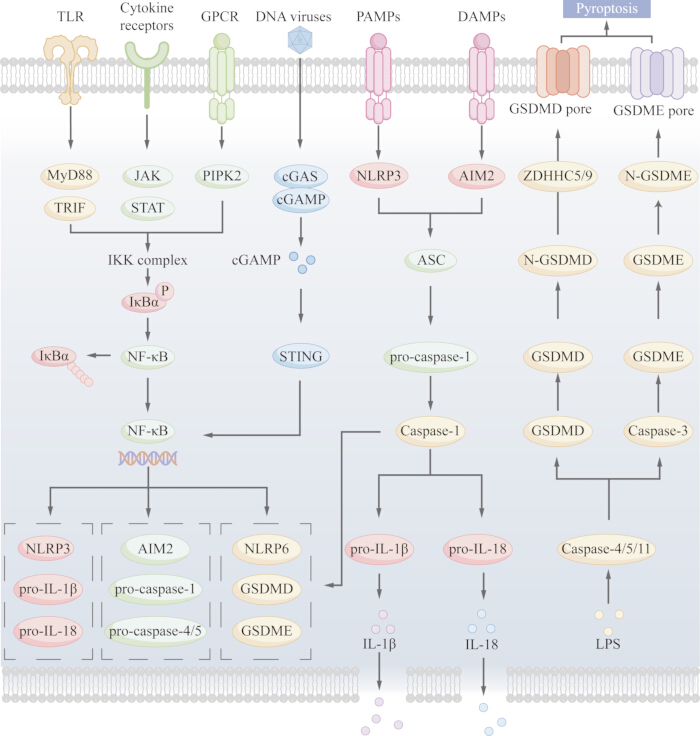
Figure 4: Pyroptotic signaling pathways. Receptor-associated inflammatory signaling and cyclic GMP-AMP synthase (cGAS)/cyclic GMP-AMP (cGAMP)/stimulator of interferon genes (STING) signaling connect innate immune sensing with nuclear factor κB (NF-κB)-associated priming. Inflammasome-mediated caspase-1 activation promotes cytokine maturation and gasdermin D (GSDMD) cleavage, while gasdermin N-terminal fragments form membrane pores. Cytosolic lipopolysaccharide (LPS) sensing by human caspase-4/5 or murine caspase-11 activates GSDMD. Caspase-3-mediated gasdermin E (GSDME) cleavage represents a distinct route and should not be interpreted as direct cleavage by caspase-4/5/11. The schematic does not establish a universal signaling sequence in ischemic cells. Table 2 summarizes cell-specific findings involving microglia, neurons, astrocytes, and brain endothelial cells, distinguishing execution evidence from association and treating C-X3-C motif chemokine ligand 1 (CX3CL1) and the CHRNA7-FAM7A fusion gene (CHRFAM7A) as modulators8,41,87–94. Original schematic created by the authors. Please click here to view a larger version of this figure.
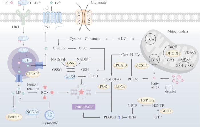
Figure 5: Ferroptotic signaling pathways. Iron transport and nuclear receptor coactivator 4 (NCOA4)-associated ferritinophagy influence the labile iron pool. Acyl-coenzyme A synthetase long-chain family member 4 (ACSL4)-dependent lipid remodeling supplies oxidation-sensitive phospholipids. System xc−/glutathione (GSH)/glutathione peroxidase 4 (GPX4), ferroptosis suppressor protein 1 (FSP1)/coenzyme Q (CoQ), dihydroorotate dehydrogenase (DHODH), and GTP cyclohydrolase 1 (GCH1)/tetrahydrobiopterin (BH4) provide complementary antioxidant defenses. Their relative contributions depend on cellular context and are not equally established in ischemic stroke. Phospholipid peroxidation is central to ferroptotic execution, whereas iron accumulation or changes in individual markers are insufficient for pathway identification. Original schematic created by the authors. Please click here to view a larger version of this figure.
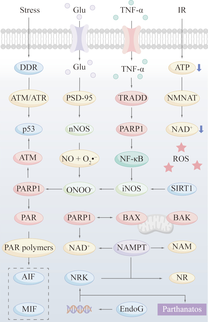
Figure 6: Parthanatos signaling. Excitotoxic and oxidative/nitrosative signals can produce DNA damage and excessive poly(ADP-ribose) polymerase 1 (PARP1) activity. Poly(ADP-ribose) (PAR) accumulation connects nuclear injury with mitochondrial apoptosis-inducing factor (AIF) release and redistribution. AIF recruits macrophage migration inhibitory factor (MIF), whose nuclease activity executes large-scale nuclear DNA cleavage. MIF-mediated cleavage is distinguished from other caspase-independent nuclear mechanisms, including endonuclease G (EndoG)-associated effects. Nicotinamide adenine dinucleotide (NAD+) consumption and bioenergetic impairment contribute to cellular injury but are not specific diagnostic features. Physiological PARP1-dependent DNA repair is distinguished from excessive death-associated PAR signaling. The supported parthanatos execution mechanism is described in the cited literature56,58,59,104. Original schematic created by the authors. Please click here to view a larger version of this figure.
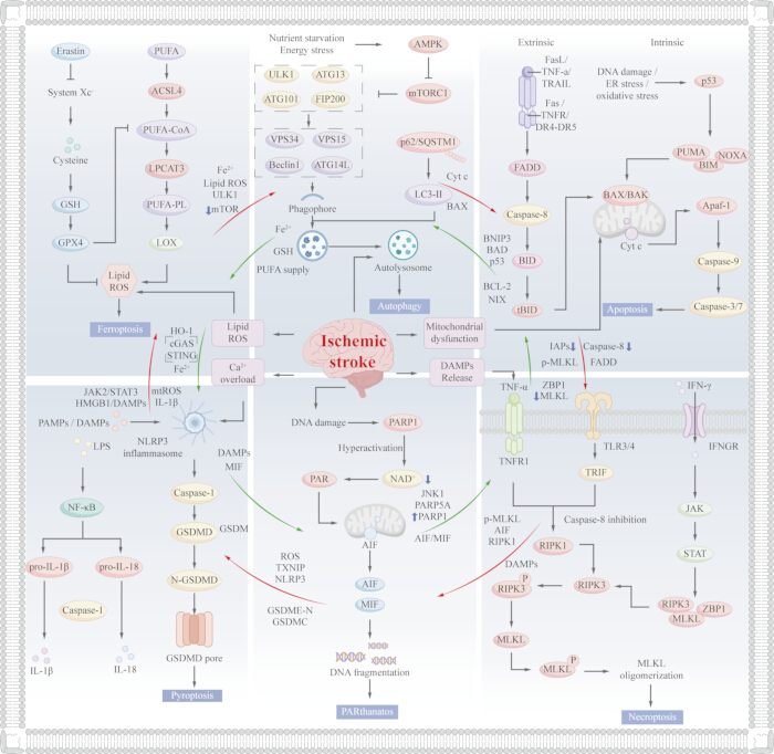
Figure 7: Interactions among cell death pathways. Shared receptor signaling, organelle dysfunction, oxidative stress, and DNA damage connect multiple regulated cell death pathways. Autophagy is represented as a regulatory process rather than evidence of autophagy-dependent cell death (ADCD). Cross-pathway arrows indicate reported or proposed regulatory interactions and do not establish that every connection operates within the same cell or that all pathways activate simultaneously. PANoptosis requires additional evidence of coordinated death machinery and relevant molecular complex interactions. Multinode therapeutic intervention remains a hypothesis requiring cell- and injury-phase-specific experimental validation. Original schematic created by the authors. Please click here to view a larger version of this figure.
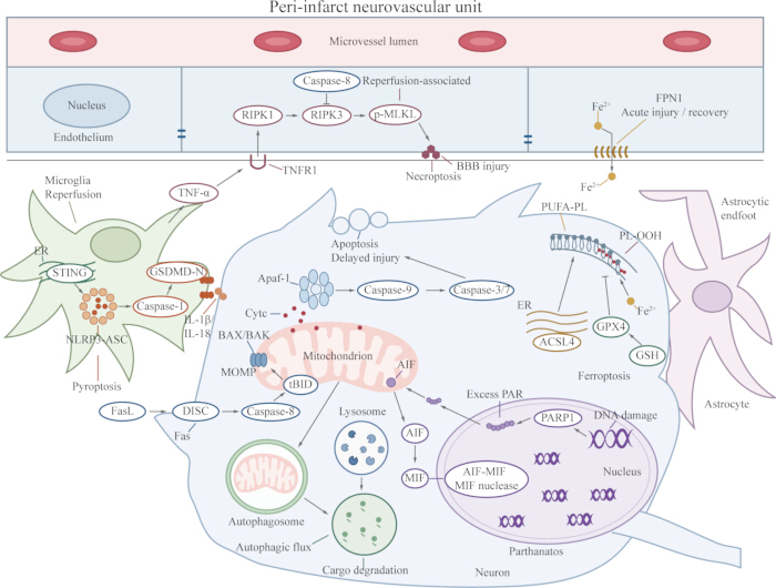
Figure 8: Cell death in the neurovascular unit. A microvessel, perivascular microglia, astrocytic endfeet, and a neuron illustrate representative mechanisms in the peri-infarct environment. Microglial tumor necrosis factor-α (TNF-α) engages endothelial TNFR1/RIPK1/RIPK3/MLKL signaling in a death-complex-permissive context, with caspase-8-mediated restraint indicated. Membrane injury is depicted locally rather than as a separate outcome panel74. Microglial STING-associated NLR family pyrin domain-containing 3 (NLRP3)/caspase-1/GSDMD signaling and cytokine release are shown at their relevant cellular sites89. Within neurons, mitochondria connect BAX/BAK-dependent permeabilization, cytochrome c/apoptosome/caspase signaling, and AIF redistribution. Nuclear PARP1/PAR signaling recruits the AIF-MIF mechanism, in which MIF executes DNA cleavage58. ACSL4-associated phospholipid remodeling, iron-dependent lipid injury, and GSH/GPX4 antioxidant defense are depicted near membrane lipids97,98. Autophagosomes and lysosomes illustrate completed cargo degradation rather than an assumed autophagy-dependent death program11. Endothelial ferroportin 1 (FPN1)-mediated iron export is represented as transport, with phase-dependent implications for acute injury and recovery107. Arrows indicate signaling or transport, potentially involving intermediate steps, while inhibition bars indicate regulatory restraint. Cell outlines organize representative findings without implying simultaneous activation of all mechanisms. Timing labels are descriptive; model-dependent chronology, core/penumbra distinctions, and evidence gaps are summarized in Table 3. Original schematic created for this review. Please click here to view a larger version of this figure.
Table 1: Evidence for cell death pathway identification. Summary of complementary molecular and functional evidence used to identify regulated cell death pathways. Rescue experiments should be interpreted alongside target engagement and appropriate controls. Terminal deoxynucleotidyl transferase dUTP nick end labeling (TUNEL) and lactate dehydrogenase (LDH) measurements are not pathway-specific8,11,47,59. Abbreviations: MDA = malondialdehyde; 4-HNE = 4-hydroxynonenal. Please click here to download this file.
Table 2: Cell-specific pyroptotic signaling. Summary of pyroptosis-associated signaling in neurons, microglia, astrocytes, and endothelial cells, distinguishing molecular associations from evidence of pathway execution. Arrows indicate supported signaling relationships or execution frameworks but do not imply that every step was demonstrated in every experimental model. Canonical caspase-1/GSDMD, noncanonical human caspase-4/5 or murine caspase-11/GSDMD, and caspase-3/GSDME signaling represent distinct routes8,41. Inflammasome assembly, cytokine maturation, gasdermin cleavage, pore formation, and terminal lysis require separate interpretation. Abbreviations: OGD/R = oxygen-glucose deprivation/reoxygenation; MCAO/R = middle cerebral artery occlusion/reperfusion. Please click here to download this file.
Table 3: Spatial and temporal patterns of cell death. Summary of representative cell-specific mechanisms across ischemic injury, reperfusion, and recovery. The stages are descriptive and may overlap. Evidence remains incomplete for pericytes, oligodendrocytes, aged or comorbid models, and human cell-resolved pathway execution. Abbreviations: I/R = ischemia/reperfusion; Cdh5 = cadherin 5; Fpn1 = ferroportin 1. Please click here to download this file.
Table 4: Therapeutic strategies targeting cell death. Summary of selected experimental interventions, treatment conditions, and molecular or functional evidence relevant to therapeutic interpretation. Findings from genetic knockout experiments establish susceptibility rather than directly deliverable treatments, and preclinical outcomes do not establish clinical efficacy or a generalizable treatment window. Abbreviations: tMCAO = transient middle cerebral artery occlusion; PC12 = rat pheochromocytoma cell line; ERK = extracellular signal-regulated kinase; IgG = immunoglobulin G; NOX2 = NADPH oxidase 2. Please click here to download this file.
Oświadczenia
The authors have no relevant financial or non-financial interests to disclose.
Podziękowania
This work was supported by the Heilongjiang Provincial Multidisciplinary Innovation Team for Traditional Chinese Medicine. The funder had no role in the conceptualization, writing, decision to publish, or preparation of the manuscript.
Bibliografia
- GBD 2021 Stroke Risk Factor Collaborators. Global, regional, and national burden of stroke and its risk factors, 1990-2021: a systematic analysis for the Global Burden of Disease Study 2021. Lancet Neurol. 2024;23(10):973-1003. doi: 10.1016/S1474-4422(24)00369-7.
- Tuo QZ, Zhang ST, Lei P. Mechanisms of neuronal cell death in ischemic stroke and their therapeutic implications. Med Res Rev. 2022;42(1):259-305. doi: 10.1002/med.21817.
- Mao R et al. Neuronal Death Mechanisms and Therapeutic Strategy in Ischemic Stroke. Neurosci Bull. 2022;38(10):1229-1247. doi: 10.1007/s12264-022-00859-0.
- Qin C et al. Signaling pathways involved in ischemic stroke: molecular mechanisms and therapeutic interventions. Signal Transduct Target Ther. 2022;7(1):215. doi: 10.1038/s41392-022-01064-1.
- Li Y et al. Cell polarization in ischemic stroke: molecular mechanisms and advances. Neural Regen Res. 2025;20(3):632-645. doi: 10.4103/NRR.NRR-D-23-01336.
- Duan WL et al. Molecular mechanisms of programmed cell death and potential targeted pharmacotherapy in ischemic stroke (Review). Int J Mol Med. 2025;56(1):103. doi: 10.3892/ijmm.2025.5544.
- Daneshpour A, Shaka Z, Rezaei N. Interplay of cell death pathways and immune responses in ischemic stroke: insights into novel biomarkers. Rev Neurosci. 2025;36(4):371-403. doi: 10.1515/revneuro-2024-0128.
- Galluzzi L et al. Molecular mechanisms of cell death: recommendations of the Nomenclature Committee on Cell Death 2018. Cell Death Differ. 2018;25(3):486-541. doi: 10.1038/s41418-017-0012-4.
- Ren K et al. Regulated necrosis pathways: a potential target for ischemic stroke. Burns Trauma. 2023;11:tkad016. doi: 10.1093/burnst/tkad016.
- Koehler RC, Dawson VL, Dawson TM. Targeting Parthanatos in Ischemic Stroke. Front Neurol. 2021;12:662034. doi: 10.3389/fneur.2021.662034.
- Klionsky DJ et al. Guidelines for the use and interpretation of assays for monitoring autophagy (4th edition). Autophagy. 2021;17(1):1-382. doi: 10.1080/15548627.2020.1797280.
- Tian X et al. Progress of Ferroptosis in Ischemic Stroke and Therapeutic Targets. Cell Mol Neurobiol. 2024;44(1):25. doi: 10.1007/s10571-024-01457-6.
- Gowtham A, Chauhan C, Rahi V, Kaundal RK. An update on the role of ferroptosis in ischemic stroke: from molecular pathways to Neuroprotection. Expert Opin Ther Targets. 2024;28(12):1149-1175. doi: 10.1080/14728222.2024.2446319.
- Zhang XY et al. Crosstalk between autophagy and ferroptosis mediate injury in ischemic stroke by generating reactive oxygen species. Heliyon. 2024;10(7):e28959. doi: 10.1016/j.heliyon.2024.e28959.
- Wang J, Lv C, Wei X, Li F. Molecular mechanisms and therapeutic strategies for ferroptosis and cuproptosis in ischemic stroke. Brain Behav Immun Health. 2024;40:100837. doi: 10.1016/j.bbih.2024.100837.
- Kerr JFR, Wyllie AH, Currie AR. Apoptosis: A Basic Biological Phenomenon with Wide-ranging Implications in Tissue Kinetics. Br J Cancer. 1972;26(4):239-257. doi: 10.1038/bjc.1972.33.
- Chinnaiyan AM, O'Rourke K, Tewari M, Dixit VM. FADD, a novel death domain-containing protein, interacts with the death domain of fas and initiates apoptosis. Cell. 1995;81(4):505-512. doi: 10.1016/0092-8674(95)90071-3.
- Muzio M et al. FLICE, A Novel FADD-Homologous ICE/CED-3-like Protease, Is Recruited to the CD95 (Fas/APO-1) Death-Inducing Signaling Complex. Cell. 1996;85(6):817-827. doi: 10.1016/s0092-8674(00)81266-0.
- Luo X et al. Bid, a Bcl2 Interacting Protein, Mediates Cytochrome c Release from Mitochondria in Response to Activation of Cell Surface Death Receptors. Cell. 1998;94(4):481-490. doi: 10.1016/s0092-8674(00)81589-5.
- Liu ZG, Hsu H, Goeddel DV, Karin M. Dissection of TNF Receptor 1 Effector Functions: JNK Activation Is Not Linked to Apoptosis While NF-κB Activation Prevents Cell Death. Cell. 1996;87(3):565-576. doi: 10.1016/s0092-8674(00)81375-6.
- Hsu H, Shu HB, Pan MG, Goeddel DV. TRADD-TRAF2 and TRADD-FADD Interactions Define Two Distinct TNF Receptor 1 Signal Transduction Pathways. Cell. 1996;84(2):299-308. doi: 10.1016/s0092-8674(00)80984-8.
- Li P et al. Cytochrome c and dATP-Dependent Formation of Apaf-1/Caspase-9 Complex Initiates an Apoptotic Protease Cascade. Cell. 1997;91(4):479-489. doi: 10.1016/s0092-8674(00)80434-1.
- Czabotar PE, Garcia-Saez AJ. Mechanisms of BCL-2 family proteins in mitochondrial apoptosis. Nat Rev Mol Cell Biol. 2023;24(10):732-748. doi: 10.1038/s41580-023-00629-4.
- Glover HL, Schreiner A, Dewson G, Tait SWG. Mitochondria and cell death. Nat Cell Biol. 2024;26(9):1434-1446. doi: 10.1038/s41556-024-01429-4.
- Ding JM et al. Apoptosis and long non-coding RNAs: Focus on their roles in ischemic stroke. Brain Res. 2025;1849:149346. doi: 10.1016/j.brainres.2024.149346.
- Degterev A et al. Identification of RIP1 kinase as a specific cellular target of necrostatins. Nat Chem Biol. 2008;4(5):313-321. doi: 10.1038/nchembio.83.
- Wang H et al. Mixed Lineage Kinase Domain-like Protein MLKL Causes Necrotic Membrane Disruption upon Phosphorylation by RIP3. Mol Cell. 2014;54(1):133-146. doi: 10.1016/j.molcel.2014.03.003.
- Cho Y et al. Phosphorylation-Driven Assembly of the RIP1-RIP3 Complex Regulates Programmed Necrosis and Virus-Induced Inflammation. Cell. 2009;137(6):1112-1123. doi: 10.1016/j.cell.2009.05.037.
- Sun L et al. Mixed Lineage Kinase Domain-like Protein Mediates Necrosis Signaling Downstream of RIP3 Kinase. Cell. 2012;148(1-2):213-227. doi: 10.1016/j.cell.2011.11.031.
- Hribljan V, Lisjak D, Petrović DJ, Mitrečić D. Necroptosis is one of the modalities of cell death accompanying ischemic brain stroke: from pathogenesis to therapeutic possibilities. Croat Med J. 2019;60(2):121-126. doi: 10.3325/cmj.2019.60.121.
- Gong YN et al. ESCRT-III Acts Downstream of MLKL to Regulate Necroptotic Cell Death and Its Consequences. Cell. 2017;169(2):286-300.e16. doi: 10.1016/j.cell.2017.03.020.
- Hill SM, Wrobel L, Rubinsztein DC. Post-translational modifications of Beclin 1 provide multiple strategies for autophagy regulation. Cell Death Differ. 2019;26(4):617-629. doi: 10.1038/s41418-018-0254-9.
- Qin C et al. Correction To: Signaling pathways involved in ischemic stroke: molecular mechanisms and therapeutic interventions. Signal Transduct Target Ther. 2022;7(1):278. doi: 10.1038/s41392-022-01129-1.
- Kim J, Kundu M, Viollet B, Guan KL. AMPK and mTOR regulate autophagy through direct phosphorylation of Ulk1. Nat Cell Biol. 2011;13(2):132-141. doi: 10.1038/ncb2152.
- Huang P et al. MCOLN/TRPML channels in the regulation of MTORC1 and autophagy. Autophagy. 2024;20(5):1203-1204. doi: 10.1080/15548627.2023.2300922.
- Fink SL, Cookson BT. Caspase-1-dependent pore formation during pyroptosis leads to osmotic lysis of infected host macrophages. Cell Microbiol. 2006;8(11):1812-1825. doi: 10.1111/j.1462-5822.2006.00751.x.
- Broz P, Dixit VM. Inflammasomes: mechanism of assembly, regulation and signalling. Nat Rev Immunol. 2016;16(7):407-420. doi: 10.1038/nri.2016.58.
- Shi J et al. Cleavage of GSDMD by inflammatory caspases determines pyroptotic cell death. Nature. 2015;526(7575):660-665. doi: 10.1038/nature15514.
- Kayagaki N et al. Caspase-11 cleaves gasdermin D for non-canonical inflammasome signalling. Nature. 2015;526(7575):666-671. doi: 10.1038/nature15541.
- Shi J et al. Inflammatory caspases are innate immune receptors for intracellular LPS. Nature. 2014;514(7521):187-192. doi: 10.1038/nature13683.
- Ye J et al. Hypoxanthine is a metabolic biomarker for inducing GSDME-dependent pyroptosis of endothelial cells during ischemic stroke. Theranostics. 2024;14(15):6071-6087. doi: 10.7150/thno.100090.
- Zhao C et al. ATP-sensitive potassium channel opener, Nicorandil, inhibits NF-κB/AIM2/GSDMD pathway activation to protect against neuroinflammation in ischemic stroke. Neurochem Int. 2024;179:105810. doi: 10.1016/j.neuint.2024.105810.
- Hu R et al. Gasdermin D inhibition ameliorates neutrophil mediated brain damage in acute ischemic stroke. Cell Death Discov. 2023;9(1):50. doi: 10.1038/s41420-023-01349-6.
- Yang WS et al. Regulation of Ferroptotic Cancer Cell Death by GPX4. Cell. 2014;156(1-2):317-331. doi: 10.1016/j.cell.2013.12.010.
- Stockwell BR et al. Ferroptosis: A Regulated Cell Death Nexus Linking Metabolism, Redox Biology, and Disease. Cell. 2017;171(2):273-285. doi: 10.1016/j.cell.2017.09.021.
- Stockwell BR. Ferroptosis turns 10: Emerging mechanisms, physiological functions, and therapeutic applications. Cell. 2022;185(14):2401-2421. doi: 10.1016/j.cell.2022.06.003.
- Chen X, Comish PB, Tang D, Kang R. Characteristics and Biomarkers of Ferroptosis. Front Cell Dev Biol. 2021;9:637162. doi: 10.3389/fcell.2021.637162.
- Mancias JD et al. Quantitative proteomics identifies NCOA4 as the cargo receptor mediating ferritinophagy. Nature. 2014;509(7498):105-109. doi: 10.1038/nature13148.
- Gao M et al. Ferroptosis is an autophagic cell death process. Cell Res. 2016;26(9):1021-1032. doi: 10.1038/cr.2016.95.
- Kagan VE et al. Oxidized arachidonic and adrenic PEs navigate cells to ferroptosis. Nat Chem Biol. 2017;13(1):81-90. doi: 10.1038/nchembio.2238.
- Sato M et al. The ferroptosis inducer erastin irreversibly inhibits system xc− and synergizes with cisplatin to increase cisplatin’s cytotoxicity in cancer cells. Sci Rep. 2018;8(1):968. doi: 10.1038/s41598-018-19213-4.
- Doll S et al. FSP1 is a glutathione-independent ferroptosis suppressor. Nature. 2019;575(7784):693-698. doi: 10.1038/s41586-019-1707-0.
- Mao C et al. DHODH-mediated ferroptosis defence is a targetable vulnerability in cancer. Nature. 2021;593(7860):586-590. doi: 10.1038/s41586-021-03539-7.
- Abdukarimov N, Kokabi K, Kunz J. Ferroptosis and Iron Homeostasis: Molecular Mechanisms and Neurodegenerative Disease Implications. Antioxidants. 2025;14(5):527. doi: 10.3390/antiox14050527.
- Yu SW et al. Mediation of Poly(ADP-Ribose) Polymerase-1-Dependent Cell Death by Apoptosis-Inducing Factor. Science. 2002;297(5579):259-263. doi: 10.1126/science.1072221.
- Fatokun AA, Dawson VL, Dawson TM. Parthanatos: mitochondrial-linked mechanisms and therapeutic opportunities. Br J Pharmacol. 2014;171(8):2000-2016. doi: 10.1111/bph.12416.
- Wang Y et al. Poly(ADP-Ribose) (PAR) Binding to Apoptosis-Inducing Factor Is Critical for PAR Polymerase-1-Dependent Cell Death (Parthanatos). Sci Signal. 2011;4(167):ra20. doi: 10.1126/scisignal.2000902.
- Wang Y et al. A nuclease that mediates cell death induced by DNA damage and poly(ADP-ribose) polymerase-1. Science. 2016;354(6308):aad6872. doi: 10.1126/science.aad6872.
- Yang L, Guttman L, Dawson VL, Dawson TM. Parthanatos: Mechanisms, modulation, and therapeutic prospects in neurodegenerative disease and stroke. Biochem Pharmacol. 2024;228:116174. doi: 10.1016/j.bcp.2024.116174.
- Ramos-Cabrer P, Campos F, Sobrino T, Castillo J. Targeting the Ischemic Penumbra. Stroke. 2011;42(Suppl 1):S7-11. doi: 10.1161/STROKEAHA.110.596684.
- Endres M et al. Attenuation of Delayed Neuronal Death after Mild Focal Ischemia in Mice by Inhibition of the Caspase Family. J Cereb Blood Flow Metab. 1998;18(3):238-247. doi: 10.1097/00004647-199803000-00002.
- Fink K et al. Prolonged Therapeutic Window for Ischemic Brain Damage Caused by Delayed Caspase Activation. J Cereb Blood Flow Metab. 1998;18(10):1071-1076. doi: 10.1097/00004647-199810000-00003.
- Martin-Villalba A et al. CD95 Ligand (Fas-L/APO-1L) and Tumor Necrosis Factor-Related Apoptosis-Inducing Ligand Mediate Ischemia-Induced Apoptosis in Neurons. J Neurosci. 1999;19(10):3809-3817. doi: 10.1523/JNEUROSCI.19-10-03809.1999.
- Meng HL et al. Neuronal Soluble Fas Ligand Drives M1-Microglia Polarization after Cerebral Ischemia. CNS Neurosci Ther. 2016;22(9):771-781. doi: 10.1111/cns.12575.
- Wang H et al. IL-17A exacerbates caspase-12-dependent neuronal apoptosis following ischemia through the Src-PLCγ-calpain pathway. Exp Neurol. 2024;379:114863. doi: 10.1016/j.expneurol.2024.114863.
- Wei L et al. Ginsenoside Rb1 reduced ischemic stroke-induced apoptosis through endoplasmic reticulum stress-associated IRE1/TRAF2/JNK pathway. Naunyn Schmiedebergs Arch Pharmacol. 2025;398(1):747-764. doi: 10.1007/s00210-024-03292-4.
- Xu B et al. Roflumilast prevents ischemic stroke-induced neuronal damage by restricting GSK3β-mediated oxidative stress and IRE1α/TRAF2/JNK pathway. Free Radic Biol Med. 2021;163:281-296. doi: 10.1016/j.freeradbiomed.2020.12.018.
- Zhao H et al. Bcl-2 overexpression protects against neuron loss within the ischemic margin following experimental stroke and inhibits cytochrome c translocation and caspase-3 activity. J Neurochem. 2003;85(4):1026-1036. doi: 10.1046/j.1471-4159.2003.01756.x.
- Saito A et al. Interaction between XIAP and Smac/DIABLO in the Mouse Brain after Transient Focal Cerebral Ischemia. J Cereb Blood Flow Metab. 2003;23(9):1010-1019. doi: 10.1097/01.WCB.0000080702.47016.FF.
- Deng XX, Li SS, Sun FY. Necrostatin-1 Prevents Necroptosis in Brains after Ischemic Stroke via Inhibition of RIPK1-Mediated RIPK3/MLKL Signaling. Aging Dis. 2019;10(4):807. doi: 10.14336/AD.2018.0728.
- Degterev A et al. Chemical inhibitor of nonapoptotic cell death with therapeutic potential for ischemic brain injury. Nat Chem Biol. 2005;1(2):112-119. doi: 10.1038/nchembio711.
- Zhang Y et al. Catalytically inactive RIP1 and RIP3 deficiency protect against acute ischemic stroke by inhibiting necroptosis and neuroinflammation. Cell Death Dis. 2020;11(7):565. doi: 10.1038/s41419-020-02770-w.
- Yang J et al. RIPK3/MLKL-Mediated Neuronal Necroptosis Modulates the M1/M2 Polarization of Microglia/Macrophages in the Ischemic Cortex. Cereb Cortex. 2018;28(7):2622-2635. doi: 10.1093/cercor/bhy089.
- Chen AQ et al. Microglia-derived TNF-α mediates endothelial necroptosis aggravating blood brain-barrier disruption after ischemic stroke. Cell Death Dis. 2019;10(7):487. doi: 10.1038/s41419-019-1716-9.
- Zhu J et al. NDRG2 attenuates ischemia-induced astrocyte necroptosis via the repression of RIPK1. Mol Med Rep. 2020;22(4):3103-3110. doi: 10.3892/mmr.2020.11421.
- Zhu YM et al. The Key Regulator of Necroptosis, RIP1 Kinase, Contributes to the Formation of Astrogliosis and Glial Scar in Ischemic Stroke. Transl Stroke Res. 2021;12(6):991-1017. doi: 10.1007/s12975-021-00888-3.
- Wan H et al. VDAC1, as a downstream molecule of MLKL, participates in OGD/R-induced necroptosis by inducing mitochondrial damage. Heliyon. 2024;10(1):e23426. doi: 10.1016/j.heliyon.2023.e23426.
- Wu Y et al. Bidirectional regulation of neuronal autophagy in ischemic stroke: Mechanisms and therapeutic potential. Ageing Res Rev. 2025;111:102842. doi: 10.1016/j.arr.2025.102842.
- Yuan Y et al. Kaempferol Mediated AMPK/mTOR Signal Pathway Has a Protective Effect on Cerebral Ischemic-Reperfusion Injury in Rats by Inducing Autophagy. Neurochem Res. 2022;47(8):2187-2197. doi: 10.1007/s11064-022-03604-1.
- Wang L et al. EGCG protects the mouse brain against cerebral ischemia/reperfusion injury by suppressing autophagy via the AKT/AMPK/mTOR phosphorylation pathway. Front Pharmacol. 2022;13:921394. doi: 10.3389/fphar.2022.921394.
- Yuyuan L et al. Downregulation of Histone H4 Lysine 16 Acetylation Ameliorates Autophagic Flux by Resuming Lysosomal Functions in Ischemic Neurons. ACS Chem Neurosci. 2023;14(10):1834-1844. doi: 10.1021/acschemneuro.3c00049.
- Kim KA et al. Autophagy-mediated occludin degradation contributes to blood-brain barrier disruption during ischemia in bEnd.3 brain endothelial cells and rat ischemic stroke models. Fluids Barriers CNS. 2020;17(1):21. doi: 10.1186/s12987-020-00182-8.
- Yue Y, Chen P, Ren C. Piezo1 Modulates Neuronal Autophagy and Apoptosis in Cerebral Ischemia-Reperfusion Injury Through the AMPK-mTOR Signaling Pathway. Neurochem Res. 2025;50(1):32. doi: 10.1007/s11064-024-04291-w.
- Wei R et al. Hydroxysafflor Yellow A Exerts Neuroprotective Effects via HIF-1α/BNIP3 Pathway to Activate Neuronal Autophagy after OGD/R. Cells. 2022;11(23):3726. doi: 10.3390/cells11233726.
- Sheng R et al. Autophagy regulates endoplasmic reticulum stress in ischemic preconditioning. Autophagy. 2012;8(3):310-325. doi: 10.4161/auto.18673.
- Zheng J et al. Endoplasmic reticulum stress and autophagy in cerebral ischemia/reperfusion injury: PERK as a potential target for intervention. Neural Regen Res. 2025;20(5):1455-1466. doi: 10.4103/NRR.NRR-D-23-00794.
- Wang K et al. Melatonin Enhances the Therapeutic Effect of Plasma Exosomes Against Cerebral Ischemia-Induced Pyroptosis Through the TLR4/NF-κB Pathway. Front Neurosci. 2020;14:848. doi: 10.3389/fnins.2020.00848.
- Wang K et al. Ablation of GSDMD Improves Outcome of Ischemic Stroke Through Blocking Canonical and Non-canonical Inflammasomes Dependent Pyroptosis in Microglia. Front Neurol. 2020;11:577927. doi: 10.3389/fneur.2020.577927.
- Li W et al. STING mediates microglial pyroptosis via interaction with NLRP3 in cerebral ischaemic stroke. Stroke Vasc Neurol. 2024;9(2):153-164. doi: 10.1136/svn-2023-002320.
- Ge Y et al. CX3CL1 inhibits NLRP3 inflammasome-induced microglial pyroptosis and improves neuronal function in mice with experimentally-induced ischemic stroke. Life Sci. 2022;300:120564. doi: 10.1016/j.lfs.2022.120564.
- Cao X, Wang Y, Gao L. CHRFAM7A Overexpression Attenuates Cerebral Ischemia-Reperfusion Injury via Inhibiting Microglia Pyroptosis Mediated by the NLRP3/Caspase-1 pathway. Inflammation. 2021;44(3):1023-1034. doi: 10.1007/s10753-020-01398-4.
- Li J et al. Lipocalin-2-mediated astrocyte pyroptosis promotes neuroinflammatory injury via NLRP3 inflammasome activation in cerebral ischemia/reperfusion injury. J Neuroinflammation. 2023;20(1):148. doi: 10.1186/s12974-023-02819-5.
- Bellut M et al. NLPR3 inflammasome inhibition alleviates hypoxic endothelial cell death in vitro and protects blood-brain barrier integrity in murine stroke. Cell Death Dis. 2022;13(1):20. doi: 10.1038/s41419-021-04379-z.
- Wang Y et al. Medioresinol as a novel PGC-1α activator prevents pyroptosis of endothelial cells in ischemic stroke through PPARα-GOT1 axis. Pharmacol Res. 2021;169:105640. doi: 10.1016/j.phrs.2021.105640.
- Wang P et al. Mitochondrial ferritin attenuates cerebral ischaemia/reperfusion injury by inhibiting ferroptosis. Cell Death Dis. 2021;12(5):447. doi: 10.1038/s41419-021-03725-5.
- Li C et al. Nuclear receptor coactivator 4-mediated ferritinophagy contributes to cerebral ischemia-induced ferroptosis in ischemic stroke. Pharmacol Res. 2021;174:105933. doi: 10.1016/j.phrs.2021.105933.
- Tuo QZ et al. Thrombin induces ACSL4-dependent ferroptosis during cerebral ischemia/reperfusion. Signal Transduct Target Ther. 2022;7(1):59. doi: 10.1038/s41392-022-00917-z.
- Cui Y et al. ACSL4 exacerbates ischemic stroke by promoting ferroptosis-induced brain injury and neuroinflammation. Brain Behav Immun. 2021;93:312-321. doi: 10.1016/j.bbi.2021.01.003.
- Jin Z et al. Astragaloside IV alleviates neuronal ferroptosis in ischemic stroke by regulating fat mass and obesity-associated-N6-methyladenosine-acyl-CoA synthetase long-chain family member 4 axis. J Neurochem. 2023;166(2):328-345. doi: 10.1111/jnc.15871.
- Ye Y et al. Nrf2 alleviates acute ischemic stroke induced ferroptosis via regulating xCT/GPX4 pathway. Free Radic Biol Med. 2025;231:153-162. doi: 10.1016/j.freeradbiomed.2025.02.040.
- Li Y et al. KDM2B regulates stroke injury by modulating OGT-mediated 0-GlcNAcylation of SLC7A11. Commun Biol. 2024;7(1):1530. doi: 10.1038/s42003-024-07251-w.
- Huang B et al. Palmitoylation-dependent regulation of GPX4 suppresses ferroptosis. Nat Commun. 2025;16(1):867. doi: 10.1038/s41467-025-56344-5.
- Hu X et al. The role of ferroptosis and its mechanism in ischemic stroke. Exp Neurol. 2024;372:114630. doi: 10.1016/j.expneurol.2023.114630.
- Liu S, Luo W, Wang Y. Emerging role of PARP-1 and PARthanatos in ischemic stroke. J Neurochem. 2022;160(1):74-87. doi: 10.1111/jnc.15464.
- Endres M et al. Ischemic Brain Injury is Mediated by the Activation of Poly(ADP-Ribose)Polymerase. J Cereb Blood Flow Metab. 1997;17(11):1143-1151. doi: 10.1097/00004647-199711000-00002.
- Naito MG et al. Sequential activation of necroptosis and apoptosis cooperates to mediate vascular and neural pathology in stroke. Proc Natl Acad Sci U S A. 2020;117(9):4959-4970. doi: 10.1073/pnas.1916427117.
- Zheng H et al. Cdh5-mediated Fpn1 deletion exerts neuroprotective effects during the acute phase and inhibitory effects during the recovery phase of ischemic stroke. Cell Death Dis. 2023;14(2):161. doi: 10.1038/s41419-023-05688-1.
- Liu T et al. Novel synergistic mechanism of 11-keto-β-boswellic acid and Z-Guggulsterone on ischemic stroke revealed by single-cell transcriptomics. Pharmacol Res. 2023;193:106803. doi: 10.1016/j.phrs.2023.106803.
- Wang J et al. Histone Methyltransferase Dot1L Contributes to RIPK1 Kinase-Dependent Apoptosis in Cerebral Ischemia/Reperfusion. J Am Heart Assoc. 2021;10(23):e022791. doi: 10.1161/JAHA.121.022791.
- Liu T et al. The protein arginine methyltransferase PRMT1 ameliorates cerebral ischemia-reperfusion injury by suppressing RIPK1-mediated necroptosis and apoptosis. Acta Pharm Sin B. 2025;15(8):4014-4029. doi: 10.1016/j.apsb.2025.06.005.
- Conos SA et al. Active MLKL triggers the NLRP3 inflammasome in a cell-intrinsic manner. Proc Natl Acad Sci U S A. 2017;114(6):E961-E969. doi: 10.1073/pnas.1613305114.
- Qi Z et al. Bcl-2 Phosphorylation Triggers Autophagy Switch and Reduces Mitochondrial Damage in Limb Remote Ischemic Conditioned Rats After Ischemic Stroke. Transl Stroke Res. 2015;6(3):198-206. doi: 10.1007/s12975-015-0393-y.
- Xingyong C et al. Upregulation of myeloid cell leukemia-1 potentially modulates beclin-1-dependent autophagy in ischemic stroke in rats. BMC Neurosci. 2013;14(1):56. doi: 10.1186/1471-2202-14-56.
- Ni Y et al. RIP1K Contributes to Neuronal and Astrocytic Cell Death in Ischemic Stroke via Activating Autophagic-lysosomal Pathway. Neuroscience. 2018;371:60-74. doi: 10.1016/j.neuroscience.2017.10.038.
- Du HP et al. RIPK1 inhibition contributes to lysosomal membrane stabilization in ischemic astrocytes via a lysosomal Hsp70.1B-dependent mechanism. Acta Pharmacol Sin. 2023;44(8):1549-1563. doi: 10.1038/s41401-023-01069-8.
- Li L et al. Oridonin ameliorates caspase-9-mediated brain neuronal apoptosis in mouse with ischemic stroke by inhibiting RIPK3-mediated mitophagy. Acta Pharmacol Sin. 2023;44(4):726-740. doi: 10.1038/s41401-022-00995-3.
- Chen W et al. Ferritin reduction is essential for cerebral ischemia-induced hippocampal neuronal death through p53/SLC7A11-mediated ferroptosis. Brain Res. 2021;1752:147216. doi: 10.1016/j.brainres.2020.147216.
- Chang X et al. YTHDF1 promotes p53 translation and induces ferroptosis during acute cerebral ischemia/reperfusion through m6A-dependent binding. Cell Biol Toxicol. 2025;41(1):112. doi: 10.1007/s10565-025-10061-3.
- Du O et al. ALPK1 signaling pathway activation by HMGB1 drives microglial pyroptosis and ferroptosis and brain injury after acute ischemic stroke. Int Immunopharmacol. 2025;149:114229. doi: 10.1016/j.intimp.2025.114229.
- Li Y et al. Myeloid-derived MIF drives RIPK1-mediated cerebromicrovascular endothelial cell death to exacerbate ischemic brain injury. Proc Natl Acad Sci U S A. 2023;120(5):e2219091120. doi: 10.1073/pnas.2219091120.
- Xu Y et al. RIP3 induces ischemic neuronal DNA degradation and programmed necrosis in rat via AIF. Sci Rep. 2016;6(1):29362. doi: 10.1038/srep29362.
- Liu H et al. Inhibition of autophagy-dependent pyroptosis attenuates cerebral ischaemia/reperfusion injury. J Cell Mol Med. 2021;25(11):5060-5069. doi: 10.1111/jcmm.16483.
- Yao M et al. LncRNA Tug1 Regulates Post-Stroke Microglial Pyroptosis via PINK1/Parkin-Mediated Mitophagy. Inflammation. 2025;48(4):2677-2691. doi: 10.1007/s10753-024-02219-8.
- Malireddi RKS, Kesavardhana S, Kanneganti TD. ZBP1 and TAK1: Master Regulators of NLRP3 Inflammasome/Pyroptosis, Apoptosis, and Necroptosis (PAN-optosis). Front Cell Infect Microbiol. 2019;9:406. doi: 10.3389/fcimb.2019.00406.
- Malireddi RKS et al. Innate immune priming in the absence of TAK1 drives RIPK1 kinase activity-independent pyroptosis, apoptosis, necroptosis, and inflammatory disease. J Exp Med. 2020;217(3):e20191644. doi: 10.1084/jem.20191644.
- Christgen S et al. Identification of the PANoptosome: A Molecular Platform Triggering Pyroptosis, Apoptosis, and Necroptosis (PANoptosis). Front Cell Infect Microbiol. 2020;10:237. doi: 10.3389/fcimb.2020.00237.
- Wang L et al. Shh-S100A10 axis curbs neuronal PANoptosis after ischemic stroke. Neural Regen Res. 2026;21(8):3579-3587. doi: 10.4103/NRR.NRR-D-24-01661.
- Tang H et al. Vagus nerve stimulation inhibits PANoptosis and promotes neurofunctional recovery of cerebral ischemic stroke in a Sirt1-dependent manner. Neurochem Int. 2025;184:105950. doi: 10.1016/j.neuint.2025.105950.
- Sundaram B et al. NLRC5 senses NAD+ depletion, forming a PANoptosome and driving PANoptosis and inflammation. Cell. 2024;187(15):4061-4077.e17. doi: 10.1016/j.cell.2024.05.034.
- Ullah I et al. Intranasal delivery of a Fas-blocking peptide attenuates Fas-mediated apoptosis in brain ischemia. Sci Rep. 2018;8(1):15041. doi: 10.1038/s41598-018-33296-z.
- Chung S et al. Systemic Treatment with Fas-Blocking Peptide Attenuates Apoptosis in Brain Ischemia. Int J Mol Sci. 2024;25(1):661. doi: 10.3390/ijms25010661.
- Peng T et al. Artemisinin attenuated ischemic stroke induced cell apoptosis through activation of ERK1/2/CREB/BCL-2 signaling pathway in vitro and in vivo. Int J Biol Sci. 2022;18(11):4578-4594. doi: 10.7150/ijbs.69892.
- Li W et al. Neuroprotective Effects of DTIO, A Novel Analog of Nec-1, in Acute and Chronic Stages After Ischemic Stroke. Neuroscience. 2018;390:12-29. doi: 10.1016/j.neuroscience.2018.07.044.
- Nikseresht S, Khodagholi F, Ahmadiani A. Protective effects of ex-527 on cerebral ischemia-reperfusion injury through necroptosis signaling pathway attenuation. J Cell Physiol. 2019;234(2):1816-1826. doi: 10.1002/jcp.27055.
- Deng Y et al. Astrocyte-derived exosomal nicotinamide phosphoribosyltransferase (Nampt) ameliorates ischemic stroke injury by targeting AMPK/mTOR signaling to induce autophagy. Cell Death Dis. 2022;13(12):1057. doi: 10.1038/s41419-022-05454-9.
- Xi ZC et al. Ginsenoside Rg1 mitigates cerebral ischaemia/reperfusion injury in mice by inhibiting autophagy through activation of mTOR signalling. Acta Pharmacol Sin. 2024;45(12):2474-2486. doi: 10.1038/s41401-024-01334-4.
- Lu N et al. HIF-1α/Beclin1-Mediated Autophagy Is Involved in Neuroprotection Induced by Hypoxic Preconditioning. J Mol Neurosci. 2018;66(2):238-250. doi: 10.1007/s12031-018-1162-7.
- Bellut M et al. Delayed NLRP3 inflammasome inhibition ameliorates subacute stroke progression in mice. J Neuroinflammation. 2023;20(1):4. doi: 10.1186/s12974-022-02674-w.
- Liang Y et al. Inhibition of Caspase-1 Ameliorates Ischemia-Associated Blood-Brain Barrier Dysfunction and Integrity by Suppressing Pyroptosis Activation. Front Cell Neurosci. 2021;14:540669. doi: 10.3389/fncel.2020.540669.
- Wu Y et al. Hydroxysafflor yellow A inhibits neuronal ferroptosis and ferritinophagy in ischemic stroke. Biochim Biophys Acta Mol Basis Dis. 2025;1871(6):167867. doi: 10.1016/j.bbadis.2025.167867.
- Jia CL et al. Rosmarinic acid liposomes suppress ferroptosis in ischemic brain via inhibition of TfR1 in BMECs. Phytomedicine. 2024;132:155835. doi: 10.1016/j.phymed.2024.155835.
- Li M et al. Baicalein ameliorates cerebral ischemia-reperfusion injury by inhibiting ferroptosis via regulating GPX4/ACSL4/ACSL3 axis. Chem Biol Interact. 2022;366:110137. doi: 10.1016/j.cbi.2022.110137.
- Liu H et al. Calycosin decreases cerebral ischemia/reperfusion injury by suppressing ACSL4-dependent ferroptosis. Arch Biochem Biophys. 2023;734:109488. doi: 10.1016/j.abb.2022.109488.
- Fan W et al. NRF2 activation ameliorates blood-brain barrier injury after cerebral ischemic stroke by regulating ferroptosis and inflammation. Sci Rep. 2024;14(1):5300. doi: 10.1038/s41598-024-53836-0.
- Abdelkarim G et al. Protective effects of PJ34, a novel, potent inhibitor of poly(ADP-ribose) polymerase (PARP) in in vitro and in vivo models of stroke. Int J Mol Med. 2001;7(3):255-260. doi: 10.3892/ijmm.7.3.255.
- Teng F et al. Neuroprotective Effects of Poly(ADP-ribose)polymerase Inhibitor Olaparib in Transient Cerebral Ischemia. Neurochem Res. 2016;41(7):1516-1526. doi: 10.1007/s11064-016-1864-6.
- Wu H et al. Crocetin antagonizes parthanatos in ischemic stroke via inhibiting NOX2 and preserving mitochondrial hexokinase-I. Cell Death Dis. 2023;14(1):50. doi: 10.1038/s41419-023-05581-x.
- Ding Y et al. Neuroprotection by Acetyl-11-Keto-β-Boswellic Acid, in Ischemic Brain Injury Involves the Nrf2/HO-1 defense Pathway. Sci Rep. 2014;4(1):7002. doi: 10.1038/srep07002.
- Ding Y et al. Author Correction: Neuroprotection by acetyl-11-keto-β-boswellic acid, in ischemic brain injury involves the Nrf2/HO-1 defense pathway. Sci Rep. 2022;12(1):11547. doi: 10.1038/s41598-022-15106-9.
- Zhao C et al. The therapeutic effects of salvianolic acids on ischemic stroke: From molecular mechanisms to clinical applications. Pharmacol Res. 2024;210:107527. doi: 10.1016/j.phrs.2024.107527.