This video demonstrates the procedure of co-culturing amoebae with mutant bacterial strains to evaluate their intracellular survival and identify virulence markers through differences in colony counts.
Для просмотра этого контента требуется подписка на JoVE. Войдите или начните свой бесплатный пробный период.
Методическая статья
Detecting Bacterial Virulence Using Amoeba as a Host System
159 просмотров
⸱
1 июля 2026 г.
В этой статье
Аннотация
Протокол
- Co-culture of amoebae-Mycobacterium abscessus Tn mutant: first screen
NOTE: This takes 3–4 months for the screening of 6,000 mutants.- Spread 5 x 10⁴ amoebae(Acanthamoeba castellanii) /well to a 96-well plate in 100 µL of AC (Acanthamoeba castellanii) buffer. Incubate for 1 h at 32 °C without shaking. Allow the floating amoeba trophozoïtes time to adhere to the wells. While incubating, thaw bacteria on ice for 1 h.
- Add 5 µL of the thawed bacteria cultures with an electronic multichannel pipette. Infect for 1.5 h. Screen around 6,000 individualized clones from the 96-well plate Tn mutants stock.
NOTE: The MOI (multiplicity of infection) is unknown at this step of the protocol; however, all the 96-well plates containing Tn mutants were cultivated in exactly the same way. This first screen aims to quickly identify intracellular deficient mutants, without considering the variations in inoculum density. - After 1.5 h of infection, invert the 96-well plate on sterile gauze placed in a sterilized metal tray to remove the AC medium under a fume hood. Refill with 200 µL of AC medium. Repeat this wash twice more.
- Add 200 µL of fresh medium supplemented with 100 µg/mL amikacin to eliminate remaining extracellular mycobacteria.
- Incubate for 2 h before proceeding with 3 additional washings. After washing, maintain the cultures with 50 µg/mL amikacin in 200 µL of AC buffer.
- Add 5 x 10⁷ heat-inactivated Escherichia coli bacteria at the end of the infection and every 24 h to prevent encystment of amoebae.
- After 48 h of co-culture, lyse the cells by adding 10 µL of 10% SDS (Sodium dodecyl sulfate) to each well and incubate for 30 min at 32 °C. Proceed with the serial dilution and add on Columbia agar plates containing 5% sheep blood (COS plates) to evaluate the number of intracellular mycobacteria. Seal the plate with the parafilm and incubate at 37 °C.
- When colonies appear on the agar plates after 3-4 days, photograph the plate and identify the mutants impaired for intracellular growth by observing the deposits with the lowest number of bacterial colonies.
NOTE: Do not mind about the shape, height or density of the colony (Figure 1A). 136/6000 mutants were identified.
Доступ ограничен. Войдите в систему или начните пробный период, чтобы просмотреть этот контент.
Результаты
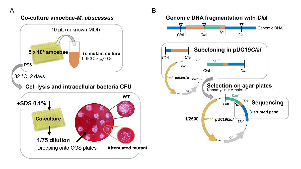
Figure 1: A. First visual screen of M. abscessus Tn mutants in amoeba. (A) large-scale screen of Tn mutants on amoebae is performed in...
Доступ ограничен. Войдите в систему или начните пробный период, чтобы просмотреть этот контент.
Материалы
| Имя | Компания | Каталожный номер | Комментарии |
|---|---|---|---|
| 24-well plates | Thermofisher | 11874235 | |
| 96-well plates | Thermofisher | 10687551 | |
| QuBit fluorometer | Thermofisher | Q33226 | |
| Acanthamoeba castellanii | ATCC | 30010 | strain |
| Amikacin | Mylan | 150927-A | powder |
| CaCl2 | Sigma-Aldrich | C1016 | >93% granular anhydrous |
| Columbia agar | Biomerieux | 43041 | 90 mm |
| D-Glucose | Sigma-Aldrich | G8270 | powder |
| DMEM | Thermofisher | 11500596 | medium |
| DNase and RNase free water | Invitrogen | 10977-035 | solution |
| Escherichia coli | Clinical isolate | personal stock | bacteria |
| Fetal Calf Serum | Gibco | 10270 | serum |
| Müller-Hinton agar | Biorad | 3563901 | powder |
| SDS 20% | Biorad | 1610418 | solution |
