В данном протоколе описывается создание и фенотипическая характеристика мышиной модели C57BL/6N-Cracdem1(c.538-83 to c.3352+255 del) с использованием технологии CRISPR/Cas9 для изучения роли CRACD в ремоделировании сердца после инфаркта миокарда.
Методическая статья
Создание и фенотипическая характеристика мышиной модели с дефицитом Cracd, созданной с помощью системы CRISPR/Cas9, для исследований ремоделирования после инфаркта миокарда
В этой статье
Краткое содержание
Аннотация
Инфаркт миокарда (ИМ) остается основной причиной заболеваемости и смертности во всем мире. В данном протоколе описывается метод создания и характеристики... Кракдлиния мышей с дефицитом [гена] на генетическом фоне C57BL/6N, созданная с помощью технологии CRISPR/Cas9. В зиготы совместно вводили мРНК Cas9, конструкцию гРНК и донорский шаблон, предназначенный для создания геномной делеции размером 3153 п.н. Наличие отредактированного аллеля подтверждали методом ПЦР-генотипирования и секвенированием по Сэнгеру. Для оценки функциональной роли CRACD в ремоделировании после инфаркта миокарда (ИМ) Пожалуйста, предоставьте исходный текст для перевода. Вы ввели только «Cracd», что похоже на опечатку. Ожидаю текст вашего научного материала.Мышам с дефицитом [ген/белок] и мышам дикого типа (WT, C57BL/6N) индуцировали инфаркт миокарда (ИМ) путем постоянного лигирования передней нисходящей ветви левой коронарной артерии. На 7-е сутки после операции оценивали функцию сердца и сократимость стенки левого желудочка с помощью трансторакальной эхокардиографии и спекл-трекинг анализа деформации, после чего проводили гистопатологическую оценку с использованием окрашивания гематоксилином и эозином (H&E).&Окрашивание по Эвансу и по Массону. Репрезентативные результаты показали, что CracdМыши с дефицитом [белка/гена] демонстрировали меньшее расширение желудочков и сохранную систолическую функцию по сравнению с контрольной группой дикого типа (WT). Данный протокол представляет собой надежную экспериментальную платформу для изучения механизмов участия CRACD в патофизиологии сердца.
Введение
Инфаркт миокарда (ИМ) остается одной из основных причин заболеваемости и смертности во всем мире, создавая значительную нагрузку на глобальное состояние сердечно-сосудистого здоровья1. Долговременный прогноз после ИМ в первую очередь определяется процессом ремоделирования левого желудочка (ЛЖ)2, который включает дилатацию желудочка, истончение стенки и интерстициальный фиброз3,4, являющиеся ключевыми факторами развития сердечной недостаточности (СН). Глобальная заболеваемость СН вследствие постинфарктного ремоделирования сердца еще больше увеличивает бремя сердечно-сосудистых заболеваний5. Несмотря на эти общепризнанные клинические наблюдения, отсутствуют систематические исследования с использованием чувствительных и воспроизводимых моделей in vivo для оценки роли специфических генов в постинфарктном ремоделировании. На данный момент нет протокола, сочетающего модель нокаута, созданную с помощью CRISPR/Cas9, с передовой визуализацией деформации (strain imaging) для оценки такого регулятора цитоскелета, как CRACD. Таким образом, существует острая необходимость в методе, обеспечивающем точные генетические манипуляции и фенотипирование сердца с высоким разрешением.
Ингибирующий регулятор актиновой динамики кэпирующего белка (CRACD), также известный как KIAA1211, представляет собой регулятор полимеризации актина, локализованный преимущественно в цитозоле и играющий критическую роль в динамике цитоскелета6. Он положительно регулирует полимеризацию актина, связываясь с кэпирующими белками актина (CAPZA1, CAPZA2, CAPZB) и препятствуя их взаимодействию с плюсовыми (barbed) концами актиновых филаментов7. В эпителиальных тканях CRACD играет важную роль в поддержании морфологии клеток и передаче сигналов, обеспечивая целостность актинового цитоскелета8,9. В опухолевых клетках его дисрегуляция связана с усилением метастазирования и пролиферации клеток6,10. Роль CRACD в сердце изучена не полностью. Исследования транскриптома сердца и данные экспериментальных вмешательств показывают, что экспрессия гена Cracd реагирует на патологический стресс и может регулировать гены, связанные с динамикой цитоскелета при ремоделировании желудочков9,11. Кроме того, его ингибирующий белок CapZ тесно связан с патологиями сердца: фосфорилирование CapZ регулирует рост миофибрилл при гипертрофии, индуцированной фенилэфрином12. Наши ранние исследования указывают на то, что умеренное снижение экспрессии CRACD обеспечивает защиту от повреждения миокарда и сохраняет сократимость миофиламентов после острой ишемии13. Эти данные позволяют предположить, что CRACD играет роль в патологическом ремоделировании желудочков после инфаркта миокарда (ИМ). Однако до сих пор отсутствовали систематические исследования потери функции in vivo, которые бы позволили определить, модулирует ли дефицит Cracd ремоделирование после ИМ, что в значительной степени было обусловлено отсутствием валидированного пошагового протокола создания и фенотипирования мышей с дефицитом Cracd в условиях ишемического стресса.
Существует несколько подходов к созданию мышей с конститутивным нокаутом. Традиционная гомологичная рекомбинация в эмбриональных стволовых (ЭС) клетках является точным методом, однако она дорогостоящая, трудоемкая (занимает от 12 до 18 месяцев) и ограничена определенными линиями мышей.14Мутагенез с помощью ENU (N-этил-N-нитрозомочевины) вызывает случайные точечные мутации, требующие проведения обширного бэккроссинга и секвенирования.15Напротив, CRISPR/Cas9 дает определенные практические преимущества для данного исследования: (i) прямая инъекция в зиготу сокращает сроки до 4–6 месяцев; (ii) метод позволяет делетировать крупный геномный фрагмент (3153 п.н.) для обеспечения полной потери функции; (iii) он работает непосредственно на генетическом фоне C57BL/6N без необходимости конверсии штамма; и (iv) он позволяет избежать использования селективного маркера, что минимизирует непреднамеренную транскрипционную интерференцию. Таким образом, CRISPR/Cas9 является наиболее эффективным выбором для создания Кракд (Cracd)дефицитная линия, подходящая для рутинного сердечно-сосудистого фенотипирования. Эта технология широко использовалась для проведения точного in vivo исследования с потерей функции16,17.
Спекл-трекинг визуализация деформации представляет собой метод постобработки, который позволяет отслеживать перемещение естественных акустических спеклов в миокарде от кадра к кадру, что дает возможность рассчитать параметры деформации миокарда, такие как деформация (strain — относительное изменение длины) и скорость деформации (strain rate)18. В отличие от традиционной эхокардиографии в M-режиме или двухмерной эхокардиографии, которые предоставляют глобальные функциональные показатели (фракция выброса, фракция укорочения, внутренний диаметр левого желудочка) и относительно нечувствительны к региональным нарушениям сократимости стенок, визуализация деформации позволяет количественно оценить сегментарную сократительную функцию. Это особенно важно в ранний постинфарктный период, когда компенсаторный гиперкинез неинфарктных сегментов может сохранять глобальную фракцию выброса, несмотря на значительную региональную дисфункцию. Спекл-трекинг обладает рядом преимуществ по сравнению со стандартными конечными точками: он позволяет определить сегментарную деформацию и скорость деформации, смещение и скорость, выявляя тем самым незначительные субклинические изменения19; он может идентифицировать диссинхронию и ранний дефицит сократимости до наступления необратимого ремоделирования; а также напрямую оценивает состояние субэндокарда — слоя, наиболее уязвимого к ишемии20. В данном исследовании CRACD рассматривается как регулятор цитоскелета, который может влиять на сократимость миофибрилл на микроскопическом уровне. Традиционные измерения, основанные на анализе полости, могут легко упустить такие локальные механические эффекты. Таким образом, интеграция анализа спекл-трекинга в данный протокол расширяет возможности обнаружения ранних защитных эффектов дефицита Cracd. Соответственно, включение спекл-трекинга в этот протокол обеспечивает чувствительный и количественный инструмент для оценки постинфарктного ремоделирования, особенно в течение первой недели после инфаркта миокарда, когда проявляются ранняя дилатация и сократительная дисфункция21,22.
Данный протокол предназначен для исследователей, изучающих функциональное влияние делеции конкретного гена на острое (7-дневное) ремоделирование после инфаркта миокарда (ИМ) с использованием комбинации конститутивного нокаута и визуализации высокого разрешения. Он подходит для исследований по выдвижению гипотез, при которых допустима глобальная утрата целевого гена, а основной конечной точкой являются ранние изменения систолической функции и структуры. Тем не менее следует учитывать несколько ограничений. Во-первых, мышь с дефицитом Cracd является глобальным нокаутом; следовательно, наблюдаемые кардиопротекторные эффекты не могут быть однозначно приписаны потере CRACD исключительно в кардиомиоцитах, так как системные факторы также могут играть роль. Для достижения кардиоспецифичной делеции в будущих исследованиях потребуется использовать стратегии условного нокаута. Во-вторых, протокол ориентирован на одну временную точку (7-й день) и не рассматривает хроническое ремоделирование или прогрессирование сердечной недостаточности. Потребуются лонгитюдные исследования за пределами острой фазы. В-третьих, анализ спекл-трекинга требует изображений высокого качества (четких границ эндокарда) и специализированного программного обеспечения, которое может быть доступно не во всех центрах общих ресурсов. Несмотря на эти ограничения, данный протокол представляет собой надежную и воспроизводимую платформу для первичной функциональной характеристики генов, участвующих в ремоделировании сердца после ИМ.
Данное исследование было направлено на использование системы CRISPR/Cas9 для создания и валидации мышиной модели с дефицитом Cracd (C57BL/6N-Cracdem1 (c.538-83 to c.3352+255 del)) с целью изучения влияния отсутствия Cracd на ремоделирование сердца и движение стенок желудочков после инфаркта миокарда (ИМ). ИМ индуцировали как у мышей дикого типа (WT, C57BL/6N), так и у мышей с дефицитом Cracd, после чего через одну неделю после ИМ проводили комплексную оценку. Анализ включал стандартные эхокардиографические параметры, анализ деформации методом трекинга спеклов (speckle-tracking strain analysis) и гистопатологическое исследование. Особое внимание было уделено ранней фазе после ИМ (день 7), чтобы зафиксировать начальные события ремоделирования и определить, влияет ли дефицит Cracd на дилатацию желудочков и систолическую функцию в острой фазе ИМ. Ожидается, что полученные результаты позволят глубже понять роль CRACD в модуляции неблагоприятного ремоделирования сердца после ИМ.
Протокол
All animal experiments were approved by the Institutional Animal Care and Use Committee of the Guangdong Institute of Biotechnology (Approval No. IACUC2025179). The study utilized 20 SPF Cracd-deficient and 20 age- and weight-matched WT mice. All mice were 10–12 weeks old and weighed 20–25 g. All male animals were housed in an AAALAC-accredited facility (License No. SYXK (Yue) 2016-0122) at the Guangdong Institute of Biotechnology (Guangdong Laboratory Animals Monitoring Institute). The housing environment was maintained at 22–25 °C with 50–70% relative humidity under a 12 h light/12 h dark cycle.
1. Generation of Cracd-deficient mice using CRISPR/Cas9
- Use CRISPR/Cas9 gene editing technology to generate Cracd-deficient mice on a C57BL/6N background. Delete the genomic sequence from c.538-83 to c.3352+255 (partial intron 5 to intron 6) of Cracd by homology-directed repair.
NOTE: The mouse Cracd gene (NCBI RefSeq: NM_001163793.1; Ensembl: ENSMUSG00000036377) is located on chromosome 5 and contains 9 exons, with the ATG start codon in exon 2 and the TAA stop codon in exon 9. - Design the sequence of gRNA targeting vector and donor oligonucleotide (with targeting sequence flanked by 120 bp homologous sequences combined on both sides).
- Log in to the NCBI website, select the Primer-BLAST function, and design PCR primers for genotyping.
- Paste the NCBI reference sequence number of Cracd (NM_001163793.1) into the PCR template field, specify the exon range for amplification, and click “Get Primers” to retrieve multiple pairs of candidate primers for PCR-based detection of the deletion and wild-type alleles.
- Design single-guide RNAs (gRNAs) to target sites flanking the intended deletion interval in the Cracd gene.
- Select four gRNA sequences (20 nt upstream of the PAM) using online CRISPR design tools.
- To minimize potential off-target effects, score the candidate gRNAs with CRISPRater and select the two optimal sequences (including the NGG PAM).
NOTE: Example selected sequences:
gRNA-A1: CGGCGCTCTTAACATCCAGAGGG (score: 0.71)
gRNA-A2: CCCTTCAGTCTGGGTAGTAGAGG (score: 0.71)
Accept gRNAs with a CRISPRater score ≥ 0.5. For in vitro validation, accept gRNAs with > 50% cleavage efficiency in a Cas9/gRNA target efficiency detection kit.
- Design a single-stranded donor oligonucleotide to promote efficient joining of the intended breakpoints. Example donor sequence: TTAACCATCAACGGGCCGCTGCAAGGGCTGCTGT
GTCTATA CTCAGGACCACCTGCCCTCTAGAGGCAGGAAGGT
AGAGTTCCAAGCTCTGCTGTGCTAGGTTAAATGCAACTTAACATC. - Co-inject Cas9 mRNA, gRNA and donor oligonucleotide into fertilized eggs to generate Cracd-deficient mice. Prepare Cas9 mRNA and gRNA by in vitro transcription.
- Preparation of gRNA.
- Transcribe gRNAs in vitro from DNA templates using the MEGAshortscript™ T7 transcription kit (see Table of Materials) following the manufacturer's protocol.
- Purify the transcribed gRNAs using an RNA purification kit (see Table of Materials) and elute them in RNase-free water.
- Assess gRNA cleavage activity using an in vitro Cas9/gRNA target efficiency detection kit (see Table of Materials).
- Perform all RNA preparation steps on ice using RNase‑free consumables (pipette tips, tubes, water). Keep RNA samples on ice at all times to prevent degradation.
- To perform the cleavage assay: mix the transcribed gRNA (25 ng/µL) with recombinant Cas9 protein (50 ng/µL) and a PCR-amplified DNA fragment containing the target sequence (50 ng).
- Incubate the reaction at 37 °C for 30 min.
- Run the products on a 2% agarose gel to visualize uncleaved and cleaved bands.
- Calculate cleavage efficiency as the ratio of cleaved band intensity to total (cleaved + uncleaved) band intensity.
- Accept gRNAs with > 50% cleavage efficiency.
- Preparation of Cas9 mRNA.
- Thaw the reagents from the T7 ARCA mRNA Kit (see Table of Materials).
- At room temperature, combine 10 µL of 2× ARCA/NTP mix, 1 µL of template DNA, 2 µL of T7 RNA polymerase mix, and nuclease-free water to 20 µL.
- Mix thoroughly, pulse-spin in a microfuge (5 s at 5000 × g), and incubate at 37 °C for 30 min.
- Remove the template DNA by adding 2 µL of DNase I (1 U/µL), mix well, and incubate at 37 °C for 15 min to obtain Cas9 mRNA. Optionally add 1 µL of RNase inhibitor (40 U/µL) (see Table of Materials) to the reaction to prevent RNA degradation.
- Perform all steps on ice with RNase‑free consumables. Keep the Cas9 mRNA on ice until use.
- Superovulate 4-week-old C57BL/6N female mice by intraperitoneal injection of 5 IU of pregnant mare serum gonadotropin (PMSG), followed 48 h later by 5 IU of human chorionic gonadotropin (hCG).
CAUTION: PMSG and hCG are hormones that may cause skin sensitization. Wear gloves and dispose of needles in a sharps container. - Approximately 14–18 h after hCG injection, euthanize the C57BL/6N female mice and one 8–12-week-old C57BL/6N male mouse by cervical dislocation. Collect oocytes from the oviducts of the females and sperm from the cauda epididymis of the male.
CAUTION: Euthanasia must be performed in accordance with institutional guidelines. Cervical dislocation should only be performed by trained personnel. - Capacitate the sperm in capacitation medium (see Table of Materials).
- Add a small volume of the sperm suspension to the oocytes for in vitro fertilization and co-culture for 3–4 h at 37 °C to obtain fertilized zygotes.
- Dilute the transcribed gRNA and Cas9 mRNA with RNase-free water to working concentrations (typically 25 ng/µL for gRNA and 50 ng/µL for Cas9 mRNA) and donor oligonucleotide at 10 ng/µL.
- Microinject 1–2 pL of the mixture into the cytoplasm of fertilized zygotes using a microinjection system (see Table of Materials).
- Identify healthy zygotes by the presence of two distinct pronuclei and an intact polar body under a stereomicroscope.
- Transplant 300 healthy fertilized zygotes into the oviducts of pseudopregnant female mice that have been mated with vasectomized males.
NOTE: This results in the generation of 2 positive founder mice (F0). - Use RNase‑free water and ice‑cold conditions during dilution to maintain RNA integrity.
- Identify F0 mice by PCR genotyping of tail biopsies (see section 1.6).
- Breed F0 founders with WT mice to generate F1 heterozygous offspring, to obtain germline transmission of the deletion.
- Intercross heterozygous F1 mice to produce F2 homozygous Cracd-deficient mice.
- Preparation of gRNA.
- Genotype by Polymerase Chain Reaction (PCR)
NOTE: The following protocol uses a commercial TIANamp Genomic DNA Kit (see Table of Materials). Buffer GL is the lysis buffer, Buffer GB is the binding buffer, and Buffer WA and Buffer WB are wash buffers.- Place approximately 2–5 mm of mouse tail tissue in a microcentrifuge tube containing 180 µL of Buffer GL, 20 µL of Proteinase K (20 mg/mL stock solution), and 10 µL of RNase A (10 mg/mL stock solution).
- Incubate the sample at 56 °C overnight to digest the tissue.
- Centrifuge the tube at 13800 × g for 2 min at room temperature to pellet tissue debris and transfer the supernatant to a new tube.
- Add 200 µL of Buffer GB and 200 µL of 100% ethanol to the supernatant, mix thoroughly, and apply the mixture to a spin column placed in a collection tube.
- Centrifuge at 13800 × g for 2 min at room temperature and discard the flow-through.
- Add 500 µL of Buffer WA to the column, centrifuge at 13800 × g for 1 min at room temperature and discard the flow-through.
- Carefully add 700 µL of Buffer WB (wash buffer containing ethanol, pre-mixed with 100% ethanol) to the spin column, centrifuge at 13800 × g for 1 min at room temperature and discard the flow-through.
- Apply Buffer WB to the inner wall of the column to wash residual salts.
- Repeat step 1.6.5 once, then transfer the column into a new collection tube and centrifuge at 13800 × g for 2 min at room temperature to dry the membrane and remove residual ethanol.
- Place the column into a new 1.5 mL microcentrifuge tube. Add 50–200 µL of sterilized water or elution buffer (preheated to 65 °C) to the center of the membrane, incubate for 5 min.
- Centrifuge the column at 13800 × g for 2 min at room temperature to elute the DNA.
- To increase DNA yield, either reapply the flow-through to the membrane or add an additional 50–200 µL of sterilized water, incubate for 5 min.
- Perform a final centrifugation at 13800 × g for 2 min.
NOTE: Expect DNA yield of 50–200 ng/µL from tail tissue; A260/A280 ratio of 1.8–2.0 indicates pure DNA.
- Place approximately 2–5 mm of mouse tail tissue in a microcentrifuge tube containing 180 µL of Buffer GL, 20 µL of Proteinase K (20 mg/mL stock solution), and 10 µL of RNase A (10 mg/mL stock solution).
- Genotyping by multiplex PCR using three primers in a single reaction.
- Design three primers to distinguish WT, heterozygous, and homozygous genotypes in one PCR reaction: Forward primer F1 (5’-CAGAAGTCATAGCAGCAAGGCAG-3’, binds upstream of the deletion), Forward primer F2 (5’-GAGGGAATTGAGAGGGAGCC-3’, binds within the deleted region, present only in the WT allele), and Reverse primer R1 (5’-GCCAATTTAGCTGCTTCAAATGTTC-3’, common reverse primer downstream of the deletion).
NOTE: The targeted allele generates a 421 bp amplicon (F1 + R1) only when the deletion is present; the WT allele generates a 357 bp amplicon (F2 + R1) only when the intact allele is present. - Set up the PCR reaction in a 25 µL total volume containing 50–100 ng genomic DNA, 0.2 µM of each primer (F1, F2, and R1), 200 µM dNTPs, 1.5 mM MgCl₂, and 1 U Taq DNA polymerase (see Table of Materials).
- Use the following thermocycling program: initial denaturation at 95 °C for 3 min; 35 cycles of denaturation at 95 °C for 15 s, annealing at 60°C for 15 s, and extension at 72 °C for 25 s; and a final extension at 72 °C for 5 min.
- Separate the PCR products by 1–2% agarose gel electrophoresis (120 V, 30 min) in 1× TAE buffer.
- Genotype interpretation based on band pattern: WT shows only a 357 bp band (F2+R1); Heterozygous shows both 421 bp (F1+R1) and 357 bp (F2+R1) bands; Homozygous shows only a 421 bp band (F1+R1).
- Design three primers to distinguish WT, heterozygous, and homozygous genotypes in one PCR reaction: Forward primer F1 (5’-CAGAAGTCATAGCAGCAAGGCAG-3’, binds upstream of the deletion), Forward primer F2 (5’-GAGGGAATTGAGAGGGAGCC-3’, binds within the deleted region, present only in the WT allele), and Reverse primer R1 (5’-GCCAATTTAGCTGCTTCAAATGTTC-3’, common reverse primer downstream of the deletion).
2. Myocardial infarction model establishment
- Anesthetize the mice via inhalation of 3% isoflurane (delivered in 100% oxygen at 1 L/min), place them in a supine position on a surgical platform, and confirm deep anesthesia by the absence of pedal withdrawal reflex.
CAUTION: Isoflurane is a volatile anesthetic. Use only in a well-ventilated area or under a fume hood to avoid inhalation. - Perform orotracheal intubation using a 20-gauge intravenous catheter sheath. Apply ophthalmic ointment to both eyes to prevent corneal drying during surgery.
- Connect the tube to a mechanical ventilator upon successful intubation to maintain ventilation and anesthesia with 2% isoflurane.
NOTE: Successful intubation is confirmed by visible chest movements synchronized with the ventilator.
- Connect the tube to a mechanical ventilator upon successful intubation to maintain ventilation and anesthesia with 2% isoflurane.
- Remove hair from the anterior chest with depilatory cream. Disinfect the surgical area by applying povidone-iodine and 70% alcohol in a circular motion for multiple times, starting from the center and moving outward.
- After fully exposing the skin, make a 1–1.5 cm incision in the left chest wall between the 2nd and 3rd intercostal spaces to open the thoracic cavity.
- Incise the pericardium to fully expose the heart.
CAUTION: Surgical procedures must be performed under sterile conditions. Use appropriate personal protective equipment (PPE), including gloves and a surgical mask.
- Using the inferior border of the left auricle as a landmark, pass a 7–0 atraumatic suture needle beneath the left anterior descending coronary artery (LAD) approximately 2–3 mm distal to its origin.
- Pass the needle to a depth of approximately 0.5–1 mm and tighten the ligature slowly to avoid myocardial tearing.
- Confirm successful occlusion by observing pallor (whitening) of the left ventricular anterior wall distal to the ligation site within 30 s.
- In sham-operated controls, open the chest and pass the suture beneath the coronary artery without tying the knot.
- After establishing the model, sequentially suture the ribs (4-0 absorbable suture, simple interrupted pattern) and skin (4-0 absorbable suture, continuous pattern) to close the thoracic cavity. Remove the endotracheal tube once the mouse resumes stable spontaneous breathing.
- Place mice on a heating pad (37 °C) until fully conscious.
- Administer postoperative analgesia (e.g., buprenorphine 0.05–0.1 mg/kg, subcutaneously, every 12 h for 48 h) according to institutional IACUC guidelines and closely monitor the postoperative health status for several days.
NOTE: Monitor for signs of distress (hunched posture, ruffled fur, reduced mobility, weight loss > 20%).
- Administer postoperative analgesia (e.g., buprenorphine 0.05–0.1 mg/kg, subcutaneously, every 12 h for 48 h) according to institutional IACUC guidelines and closely monitor the postoperative health status for several days.
- Mortality and exclusion criteria.
- Record the number of mice that die within the first 7 days after MI surgery.
NOTE: In a typical cohort, expect a mortality rate of approximately 10–20% by postoperative day 7, with no significant difference between WT and Cracd‑deficient mice. Common causes of death include extensive infarction leading to acute heart failure, surgical bleeding, or pneumothorax. Exclude any mouse with persistent arrhythmia (heart rate < 350 bpm) or signs of severe distress (e.g., hunched posture, ruffled fur, weight loss > 20%) from subsequent echocardiographic and histological analyses. For sham‑operated mice, mortality should be < 5%.
- Record the number of mice that die within the first 7 days after MI surgery.
3. Echocardiographic assessment
- Animal Preparation and Anesthesia
- Power on the high-resolution small animal ultrasound imaging system (see Table of Materials) and its integrated heated stage.
- Ensure that the appropriate high-frequency transducer is securely connected to the mounting apparatus following the manufacturer’s instructions. Set the temperature of the heated stage to 37 °C and allow it to stabilize.
- Place mice in an induction chamber and anesthetize them with 3% isoflurane in oxygen (1–2 L/min) until the pedal withdrawal reflex is absent. Transfer anesthetized mice to the heated platform in a supine position.
- Maintain anesthesia with 1–1.5% isoflurane delivered via a nose cone, adjusting to maintain heart rate between 450–550 beats per min.
- Apply ophthalmic ointment to both eyes to prevent corneal drying.
- Shave the thoracic region, evenly apply depilatory cream for no longer than 3 min, then remove the depilatory cream thoroughly with wet gauze.
- Rinse the skin gently with warm saline if necessary and dry the area. Attach surface ECG electrodes to the limbs using electrode gel at the contact sites and secure them with tape.
NOTE: Confirm stable ECG and heart rate within 450–550 bpm before proceeding to ultrasound imaging.
- Power on the high-resolution small animal ultrasound imaging system (see Table of Materials) and its integrated heated stage.
- Conventional Echocardiography Image Acquisition
- Position the mouse in a horizontal, supine posture on the heated stage, with a slight leftward tilt if needed to optimize cardiac imaging.
- Apply a generous amount of pre-warmed ultrasound coupling gel to the hairless left precordial region, avoiding air bubbles.
- Place the transducer gently on the chest wall at the left parasternal position. Adjust its orientation so that the index mark points toward the mouse’s right shoulder and fine-tune the angle and pressure to optimize image quality.
- Switch to B-mode (two-dimensional) imaging, adjust depth, focus zone, and frame rate to obtain a clear parasternal long-axis view of the left ventricle. Acceptable image quality requires clear visualization of the endocardial border with no dropouts.
- Once the heart rate stabilizes (450–550 bpm), acquire three separate parasternal long-axis B-mode cine loops, each containing at least 3–5 consecutive cardiac cycles.
- Save each loop by pressing “Cine Store”, and store representative still frames using “Frame Store”.
- From the long‑axis view, rotate the transducer 90° clockwise to obtain a short‑axis view at the mid‑papillary level.
- Acquire three separate short‑axis B‑mode cine loops (each with ≥ 3–5 cycles).
- Activate M‑mode, position the cursor perpendicular to the interventricular septum and posterior wall, and record three separate M‑mode tracings.
NOTE: Acceptable M‑mode tracings require clear endocardial borders throughout the cardiac cycle.
- Use the VevoLab 3.2.0 software (see Table of Materials) to measure the following parameters from M‑mode tracings according to the American Society of Echocardiography leading‑edge method: interventricular septum thickness at diastole and systole (IVSd, IVSs), left ventricular internal diameter at diastole and systole (LVIDd, LVIDs), and left ventricular posterior wall thickness at diastole and systole (LVPWd, LVPWs).
- Calculate left ventricular ejection fraction (EF) and fractional shortening (FS) using the Teichholz formula: EF (%) = [(LV Vold - LV Vols) / LV Vold] × 100, LV Vold = ((7 / (2.4 + LVIDd)) × LVIDd3), LV Vol;s = ((7 / (2.4 + LVIDs)) × LVIDs3), FS (%) = [(LVIDd – LVIDs) / LVIDd] × 100.
NOTE: For each mouse, measurements were averaged from three consecutive cardiac cycles per M‑mode recording, and the average was taken across three separate recordings, resulting in a total of 9 measurements per parameter per mouse.
- Calculate left ventricular ejection fraction (EF) and fractional shortening (FS) using the Teichholz formula: EF (%) = [(LV Vold - LV Vols) / LV Vold] × 100, LV Vold = ((7 / (2.4 + LVIDd)) × LVIDd3), LV Vol;s = ((7 / (2.4 + LVIDs)) × LVIDs3), FS (%) = [(LVIDd – LVIDs) / LVIDd] × 100.
- Position the mouse in a horizontal, supine posture on the heated stage, with a slight leftward tilt if needed to optimize cardiac imaging.
- Speckle-Tracking Strain Analysis
- Perform speckle-tracking analysis using the dedicated analysis software (VevoStrain module within VevoLab 3.2.0).
- Open a clear parasternal long-axis B-mode cine loop, select "Vevo Strain" to enter the strain analysis mode, retain only complete cardiac cycles, and choose "Long Axis".
- Manually trace the endocardial border at the end‑diastolic frame. After the endocardial border is drawn, the software automatically generates an epicardial border (adjust as needed). Then start the strain analysis and activate the "toggle contour/vector/orbit line/B mode" function to visualize the motion trajectory.
- Assess tracking quality via the colour‑coded mesh: it should appear green, closely follow the endocardial and epicardial borders without jerking, and move synchronously with myocardial contraction/relaxation. If not, manually refine the borders and re‑analyze.
- Activate "Time‑to‑peak analysis" to divide the left ventricle into six segments (basal anterior, mid anterior, apical anterior, apical posterior, mid posterior, basal posterior), with each colour representing an individual segment. Once satisfactory tracking is achieved, export data for wall motion parameters: velocity (cm/s), displacement (cm), strain (%), strain rate (1/s) for both longitudinal and radial.
NOTE: If tracking quality remains poor after three acquisition attempts (e.g., erratic strain curves, or inability to visualise clear motion trajectories), exclude the mouse from strain analysis.
- Perform speckle-tracking analysis using the dedicated analysis software (VevoStrain module within VevoLab 3.2.0).
4. Histopathological analysis
- After echocardiography, administer a lethal dose of isoflurane to deeply anesthetized mice and apply a secondary physical method (e.g., cervical dislocation) according to institutional guidelines.
- Open the thoracic cavity, rapidly harvest the hearts and rinse the hearts gently in cold phosphate-buffered saline (PBS).
CAUTION: Euthanasia must be performed according to institutionally approved protocols.
- Open the thoracic cavity, rapidly harvest the hearts and rinse the hearts gently in cold phosphate-buffered saline (PBS).
- Trim the hearts, remove the great vessels and cut transversely from apex to base into 2–3 slices (approximately 2–3 mm thick).
- Immerse the tissue slices in 4% paraformaldehyde in PBS and fix them at 4 °C for 12–24 h.
NOTE: Use a volume ratio of fixative to tissue of at least 10:1.
CAUTION: Paraformaldehyde is toxic and a suspected carcinogen. Handle in a fume hood and wear appropriate PPE (gloves, lab coat, safety goggles). Dispose of paraformaldehyde waste as hazardous chemical waste according to institutional guidelines.
- Immerse the tissue slices in 4% paraformaldehyde in PBS and fix them at 4 °C for 12–24 h.
- Dehydrate tissue through a graded ethanol series (50%, 70%, 80%, 95%, 100% ethanol, each for 1 h), clear them in xylene (2 changes, 30 min each), embed them in paraffin, and section them at 4–5 µm thickness using a microtome.
NOTE: Sections should be free of folds and tears.
CAUTION: Xylene is flammable and toxic. Use only in a fume hood and avoid skin contact. Collect xylene waste in a designated container and dispose of as hazardous chemical waste. - Deparaffinize the sections in xylene (2 changes, 5–10 min each), rehydrate them through descending ethanol series (100%, 95%, 80%, 70%, each for 3 min) to water.
- Stain them using standard Hematoxylin and Eosin (H&E) (see Table of Materials) and Masson’s trichrome protocols (see Table of Materials).
CAUTION: Xylene is toxic. Use only in a fume hood and avoid skin contact. Dispose of xylene waste as hazardous material.
- Stain them using standard Hematoxylin and Eosin (H&E) (see Table of Materials) and Masson’s trichrome protocols (see Table of Materials).
- Examine the stained sections under a light microscope (see Table of Materials).
- For quantitative analysis, capture digital images at 4×, 20×, and 40× magnification using a digital camera attached to the microscope. Measure infarct size and fibrotic area using ImageJ.
NOTE: Infarct size (%) = (infarct area / total LV area) × 100; collagen content (%) = (blue-stained area / total myocardial area) × 100.
- For quantitative analysis, capture digital images at 4×, 20×, and 40× magnification using a digital camera attached to the microscope. Measure infarct size and fibrotic area using ImageJ.
Результаты
Generation of Cracd-deficient mice
A heritable mouse model carrying a targeted deficiency in the Cracd gene was generated using CRISPR/Cas9-mediated genome editing (Figure 1A, Figure 1B). To distinguish WT, heterozygous, and homozygous genotypes, a multiplex PCR was performed using three primers (F1, F2, R1) in a single reaction. Mice homozygous for the deletion (Cracd‑deficient) displayed only the 421 bp band, WT mice showed only the 357 bp band (Figure 1C). Sanger sequencing of genomic DNA from Cracd-deficient mice confirmed the predicted deletion in the target gene compared to WT controls (Figure 1D). Western blot analysis further demonstrated a marked reduction in CRACD protein levels in cardiac tissues from Cracd-deficient mice compared with WT controls (Figure 1E). Together, these data confirm the successful establishment of the Cracd-deficient mouse line.
Echocardiography
Echocardiographic assessment on postoperative day 7 revealed pronounced pathological cardiac remodeling in WT mice after MI (WT-MI) compared with sham-operated controls (Figure 2A). The WT-MI group showed thinning of the interventricular septum (Figure 2B, C), significant left ventricular (LV) dilation as reflected by increased LVIDd (Figure 2D) and LVIDs (Figure 2E), and thinning of the posterior wall (Figure 2F, G). These structural alterations were associated with a pronounced decline in global systolic function, as demonstrated by significantly reduced EF (Figure 2H) and FS (Figure 2I). In contrast, Cracd-deficient mice displayed a cardioprotective phenotype following MI. LV dilation was significantly attenuated, with notably smaller LVIDd and LVIDs compared with the WT-MI group. Furthermore, these mice exhibited improved systolic thickening of the posterior wall and maintained significantly higher EF and FS values. These findings indicate that Cracd deficiency effectively attenuates post-infarction LV dilation and preserves global systolic function.
Speckle-tracking strain analysis
Left ventricular myocardial motion and deformation were further evaluated by speckle‑tracking echocardiography using only parasternal long‑axis views (Figure 3A). To ensure data reliability, we defined pragmatic criteria for acceptable output based on the capabilities of the VevoStrain software. Acceptable strain analysis required: (i) a clear parasternal long‑axis view with endocardial borders visible throughout the cardiac cycle; (ii) a stable heart rate between 450–550 bpm during acquisition; (iii) after automatic tracking, the colour‑coded mesh (activated via “toggle contour/vector/orbit line/B mode”) had to closely follow the endocardial and epicardial borders without jerking or losing contact; and (iv) strain curves had to be smooth, show consistent waveforms across segments, and lack sudden spikes or arrhythmic interruptions. Suboptimal output—most commonly erratic strain curves or inability to visualise clear motion trajectories—led to manual adjustment of endocardial or epicardial contours and re‑analysis. If quality remained poor after three attempts, the mouse was excluded. Using these criteria, we obtained reliable strain data in WT‑MI mice and Cracd‑deficient MI mice.
Compared with the sham group, WT-MI mice exhibited significantly reduced radial velocity (Figure 3B) and longitudinal velocity (Figure 3C), as well as radial displacement (Figure 3D) and longitudinal displacement (Figure 3E), indicating impaired global myocardial motion. The absolute values of radial strain (Figure 3F), radial strain rate (Figure 3G), longitudinal strain (Figure 3H), and longitudinal strain rate (Figure 3I) were also markedly decreased, reflecting severely compromised regional myocardial deformation and function. In contrast, Cracd-deficient mice showed significant improvement in these parameters. Compared with the WT-MI group, they displayed better recovery of radial and longitudinal displacements. Furthermore, radial velocity, radial strain, and radial strain rate were maintained at higher levels, while longitudinal strain and longitudinal strain rate were significantly improved. These results suggest that Cracd deficiency not only preserves global systolic function but also enhances regional LV myocardial deformation after MI.
The speckle‑tracking imaging analysis also included visualization of segmental strain distribution and cardiac motion trajectories. Parasternal long‑axis B‑mode images with segmented six regions (BA, MA, AA, AP, MP, BP) and color gradient mapping are displayed in Figure 4A. Cardiac motion trajectories, time‑to‑peak curves for radial and longitudinal strain, parametric distribution maps of strain synchrony, and three‑dimensional reconstructions are presented in Figure 4B. These data further support the functional improvements observed in Cracd‑deficient mice.
Histological examination
To assess the protective effect of Cracd deficiency against MI, histopathological examination was performed on heart sections from WT and Cracd-deficient mice at 7 days post-MI using H&E and Masson's trichrome staining. Whole‑heart longitudinal sections are shown in Figure 5A, with quantitative analysis of infarct size and collagen content presented in Figure 5B and Figure 5C, respectively. High‑magnification views of the infarct zone, border zone, and remote zone are displayed in Figure 5D. In WT-MI hearts, H&E staining showed pronounced left ventricular wall thinning and a well-demarcated infarct zone. Within the infarct region, extensive inflammatory cell infiltration, myocardial necrosis, and marked fibrosis with scar formation were observed. In the border zone, inflammatory infiltration, myocardial necrosis, fibrosis and cardiomyocyte hypertrophy were evident, while the remote zone displayed prominent hypertrophic changes. Corresponding Masson's trichrome staining further demonstrated extensive interstitial fibrosis within both the infarct and border zones of WT hearts following MI. In contrast, Cracd-deficient mice exhibited substantially attenuated pathological alterations, including reduced wall thinning, diminished inflammatory infiltration and myocardial necrosis, less pronounced cardiomyocyte hypertrophy, and markedly decreased interstitial fibrosis compared with WT-MI hearts. These histological findings indicate that Cracd deficiency mitigates post-infarction myocardial damage and adverse remodeling.
Successful protocol execution should yield: (i) a well‑demarcated infarct zone with extensive collagen deposition in WT-MI hearts; (ii) clear cellular morphology without staining artifacts; and (iii) significantly attenuated pathology in Cracd‑deficient MI hearts. Although infarct size may vary with ligation precision, the genotype‑dependent difference is a reliable indicator of successful performance.
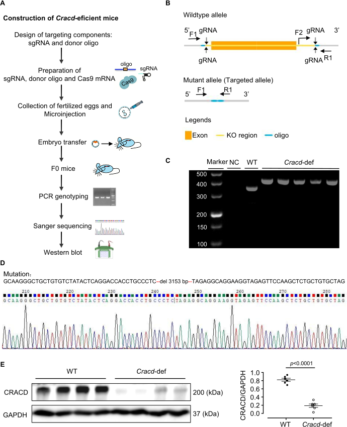
Figure 1: Generation of Cracd-deficient mice. (A) Construction of Cracd-deficient mice. (B) Schematic of the Cracd gene modification strategy. (C) PCR genotyping of tail genomic DNA. WT mice show a single 357 bp band, while homozygous (Cracd‑deficient) mice show a single 421 bp band. (D) Sanger sequencing of genomic DNA confirmed the predicted deletion in the Cracd locus of deficient mice compared to WT controls. (E) Western blot analysis showed significantly reduced CRACD protein levels in the hearts of Cracd-deficient mice compared to WT controls. n = 6 mice per group. Data are presented as mean ± SEM. Statistical significance was calculated with a two-tailed unpaired Student's t-test. Please click here to view a larger version of this figure.
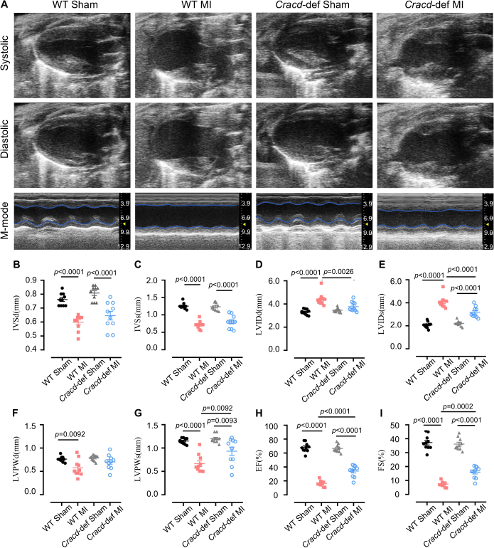
Figure 2: Echocardiographic assessment of cardiac function. (A) Representative B-mode and M-mode echocardiographic images of mouse hearts at diastole and systole. Quantitative analysis of (B) interventricular septum thickness at diastole (IVSd) and (C) systole (IVSs), (D) left ventricular internal diameter at diastole (LVIDd) and (E) systole (LVIDs), (F) left ventricular posterior wall thickness at diastole (LVPWd) and (G) systole (LVPWs), (H) ejection fraction (EF), and (I) fractional shortening (FS). n = 10 mice per group. Data are presented as mean ± SEM. Statistical significance was determined by one-way ANOVA followed by Tukey's multiple comparisons test. Please click here to view a larger version of this figure.
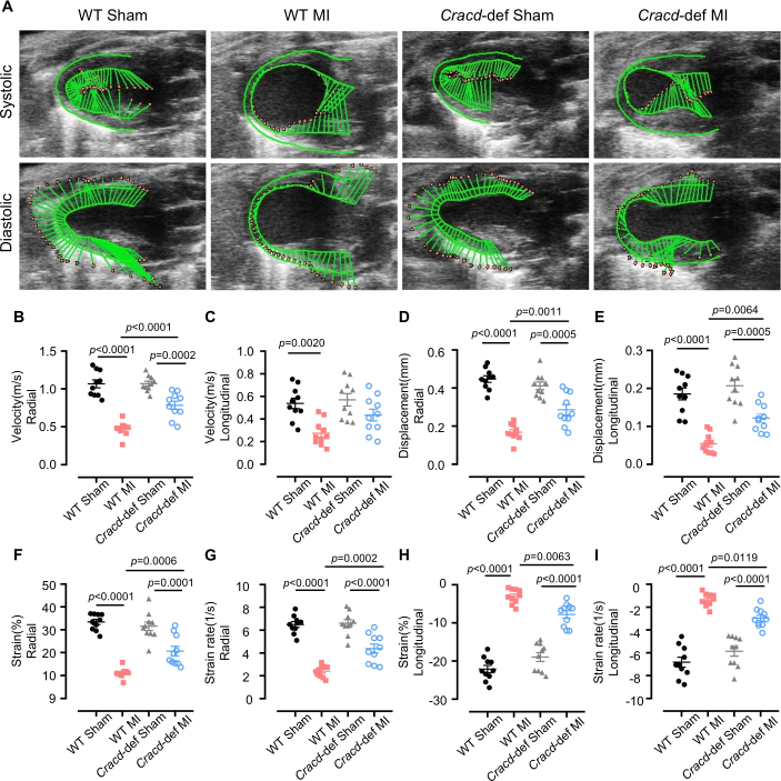
Figure 3: Speckle-tracking strain analysis. (A) Representative images of left ventricular wall motion and deformation at diastole and systole. Quantitative analyses of the following parameters: (B) radial velocity, (C) longitudinal velocity, (D) radial displacement, (E) longitudinal displacement, (F) radial strain, (G) radial strain rate, (H) longitudinal strain, and (I) longitudinal strain rate. n = 10 mice per group. Data are presented as mean ± SEM. Statistical significance was determined by one-way ANOVA followed by Tukey's multiple comparisons test. Please click here to view a larger version of this figure.
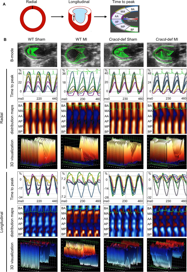
Figure 4: Speckle-tracking imaging in WT-Sham, WT-MI, Cracd-deficient Sham, and Cracd-deficient MI groups at 7 days post-MI. (A) Parasternal long‑axis B‑mode images. Left ventricular wall was segmented into six regions: BA (basal anterior), MA (mid anterior), AA (apical anterior), AP (apical posterior), MP (mid posterior), and BP (basal posterior). Color gradient from blue to red represents increasing strain magnitude. (B) Cardiac motion trajectories; time-to-peak curves for radial and longitudinal strain values along with corresponding peak times. Parametric distribution maps of strain synchrony (blue/red indicate direction: radial – away from/toward center; longitudinal – away from/toward apex). White dots = highest velocity regions, black dots = lowest velocity. Three-dimensional reconstructions (X: time, Y: spatial position, Z: strain magnitude). Please click here to view a larger version of this figure.
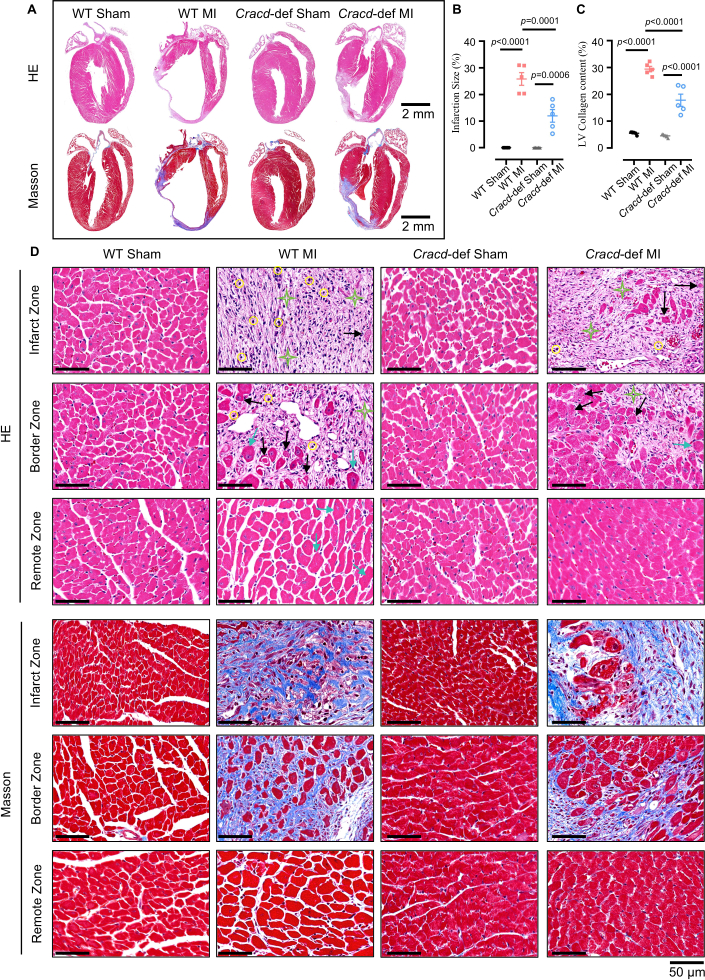
Figure 5: Histopathological analysis of cardiac remodeling after MI. (A) Whole-heart longitudinal sections with H&E and Masson's trichrome staining. Scale bars: 2 mm. (B) Quantitative analysis of infarct size and (C) collagen content. (D) High‑magnification views of infarct zone (IZ), border zone (BZ), and remote zone (RZ). H&E: yellow circles = inflammatory infiltration; black arrows = myocardial necrosis; green stars = fibrosis; blue arrows = hypertrophy. Masson’s trichrome: blue indicates collagen deposition. Scale bar: 50 μm. n = 5 mice per group. Data are presented as mean ± SEM. Statistical significance was determined by one-way ANOVA followed by Tukey's multiple comparisons test. Please click here to view a larger version of this figure.
Обсуждение
В данном исследовании мы создали линию мышей с дефицитом Cracd, разработанную с помощью системы CRISPR/Cas9, и разработали рабочий процесс для их фенотипирования после инфаркта миокарда (ИМ) с использованием традиционной эхокардиографии, метода спекл-трекинга (strain imaging) и гистологии. Согласно данному протоколу были получены нокаутные животные с подтвержденной потерей белка CRACD. Мыши с дефицитом Cracd после ИМ демонстрировали меньшее расширение желудочков, более высокую фракцию выброса (EF) и снижение степени фиброза по сравнению с контрольной группой дикого типа (WT), что подтверждает применимость данной модели для изучения ремоделирования сердца.
Успех данной модели зависит от нескольких критических этапов. Дизайн gRNA и валидация in vitro напрямую влияют на эффективность редактирования зиготы23. Мы используем CRISPick или Benchling для выбора gRNA с показателем CRISPRater ≥ 0,5, после чего тестируем эффективность расщепления с помощью анализа Cas9/gRNA in vitro. Для микроинъекций используются только те gRNA, которые обеспечивают расщепление > 50%. Столь же важным является правильное лигирование LAD: шов 7‑0 должен проходить на расстоянии 2–3 мм от начала LAD на глубине 0,5–1 мм, чтобы избежать прокола правого желудочка (слишком глубоко) или неполной окклюзии сосуда (слишком поверхностно); мгновенное побеление передней стенки подтверждает окклюзию. Также необходим контроль частоты сердечных сокращений во время эхокардиографии24; поддержание ЧСС на уровне 450–550 bpm путем регулировки концентрации изофлурана (обычно 1–2%) повышает воспроизводимость стандартных измерений и анализа деформации. Наконец, для анализа трекинга спеклов необходима визуальная оценка цветовой сетки25. Сетка должна точно следовать границам миокарда без резких скачков, в противном случае требуется ручная корректировка; если качество остается низким после трех попыток, мышь исключается из исследования.
Даже при тщательном соблюдении протокола могут возникнуть определенные технические трудности. Систематический поиск и устранение неисправностей позволяют решить большинство из них. Низкие показатели передачи в зародышевой линии (ниже 10%), как правило, указывают на плохую интеграцию донорного олигонуклеотида или слабую активность gRNA26. Следовательно, увеличение концентрации донорного олиго до 20 ng/µL, увеличение длины плеч гомологии до 120–200 bp и повторная проверка расщепления gRNA (> 50%) могут улучшить передачу. Высокая летальность после MI (выше 30%) часто обусловлена пневмотораксом, кровотечением и обширным инфарктом27. Таким образом, нахождение мышей на постоянном кислороде до их пробуждения и использование нагревательного коврика для поддержания температуры тела заметно снижают показатели смертности. Если передняя стенка не бледнеет после лигирования, вероятно, ПМЖВ (LAD) была определена неверно или шов слишком поверхностный. В этом случае повторный поиск ПМЖВ (ярко-красный диагональный сосуд) и повторное проведение иглы на глубину 0.5–1 mm, иногда с использованием диссекционного микроскопа, позволяют решить эту проблему. При низком качестве трекинга спеклов (speckle-tracking) обычными причинами являются низкая частота кадров, размытые границы или артефакты движения28. Соответственно, помогает увеличение частоты кадров, точная настройка усиления, ручная корректировка эндокардиального контура и исключение циклов с аритмией.
Помимо этих операционных аспектов, данный метод имеет явные ограничения. Нокаут Cracd является глобальным, поэтому белок CRACD отсутствует во всех тканях. Следовательно, наблюдаемый защитный эффект нельзя приписывать исключительно кардиомиоцитам, так как системные факторы также могут играть определенную роль. Для разграничения функций конкретных типов клеток потребуется создание условного нокаута. Другим ограничением является использование единственной временной точки на 7-й день, что не позволяет судить о хроническом ремоделировании. Таким образом, для долгосрочных исследований следует добавить более поздние временные точки (4 или 8 недель). Воспроизводимость также зависит от качества изображений: не каждая система ультразвукового исследования может обеспечить четкие границы эндокарда, и значительную роль играет опыт оператора. В связи с этим рекомендуется, чтобы все операции и измерения проводились профессионально подготовленным специалистом.
Учитывая эти ограничения, полезно сравнить наш подход с альтернативными стратегиями изучения функции генов при ремоделировании после инфаркта миокарда (MI). Двумя распространенными альтернативами являются манипуляции с генами с помощью AAV и антисмысловые олигонуклеотиды (ASO)29,30. AAV9 позволяет обеспечить специфическую для сердца транзиентную гиперэкспрессию или нокдаун без необходимости скрещивания, однако его недостатками являются лимит упаковки (~4.7 kb), вариабельная эффективность трансдукции и побочные эффекты31. Напротив, наша модель нокаута на основе CRISPR/Cas9 обеспечивает перманентную и полную потерю функции, что является преимуществом для долгосрочных исследований. ASOs обеспечивают обратимый дозозависимый нокдаун с быстрым началом действия, что делает их подходящими для острых вмешательств; однако они требуют повторного введения и не обладают тканеспецифичностью32. Таким образом, описанный здесь конститутивный нокаут лучше всего подходит для первоначального поиска и хронического фенотипирования.
Наконец, данный протокол может быть применен значительно шире, чем только для CRACD. Тот же рабочий процесс — создание нокаутных мышей с помощью CRISPR/Cas9 в сочетании с эхокардиографией, анализом деформации миокарда (strain imaging) и гистологией — позволяет оценивать любой ген-кандидат в процессе ремоделирования после инфаркта миокарда. Эта схема фенотипирования также подходит для других моделей сердечно-сосудистых заболеваний, таких как поперечное сужение аорты (перегрузка давлением) или ишемически-реперфузионное повреждение. Кроме того, образцы тканей, полученные в ходе данного протокола, могут быть использованы для мультиомиксного анализа (транскриптомики, протеомики, метаболомики) с целью выявления молекулярных механизмов. CracdДефицитная модель может служить инструментом для тестирования лекарственных препаратов: с ее помощью можно оценить соединения, которые, как предполагается, действуют через путь CRACD. Таким образом, данный протокол представляет собой платформу, адаптируемую для широкого спектра механистических и трансляционных исследований в области сердечно-сосудистых заболеваний.
Раскрытие информации
Авторы заявляют об отсутствии конфликта интересов.
Благодарности
Данная работа была поддержана Фондом фундаментальных и прикладных базовых исследований провинции Гуандун (2023A1515011278), проектом с собственным финансированием Провинциального института биотехнологических исследований Гуандуна (GDBRI-ZL202602), а также программой китайско-канадского академического обмена и сотрудничества по моделям и патогенезу сердечно-сосудистых заболеваний (MS202500058).
Материалы
| Имя | Компания | Каталожный номер | Комментарии |
|---|---|---|---|
| CRISPR/Cas9 | |||
| Раствор гидрохлорида тризма | Sigma-Aldrich, USA | T2663 | Используется для приготовления буфера для экстракции ДНК |
| Протеиназа K | Sigma-Aldrich, USA | 539480 | Используется для переваривания тканей хвоста мыши |
| Green Taq Mix | Vazyme, China | P131 | Используется для ПЦР-амплификации |
| Агароза | BioFroxx, Germany | 1110GR500 | Используется для гель-электрофореза ДНК при концентрации 1–2% |
| Маркер ДНК | Thermo Scientific, USA | SM0242 | Используется в качестве лестницы ДНК для определения размера продуктов ПЦР |
| TIANamp Genomic DNA Kit | Tiangen Biotech, China | DP304 | Используется для экстракции геномной ДНК из тканей хвоста мыши |
| MEGAshortscript™ T7 transcription kit | Thermo Scientific, USA | AM1354 | Используется для транскрипции гРНК in vitro |
| Набор для очистки РНК | Zymo Research, USA | R1016 | Используется для очистки транскрибированной гРНК |
| BeyoCRISPR™ sgRNA Screening Kit | Beyotime, China | D8412S | Используется для оценки активности расщепления гРНК in vitro |
| Ингибитор РНКазы | NEB, USA | M0314 | Используется для предотвращения деградации РНК во время транскрипции in vitro |
| TaKaRa Taq | Takara Bio, Japan | R001A | Используется для ПЦР-генотипирования ДНК из хвоста мыши |
| FERTIUP® Mouse Sperm Preincubation Medium: PM | Cosmo Bio, Japan | KYD-002-EX | Используется для капацитации сперматозоидов перед оплодотворением in vitro |
| T7 ARCA mRNA Kit | NEB, USA | E2060 | Используется для транскрипции мРНК Cas9 in vitro |
| Гонадотропин сыворотки беременной кобылы | MCE, USA | 9002-70-4 | Используется для суперовуляции, вводится путем внутрибрюшинной инъекции |
| Хорионический гонадотропин человека | ProSpec, Israel | HOR-250 | Используется для индукции овуляции, вводится путем внутрибрюшинной инъекции |
| Вода, свободная от РНКаз | Aladdin, China | R665526 | Используется для растворения РНК и подготовки ПЦР |
| Праймеры | Sangon Biotech, China | / | Используются для генотипирования |
| Хирургия | |||
| Изофлуран | RWD Life Science, China | R510-22-10 | Используется для индукции и поддержания анестезии во время операции |
| Крем для удаления волос | Veet, France | 1000207 | Наносится на область грудной клетки для удаления шерсти перед операцией и УЗИ |
| Шовный материал 7-0 | Lingqiao, China | LQ7022380206 | Используется для постоянного лигирования левой передней нисходящей (ЛПН) коронарной артерии |
| Шовный материал 4-0 | Lingqiao, China | LQ4022380412 | Используется для закрытия кожного разреза после операции |
| Анестезиологический вентилятор | Harvard Apparatus, USA | Tabletop | Используется для подачи изофлурана и поддержки дыхания |
| Вентилятор для мелких животных | Taimeng, China | HX-101E | Используется для обеспечения вентиляции с положительным давлением во время операции на открытой грудной клетке |
| Термостатическая грелка | TigerGene, USA | TG-TP-GS | Используется для поддержания температуры тела мыши во время операции и восстановления |
| Ребрасширитель | Shenzhen Huayon Biotech, China | LN-18-4101 | Используется для разведения ребер и обнажения сердца для лигирования ЛПН |
| Ножницы | Shenzhen Huayon Biotech, China | 18-0551 | Используются для выполнения разрезов кожи и рассечения тканей |
| Стереоскопический микроскоп | Nikon, Japan | SMZ 745 | Используется для обеспечения увеличенного обзора при проведении операции по лигированию ЛПН артерии |
| Микрохирургический иглодержатель | Shenzhen Huayon Biotech, China | 18-2211 | Используется для манипуляций с малой иглой шовного материала 7-0 под микроскопом |
| Химические вещества и реагенты | |||
| Ультразвуковые контактные гели | Tianjin Jinya Technology Development, China | TM-100 | Наносятся на грудную клетку для эхокардиографии, обеспечивают надлежащий контакт с датчиком |
| Параформальдегид | Sigma-Aldrich, USA | V900894 | Используется для фиксации тканей сердца после забора |
| Ксилол | Macklin, China | 1330-20-7 | Используется для удаления парафина и просветления тканей перед окрашиванием |
| Гематоксилин | Sigma-Aldrich, USA | H3136 | Ядерный краситель, используется при окрашивании Г&Э |
| Эозин | Sigma-Aldrich, USA | E4009 | Цитоплазматический краситель, используется при окрашивании Г&Э |
| Понсо S | Sigma-Aldrich, USA | P3504 | Используется при окрашивании по Массону для окрашивания цитоплазмы |
| Фосформолибденовая кислота | Sigma-Aldrich, USA | 221856 | Используется при окрашивании по Массону в качестве протравы и для дифференцировки |
| Раствор анилинового синего | Sigma-Aldrich, USA | 415049 | Используется при окрашивании по Массону для окрашивания коллагеновых волокон (в синий цвет) |
| Антитело к CRACD | Absea, China | KC-35356 | Первичное антитело для детекции белка CRACD методом Вестерн-блоттинга, разведение 1:2000 |
| Антитело к GAPDH | CST, USA | 2118S | Контрольное антитело для Вестерн-блоттинга, разведение 1:5000 |
| Этанол | Macklin, China | 64-17-5 | Используется для дегидратации и приготовления реагентов (марки 70%, 80%, 95%, 100%) |
| Лабораторное оборудование | |||
| Центрифуга Sorvall™ Legend™ Micro 17 | Thermo Scientific, USA | 75002541 | Используется для рутинной подготовки образцов |
| ПЦР-термоциклер | Bio-Rad Laboratories, USA | 1861096 | Используется для амплификации ДНК |
| Система микроинъекций | Eppendorf, Germany | 5193000020 | Используется для микроинъекции реагентов CRISPR/Cas9 в зиготы |
| Система визуализации Vevo 2100 | VisualSonics, Canada | Vevo2100 | Высокочастотная система визуализации для неинвазивной оценки функции и структуры сердца после инфаркта миокарда (ИМ) |
| Световой микроскоп | Leica, Germany | DM2500 | Используется для наблюдения за срезами сердца, окрашенными Г&Э и по Массону |
| Программное обеспечение для анализа | |||
| ImageJ | National Institutes of Health, USA | / | Используется для анализа изображений, включая измерение площади, количественную оценку сигнала и обработку гистологических снимков (окрашивание Г&Э и по Массону) |
| VevoLab 3.2.0 / VevoStrain | VisualSonics, Canada | / | ПО VevoLab для анализа функции сердца; модуль VevoStrain используется для анализа деформации миокарда с целью оценки сердечной деятельности после ИМ |
| GraphPad Prism 8 | GraphPad Software, USA | / | Используется для статистического анализа и построения графиков |
Ссылки
- Fauconnier, J., Meli, A. C., Thireau, J., Roberge, S., Shan, J., et al. Ryanodine receptor leak mediated by caspase-8 activation leads to left ventricular injury after myocardial ischemia-reperfusion. Proc Natl Acad Sci U S A. 108 (32), 13258-13263 (2011).
- Cohn, J. N., Ferrari, R., Sharpe, N. Cardiac remodeling-concepts and clinical implications: a consensus paper from an international forum on cardiac remodeling. J Am Coll Cardiol. , (2000).
- Liu, D., Tian, X., Liu, Y., Song, H., Cheng, X., et al. CREG ameliorates the phenotypic switching of cardiac fibroblasts after myocardial infarction via modulation of CDC42. Cell Death Dis. 12 (4), (2021).
- Contessotto, P., Pandit, A. Therapies to prevent post-infarction remodelling: from repair to regeneration. Biomaterials. 275, (2021).
- Zhu, R., Sun, H., Yu, K., Zhong, Y., Shi, H., et al. Interleukin-37 and dendritic cells treated with interleukin-37 plus troponin I ameliorate cardiac remodeling after myocardial infarction. J Am Heart Assoc. 5 (12), (2016).
- Chang, Y., Zhang, J., Huo, X., Qu, X., Xia, C., et al. Substrate rigidity dictates colorectal tumorigenic cell stemness and metastasis via CRAD-dependent mechanotransduction. Cell Rep. 38 (7), (2022).
- Seo, Y., Jang, J., Ko, K. P., Zou, G., Huang, Y., et al. Actin dysregulation induces immune evasion via oxidative stress-activated PD-L1 in gastric cancer. bioRxiv. , (2024).
- Kim, B., Zhang, S., Huang, Y., Ko, K. P., Jung, Y. S., et al. CRACD loss induces neuroendocrine cell plasticity of lung adenocarcinoma. Cell Rep. 43 (6), (2024).
- Jung, Y. S., Wang, W., Jun, S., Zhang, J., Srivastava, M., et al. Deregulation of CRAD-controlled cytoskeleton initiates mucinous colorectal cancer via β-catenin. Nat Cell Biol. 20 (11), 1303-1314 (2018).
- Liu, Z., Cao, H., Shi, Y., Yang, R. KIAA1211 plays an oncogenic role in human non-small cell lung cancer. J Cancer. 10 (26), 6747-6753 (2019).
- Snider, P. L., Snider, E., Simmons, O., Lilly, B., Conway, S. J. Analysis of uncharacterized mKiaa1211 expression during mouse development and cardiovascular morphogenesis. J Cardiovasc Dev Dis. 6 (2), (2019).
- Lin, Y. H., Warren, C. M., Li, J., McKinsey, T. A., Russell, B. Myofibril growth during cardiac hypertrophy is regulated through dual phosphorylation and acetylation of the actin capping protein CapZ. Cell Signal. 28 (8), 1015-1024 (2016).
- Yang, F. H., Pyle, W. G. Reduced cardiac CapZ protein protects hearts against acute ischemia-reperfusion injury and enhances preconditioning. J Mol Cell Cardiol. 52 (3), 761-772 (2012).
- Tong, C., Huang, G., Ashton, C., Wu, H., Yan, H., et al. Rapid and cost-effective gene targeting in rat embryonic stem cells by TALENs. J Genet Genomics. 39 (6), 275-280 (2012).
- Cordes, S. P. N-ethyl-N-nitrosourea mutagenesis: boarding the mouse mutant express. Microbiol Mol Biol Rev. 69 (3), 426-439 (2005).
- Cong, L., Ran, F. A., Cox, D., Lin, S., Barretto, R., et al. Multiplex genome engineering using CRISPR/Cas systems. Science. 339 (6121), 819-823 (2013).
- Kanke, K. L., Rayner, R. E., Bozik, J., Abel, E., Venugopalan, A., et al. Single-stranded DNA with internal base modifications mediates highly efficient knock-in in primary cells using CRISPR-Cas9. Nucleic Acids Res. 52 (22), 13561-13576 (2024).
- Mihos, C. G., Liu, J. E., Anderson, K. M., Pernetz, M. A., O'Driscoll, J. M., et al. Speckle-tracking strain echocardiography for the assessment of left ventricular structure and function: a scientific statement from the American Heart Association. Circulation. 152 (10), (2025).
- Garg, P. K., Biggs, M. L., Kizer, J. R., Shah, S. J., Psaty, B., et al. Glucose dysregulation and subclinical cardiac dysfunction in older adults: the Cardiovascular Health Study. Cardiovasc Diabetol. 21 (1), (2022).
- Asanuma, T., Fukuta, Y., Masuda, K., Hioki, A., Iwasaki, M., et al. Assessment of myocardial ischemic memory using speckle tracking echocardiography. JACC Cardiovasc Imaging. 5 (1), 1-11 (2012).
- Chong, S. Y., Zharkova, O., Yatim, S. M. J. M., Wang, X., Lim, X. C., et al. Tissue factor cytoplasmic domain exacerbates post-infarct left ventricular remodeling via orchestrating cardiac inflammation and angiogenesis. Theranostics. 11 (19), 9243-9261 (2021).
- Hung, C. L., Verma, A., Uno, H., Shin, S. H., Bourgoun, M., et al. Longitudinal and circumferential strain rate, left ventricular remodeling, and prognosis after myocardial infarction. J Am Coll Cardiol. 56 (22), 1812-1822 (2010).
- Labuhn, M., Adams, F. F., Ng, M., Knoess, S., Schambach, A., et al. Refined sgRNA efficacy prediction improves large- and small-scale CRISPR-Cas9 applications. Nucleic Acids Res. 46 (3), 1375-1385 (2018).
- Wu, J., Bu, L., Gong, H., Jiang, G., Li, L., et al. Effects of heart rate and anesthetic timing on high-resolution echocardiographic assessment under isoflurane anesthesia in mice. J Ultrasound Med. 29 (12), (2010).
- Voigt, J. U., Pedrizzetti, G., Lysyansky, P., Marwick, T. H., Houle, H., et al. Definitions for a common standard for 2D speckle tracking echocardiography: consensus document of the EACVI/ASE/Industry Task Force to standardize deformation imaging. J Am Soc Echocardiogr. 28 (2), 183-193 (2015).
- Port, F., Chen, H. M., Lee, T., Bullock, S. L. Optimized CRISPR/Cas tools for efficient germline and somatic genome engineering in Drosophila. Proc Natl Acad Sci U S A. 111 (29), (2014).
- Gao, E., Lei, Y. H., Shang, X., Huang, Z. M., Zuo, L., et al. A novel and efficient model of coronary artery ligation and myocardial infarction in the mouse. Circ Res. 107 (12), 1445-1453 (2010).
- Rösner, A., Barbosa, D., Aarsæther, E., Kjønås, D., Schirmer, H., et al. The influence of frame rate on two-dimensional speckle-tracking strain measurements: a study on silico-simulated models and images recorded in patients. Eur Heart J Cardiovasc Imaging. 16 (10), 1137-1147 (2015).
- Argiro, A., Bui, Q., Hong, K. N., Ammirati, E., Olivotto, I., et al. Applications of gene therapy in cardiomyopathies. JACC Heart Fail. 12 (2), 248-260 (2024).
- Micheletti, R., Plaisance, I., Abraham, B. J., Sarre, A., Ting, C. C., et al. The long noncoding RNA Wisper controls cardiac fibrosis and remodeling. Sci Transl Med. 9 (395), (2017).
- Wu, Z., Yang, H., Colosi, P. Effect of genome size on AAV vector packaging. Mol Ther. 18 (1), 80-86 (2010).
- Juliano, R. L. The delivery of therapeutic oligonucleotides. Nucleic Acids Res. 44 (14), 6518-6548 (2016).
Перепечатки и разрешения
