This protocol is an excerpt from Hsu et al, In Vitro Reconstitution of Spatial Cell Contact Patterns with Isolated Caenorhabditis elegans Embryo Blastomeres and Adhesive Polystyrene Beads, J. Vis. Exp. (2019).
- Isolation of embryo blastomere
- Wear gloves and lab coat to avoid cut and contact with the bleaching solution.
- Hold each end of a microcapillary (capacity; 10 µL) with right and left hand.
- Pull the microcapillary towards both ends to apply tension and bring the center of the capillary over a burner to make two hand-pulled capillaries (Figure 1A).
- Trim the tips of the hand-pulled capillaries with forceps under the dissecting microscope and attach the pulled capillary into a mouth pipetting apparatus. Prepare two types of pipettes. The tip opening sizes for the pipettes should be approximately 2x and 1x the short axis length of C. elegans embryos (30 µm) for the embryo transfer and eggshell removal, respectively Figure 1B-D).
- Pipette 45 μL of egg salt solution onto a well of a multiwell slide (Figure 2A; bottom).
- Place 5-10 adult C. elegans onto a well containing egg salt solution.
- To obtain early C. elegans embryos, cut adults into pieces by positioning two needles to the right and left of C. elegans body and sliding the needles past each other (Figure 2A; upper schematics).
- Pipette 45 μL of hypochlorite solution onto a well next to the well containing egg salt solution (Figure 2B).
- Pipette 45 μL of Shelton's growth medium onto the subsequent three wells next to the well containing hypochlorite solution (Figure 2B).
- Transfer 1-cell stage and early 2-cell stage embryos into the hypochlorite solution by mouth pipetting with the hand-drawn capillary for embryo transfer (Figure 2B).
- Wait for 40–55 s.
- Wash the embryos by transferring the embryos from hypochlorite solution into Shelton's growth medium by mouth pipetting with the hand-drawn capillary for embryo transfer (Figure 2B).
- Wash the embryos again by transferring the embryos into a new well of Shelton's growth medium by mouth pipetting with the hand-drawn capillary for embryo transfer (Figure 2B).
- Transfer the washed embryos into a new well of Shelton's growth medium by mouth pipetting with the hand-drawn capillary for embryo transfer. Using the hand-drawn capillary for eggshell removal, carefully repeat the pipetting (Figure 2C; middle schematics). If the eggshell is successfully removed, the embryonic cells will become more spherical (Figure 2C; right).
- Separate the 2-cell stage embryonic blastomeres by gently and continuously pipetting with the hand-drawn capillary for eggshell removal (Figure 2D).
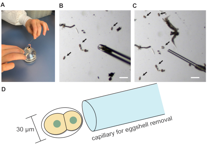
Figure 1: Blastomere isolation. (A) Hand pulling of glass capillary. (B) Hand-drawn glass capillary for embryo transfer. (C) Hand-drawn glass capillary for eggshell removal. (D) Schematics showing the appropriate size of capillary opening for the eggshell removal. Arrows indicate embryos. Scale bars show 100 µm.
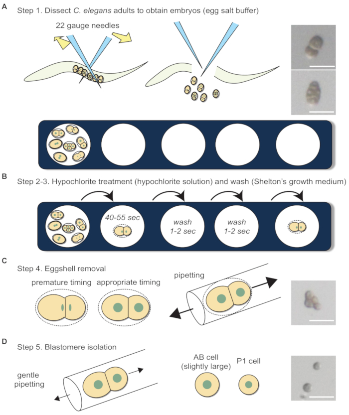
Figure 2: Blastomere isolation workflow. (A) Dissection of adult C. elegans in egg salt buffer to obtain embryos. Photographs show 2-cell and 4-cell stage embryos before eggshell removal. (B) Hypochlorite treatment and washing. (C) Schematics depict the appropriate timing for eggshell removal. Photograph shows a 4-cell stage embryo after eggshell removal. (D) Blastomere separation. Photograph shows a separated 2-cell stage embryo. Sizes of the arrows in C and D indicate the relative forces required during pipetting. Scale bars show 50 µm.
