A subscription to JoVE is required to view this content. Sign in or start your free trial.

Method Article

Tissue Immunostaining for Imaging Mass Cytometry

1.2K views

⸱

January 15th, 2026

In This Article

Abstract

Source:
Jake Griner1, Arwen Wedgeworth2, Brenton Puckett2, Kacey Idouchi1, Slivia Guglietta2, Carsten Krieg1

1Department of Pathology and Laboratory Medicine, Medical University of South Carolina.

2Department of Regenerative Medicine and Cell Biology, Medical University of South Carolina.

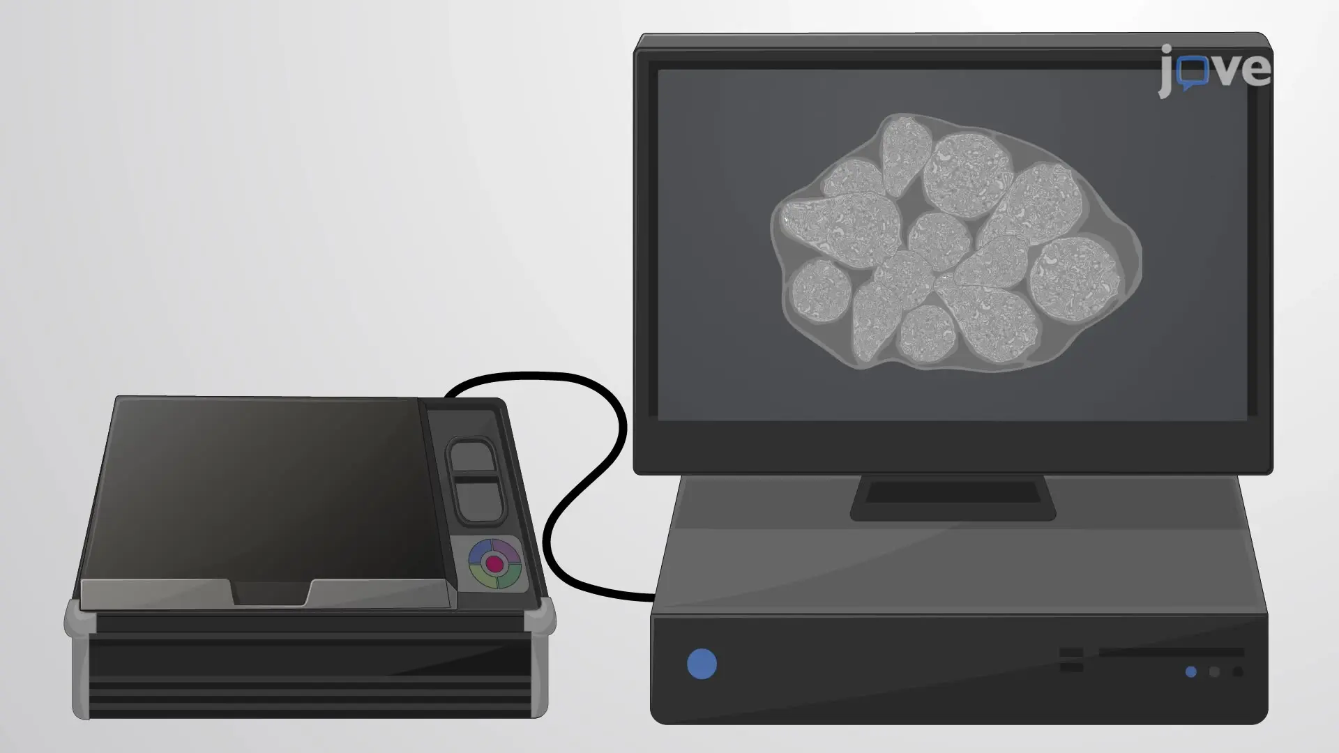
The video describes the preparation of a tissue sample for imaging mass cytometry. The tissue is first processed to unmask the antigen epitopes, permeabilize the cells, and block non-specific antibody binding sites. Then, the tissue is incubated with a metal-conjugated antibody cocktail and a nuclear stain. Finally, the treated tissue sample is imaged using an optical scanner.
Protocol

1. Tissue pretreatment:

For Fresh Frozen tissues:

  1. Place a glass Coplin jar in the -20°C freezer for at least one hour before use.
  2. Remove the slides from the -80°C freezer and place them into the glass Coplin jar in the -20°C freezer for at least 15 minutes.
  3. Under the fume hood, use an ampule cutter to open the 16% paraformaldehyde (PFA) glass ampule. Discard the broken glass appropriately.
  4. Add 10 mL of 16% PFA and 30 mL phosphate-buffered saline (PBS) to a plastic Coplin jar. Ensure that the PBS is pre-chilled to 4°C.
  5. Transfer this solu....

Access restricted. Please log in or start a trial to view this content.

Explore More Articles

Antigen RetrievalPermeabilization BlockingMetal Conjugated AntibodyNuclear StainOptical ScannerHumidity ChamberHydrophobic BarrierSlide Mailer