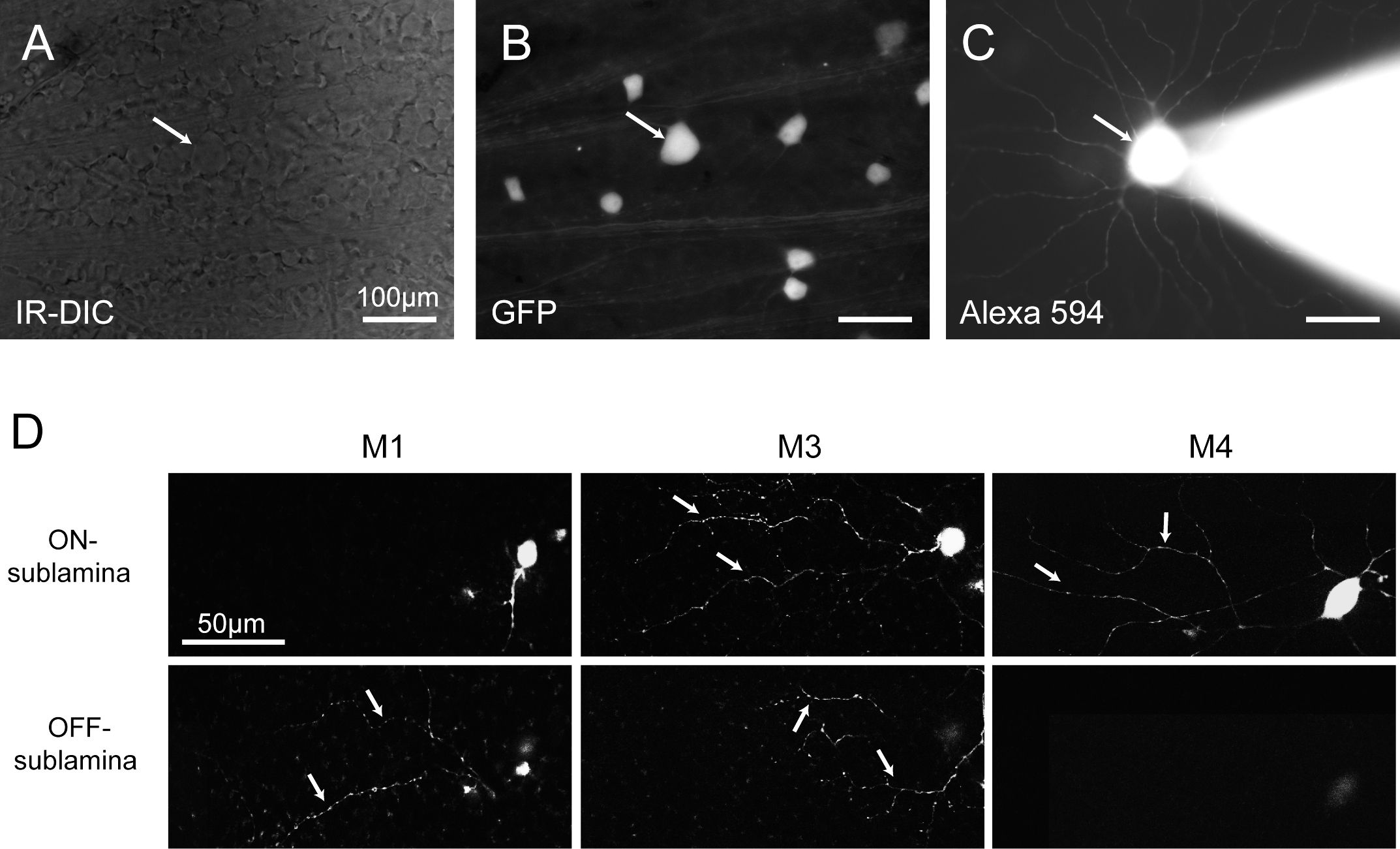
This video demonstrates a fluorescent tracing technique to identify retinal ganglion cell (RGC) subtypes in a transgenic mouse retina. The process involves securing the isolated retina in a recording chamber, maintaining cell viability with oxygenated perfusion, and targeting fluorescently labeled RGCs using a pipette with a tracer. By analyzing dendritic morphology and stratification, the method enables classification of distinct RGC subtypes.
A subscription to JoVE is required to view this content. Sign in or start your free trial.
Method Article
Identification of Mouse Retinal Ganglion Cell Subtypes Through Fluorescent Tracing
771 views
⸱
May 29th, 2025
In This Article
Abstract
Protocol
All procedures involving animal samples have been reviewed and approved by the appropriate animal ethical review.
Access restricted. Please log in or start a trial to view this content.
Results
Access restricted. Please log in or start a trial to view this content.
Materials
| Name | Company | Catalog Number | Comments |
|---|---|---|---|
| Diethyl pyrocarbonate (DEPC) | |||
| Alexa Fluor 594 Hydrazide | |||
| Petri dish (35mm diameter) | |||
| Ophthalmologic scissors | |||
| #5 Forceps | |||
| Glass Micropipette | |||
| Micropipette Puller | |||
| 1mL syringe |
