This video demonstrates a minimally invasive endoscopic procedure to treat lumbar disc herniation by removing the protruded nucleus pulposus and ruptured annulus fibrosus. The technique decompresses the affected nerve root, alleviates pain, and restores nerve function through targeted tissue resection and radiofrequency modulation.
A subscription to JoVE is required to view this content. Sign in or start your free trial.
Method Article
Full-Endoscopic Isolation Zone Technique for the Treatment of Lumbar Disc Herniation
624 views
⸱
August 29th, 2025
In This Article
Abstract
Protocol
All procedures involving human participants have been performed in compliance with the institutional, national, and international guidelines for human welfare and have been reviewed by the local institutional review board.
1. "Isolation zone" technique of spinal endoscopy
- Percutaneously and transforaminally place a 7.5 mm diameter spinal endoscope with a 3.7 mm diameter working channel at the lumbar disc herniation at the appropriate level to explore and clean the ligaments and the residual bone fragments in the intervertebral foramen area, the soft tissues in the spinal canal, and the protruding lumbar intervertebral disc.
- Remove the proliferated, inflamed, and red soft tissues scattered in the vertebral canal with microscopic surgical instruments. Use nucleus pulposus forceps to explore the torn area of the annulus fibrosus, and remove the severely degenerated and inelastic tissue.
- Remove the ruptured part of the annulus fibrosus and the protruding nucleus pulposus using miniature surgical forceps. Use flexible bipolar radiofrequency to coagulate the annulus fibrosus and the nucleus pulposus to stop the bleeding and shrink and denervate them.
- When there is enough space around the nerve root and the dural sac during the operation, use flexible bipolar radiofrequency to perform an annuloplasty and nucleoplasty on the residual annulus fibrosus and nucleus pulposus, respectively.
- Explore the nerve root with a miniature probe hook to ensure that it has sufficient space and spontaneous pulsation (Figure 1E). Remove the endoscope and its working channel, and suture the skin with a 4-0 nonabsorbent surgical thread suture.
Access restricted. Please log in or start a trial to view this content.
Results
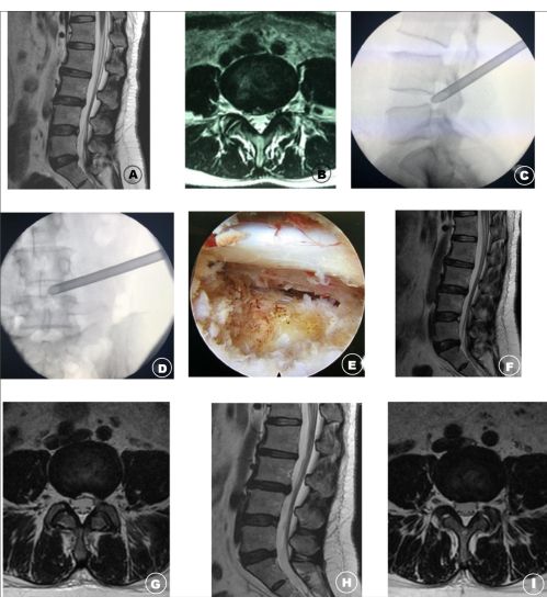
Figure 1: Image data of Lumbar Disc Herniation patients before, during, and after the oper...
Access restricted. Please log in or start a trial to view this content.
Materials
| Name | Company | Catalog Number | Comments |
|---|---|---|---|
| 3.7 mm spinal endoscope | Joimax | FS6342181C | |
| 4-0 nonabsorbent surgical suture | Johnson & Johnson | WB761 | |
| 7.5 mm working channel | Maxmore | 1001-ES04 | |
| Flexible bipolar radiofrequency | Tian song | G8002.2 | |
| Nucleus pulposus forceps | Maxmore | 1001-EF 001 003 | 0°and 15° |
