This video demonstrates the procedure for generating cytotoxic supernatant from pulmonary endothelial cells following infection with Pseudomonas aeruginosa. It outlines how bacterial effector proteins disrupt host cell structure and induce the release of cytotoxic amyloid oligomers into the supernatant.
A subscription to JoVE is required to view this content. Sign in or start your free trial.
Method Article
Obtaining Cytotoxic Amyloids Following Bacterial Infection of Rat Pulmonary Endothelial Cells
369 views
⸱
September 26th, 2025
In This Article
Abstract
Protocol
- Generation of Cytotoxic Supernatants:
Primary cultures of rat pulmonary microvascular endothelial cells (PMVECs) were obtained from the Cell Culture Core Facility at the University of South Alabama’s Center for Lung Biology.
- For generation of cytotoxic supernatants, plate 2 x 10⁶ cells into individual 150 cm dishes and grow for 3 - 4 days until confluence is achieved. Use two plates per experiment.
- To produce supernatant, wash one of the two 150 cm dishes of PMVECs with phosphate-buffered saline, trypsinize, and then count the cells. Wash the other dish of PMVECs with Hank’s Balanced Salt Solution (HBSS) and then infect with either strain of Pseudomonas aeruginosa at a multiplicity of infection (MOI) of 20:1. This is achieved as follows:
- For P. aeruginosa, OD540 0.25 = 2 x 10⁸ CFUs/mL. To prepare this dilution, add 1 mL HBSS to a 1 mL disposable cuvette. Scrape enough bacteria off of the plate until an OD540 of 0.25 is achieved when measured using a spectrophotometer. The next step will provide a calculation of how much bacteria will be needed.
- Once the number of PMVECs in the culture dish is determined, determine the appropriate number of bacteria to be added to the second, non-trypsinized culture dish containing the PMVECs. For example, if it is determined that the culture dish of PMVECs contains 1.3 x 10⁷ cells, then prepare 1.3 x 10⁷ cells x 20 bacteria/cell x 1 mL/2 x 108 CFUs = 1.3 mL bacteria.
- Dilute the 1.3 mL of bacteria into HBSS to a final volume of 20 mL, so that the entire surface of a 150 cm dish can be covered with fluid, then add the diluted bacteria to the plate of PMVECs.
- Incubate the PMVECs inoculated with the bacteria at 37 °C in a 5% CO2 incubator for 4 - 5 h, until gaps can be seen forming in the cell monolayer when the plate is observed microscopically (See Figure 1 for proper level of gap formation).
- Collect the supernatant, then centrifuge for 10 min at 2,000 x g in a tabletop centrifuge to remove cellular debris.
Note: Any table top centrifuge capable of generating sufficient g force to pellet unlysed cells and large cell fragments can be used for this step. - Pour the supernatant into a syringe that has a 0.2 µm filter attached to its end, then pass the supernatant through the filter to remove bacteria.
- Use an aliquot of the sterile supernatant to test cytotoxicity and freeze the rest of the supernatant at -80 °C for future use.
Access restricted. Please log in or start a trial to view this content.
Results
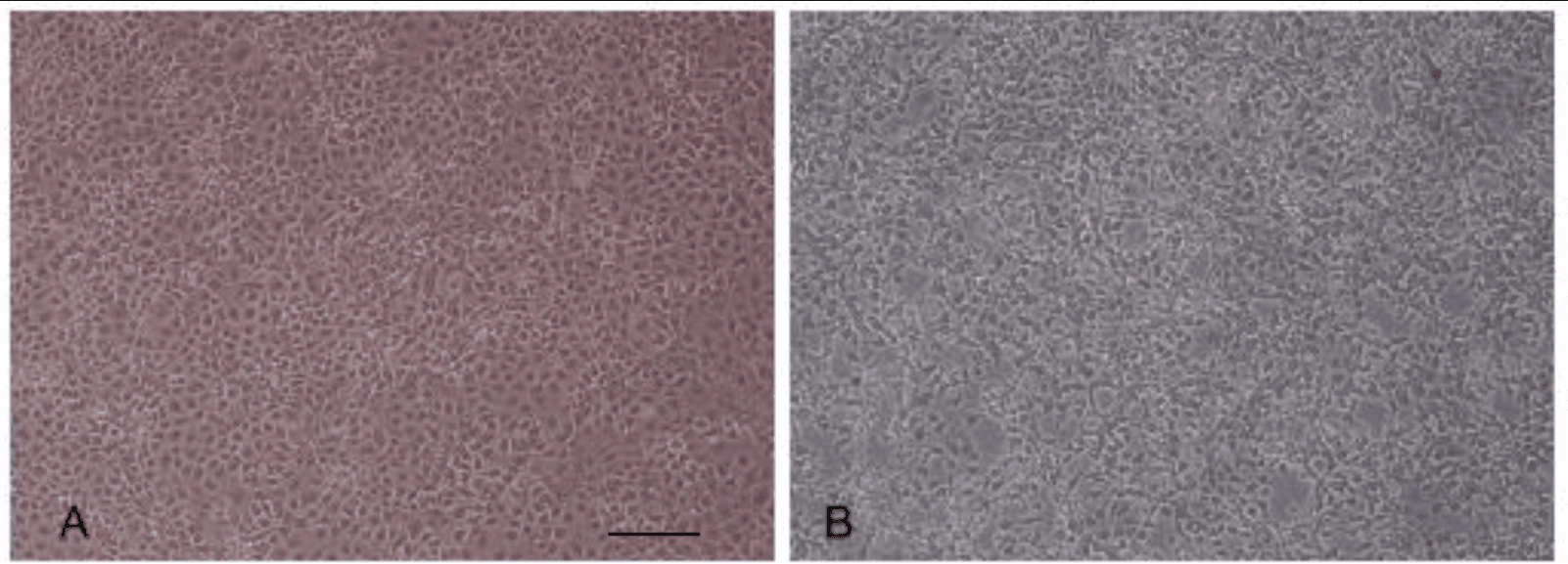
Figure 1. Effects of P. aeruginosa inoculation on PMVEC morphology. A confluent monolayer ...
Access restricted. Please log in or start a trial to view this content.
Materials
| Name | Company | Catalog Number | Comments |
|---|---|---|---|
| HBSS | Gibco | 14025-092 | |
| PBS | Gibco | 10010-023 | |
| 0.22 micron syringe filters | Millipore | SLGP033RS | |
| DMEM | Gibco | 11965-092 | |
| Countess Automated Cell Counter | Invitrogen | C10277 | |
| Microcetrifugation concentrators- 10,000 MW cut-off | Millipore | UFC801008 | |
| Strains PA103 and ΔPcrV | These strains of P. aeruginosa were obtained from Dr. Dara Frank, University of Wisconsin Medical College, Milwaukee, WI | ||
| Agar | Fisher | BP1423-500 | |
| Fetal bovine serum | Hyclone | SH30898.03IH | |
| PenStrep | Gibco | 15070063 |
