This video demonstrates a luminescence-based assay for assessing the antibacterial efficacy of an antibiotic-loaded polymeric strip by quantifying ATP-driven luminescence, which decreases in bacterial cultures exposed to the antibiotic.
A subscription to JoVE is required to view this content. Sign in or start your free trial.
Method Article
Determining the Antibacterial Efficacy of an Antibiotic-Loaded Polymeric Strip
377 views
⸱
September 26th, 2025
In This Article
Abstract
Protocol
Time-dependent antibacterial activity study setup
- Culture the bacteria overnight in 1 mL of TSB (Tryptic soy broth) broth.
- Dilute the overnight-grown bacterial suspension to 105 CFU/mL in sterile MHB (Mueller-Hinton broth), and verify the viable bacterial count using the luminescence units before starting the experiment.
- Place the virgin UHMWPE (Ultrahigh molecular weight polyethylene) and drug-loaded UHMWPE strips (3 mm x 5 mm x 10 mm) within a 3 mL syringe.
- Draw MHB containing 105 CFU/mL into the syringe through the attached needle up to the 1.5 mL mark, which is equivalent to 1.35 mL of MHB (Figure 1: Step 1).
- Place the syringe setup in a shaking incubator (100 rpm) at 37 °C until the indicated time points: 6 h, Day 1, Day 2, Day 3, Day 4, Day 5, Day 6, and Day 7 (Figure 1: Step 2).
- At each indicated time point, take out the syringe setup, and perform a real-time microbial viability assay on 100 µL of bacterial suspension (Figure 1: Step 3-4).
- Perform the luminescence-based assay using white opaque-bottom 96-well plates.
- Mix 100 µL of diluted bacterial suspensions with 100 µL of assay reagent.
- Place a lid covered with aluminum foil over the prepared 96-well plate, and incubate for 5 mins at 100 rpm shaking.
- Measure the luminescence immediately with a microplate reader using the following settings in Gen5 3.11 software.
- Click New to set up a protocol in the Task Manager window.
- Select Costar 96 white opaque in the drop-down menu for Plate Type.
- Click on Read under the Select Steps > Actions menu.
- Click on Luminescence within the Read Method window. Keep the Endpoint/kinetic and Luminescence fiber options selected under read type and optics type, respectively. Click on OK.
- Keep the <default> settings in the Read Step window (gain = 135; integration time = 1 sec; read height = 4.5 mm). Click on OK.
- The plate layout window appears ready for the run. Check the Use Lid box in the load plate window, and click on OK.
- Place the 96-well plate into the machine to read the luminescence values.
- Determine the viable bacteria count for the 6 h, Day 1, Day 2, Day 3, and Day 7 time points. Determine the CFU (Colony forming units)/mL from the luminescence units using the corresponding standard curve.
- Verify the absence of viable bacteria in the samples that showed luminescence values below the limit of detection by performing the spread-plate method on TSA (Tryptic soy agar) plates and incubating at 35 °C overnight. Check for the presence of colonies the next day.
- Centrifuge the remaining bacterial suspension at 10,000 × g for 10 min, and gently aspirate the spent media. For tubes in which pellets are not visually observed due to the antibacterial activity of the drugs, leave 100 µL in the tube to ensure the unseen pellet is not disturbed (Figure 1: Step 5).
- Resuspend the pelleted bacteria in fresh MHB, and draw back the bacterial suspension into the same syringe setup through the attached needle (Figure 1: Step 6).
Access restricted. Please log in or start a trial to view this content.
Results
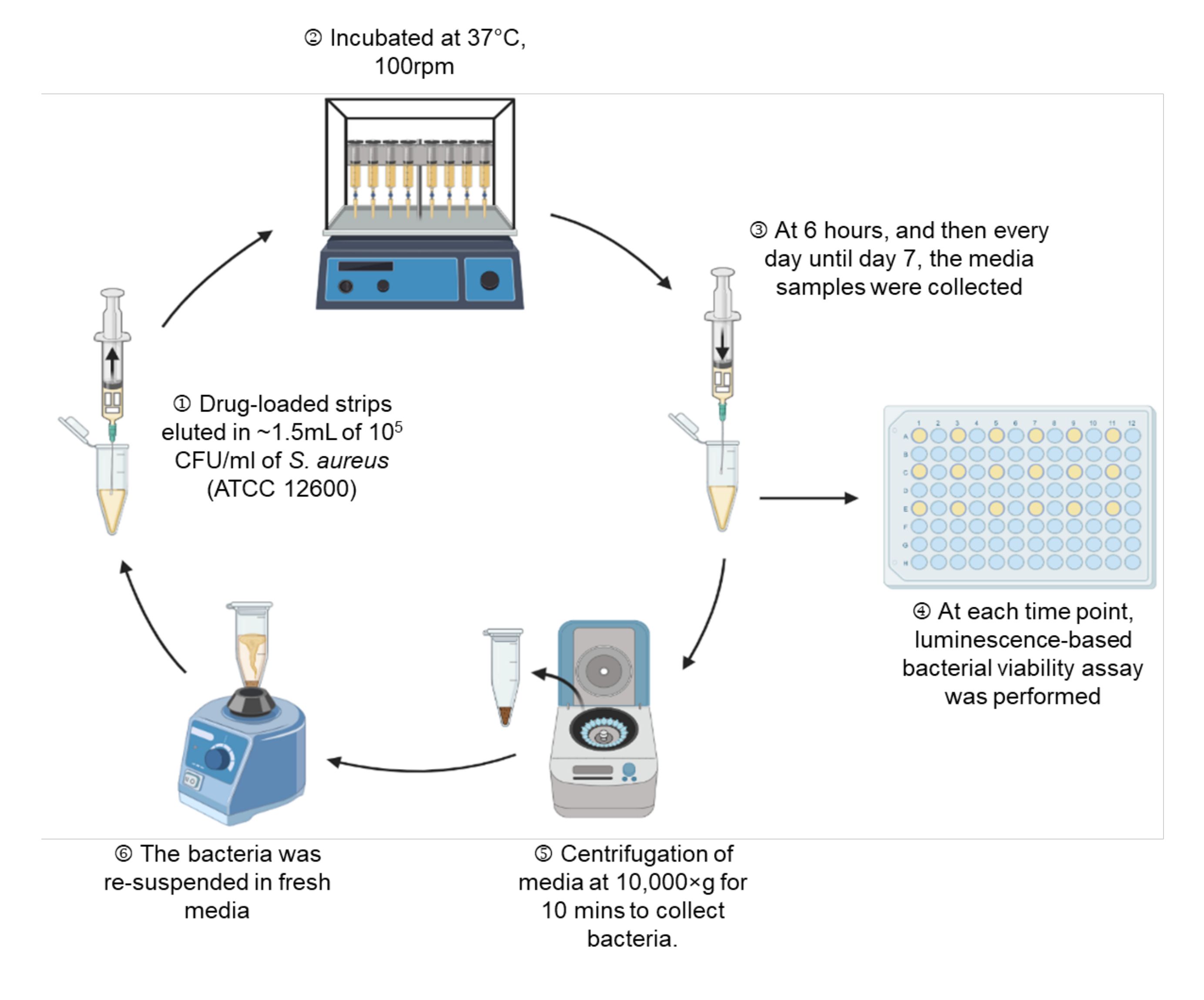
Figure 1: A schematic representation of the experimental setup.
Access restricted. Please log in or start a trial to view this content.
Materials
| Name | Company | Catalog Number | Comments |
|---|---|---|---|
| ATCC 12600 | American Type culture Collection, VA, USA | ||
| BacTiter-Glo Microbial Cell Viability Assay | Promega Corporation, USA | G8231 | |
| BD Bacto Tryptic Soy Broth (Soybean-Casein Digest Medium) | Becton-Dickinson, USA | BD 211825 | Purchased from Fisher Scientific, USA |
| BD Luer-Lok Syringe sterile, single use, 3 mL | BD, USA | 309657 | |
| BD Needle 5/8 in. single use, sterile, 25 G | BD, USA | 305122 | |
| BD BBL Dehydrated Culture Media: Mueller Hinton II Broth (Cation-Adjusted) | Becton-Dickinson, USA | B12322 | Purchased from Fisher Scientific, USA |
| BD Difco Dehydrated Culture Media: Tryptic Soy Agar (Soybean-Casein Digest Agar) | Becton-Dickinson, USA | DF0369-17-6 | Purchased from Fisher Scientific, USA |
| Gentamicin Sulfate | Fujian Fukang Pharmaceutical Co., Fuzhou, China | ||
| LSE benchtop shaking incubator | Corning, NY, USA | ||
| Plate reader (Synergy H1 | Biotek, VT, USA | ||
| Shaker Innova 2100 | New Brunswick Scientific, NJ, USA | ||
| UHMWPE | GUR1020, Celanese, TX, USA | ||
| Vancomycin Hydrochloride | Fagron, The Netherlands | 804148 |
