This video demonstrates the manual isolation of single epithelial cells containing intracellular bacterial communities from infected mouse bladder using mouth micropipetting, enabling downstream single-cell analysis of viable host-associated E. coli populations in urinary tract infection models.
A subscription to JoVE is required to view this content. Sign in or start your free trial.
Method Article
Isolation of Mouse Bladder Epithelial Cells Containing Intracellular Bacterial Communities
471 views
⸱
February 2nd, 2026
In This Article
Abstract
Protocol
All procedures involving animal models have been reviewed by the local institutional animal care committee and the JoVE veterinary review board.
1. Intracellular Bacterial Community (IBC) isolation: Mouth Pipetting of IBCs
NOTE: All methods described in this section have undergone an institutional risk assessment. Mouth pipetting carries the inherent risk of ingestion of the solution that is being transferred. This risk is largely mitigated by the nanoliter volumes that this protocol uses, and we recommend that all users of the protocol pay heed to the precautionary and practice notes listed here and in the discussion.
- After the cells have been scraped into the PBS (phosphate-buffered saline), set up the mouth micropipetting apparatus (Figure 1C).
- Insert the thicker end of the pulled glass capillary (the unpulled end) into the rubber plug (white end) of the aspirator tube.
- Insert the narrow end of a 1 mL pipette tip into the other open (red) end of the aspirator tube, ensuring a tight fit.
- Insert the narrow end of a 2 mL aspirating pipette into the open, wider end of the 1 mL pipette tip, again ensuring a tight fit.
NOTE: The resulting setup allows the researcher to mouth pipette from the wider, open end of the aspirating pipette to create a gentle suction force from the narrow end of the capillary tube at the other end of the apparatus. - Test the final mouth micropipetting apparatus using a 100 mm Petri dish containing fresh deionized water. A slight suction action on the open end of the aspirating pipette (similar to sipping on a drink through a straw) should increase the level of liquid in the capillary, but not cause the deionized water to overflow into the aspirator tube. Use one hand to control the capillary tube, while using the other hand to adjust the position of the Petri dish.
NOTE: The strength of suction required for mouth-pipetting a single IBC will vary among researchers. However, it is recommended that each researcher attempting this technique start with a weak suction and slowly increase it if no liquid is flowing up the capillary. There is no need for a force greater than sucking on a straw for drinking. If the capillary does not appear to be picking up liquid during the test in step 1.1.4, it is possible that either the capillary or the aspirating tube is occluded and needs to be replaced. It is further recommended that all new researchers first practice controlling suction in the mouth pipetting apparatus using sterilized water. Additionally, note that the control of the volume taken up by the mouth pipetting apparatus is through the use of the researcher's tongue. The tongue can finely adjust the strength of suction applied, as well as act as an emergency stop. - After achieving successful uptake of liquid, test the ability to expel it from the capillary by gently blowing into the open end of the aspirating pipette. Ensure that no bubbles are created in the process of expelling the liquid to prevent contamination of the IBCs during step 1.5.
NOTE: As with suction, the strength of positive pressure applied by the researcher to expel the IBC into the centrifuge tube will vary among researchers. It is recommended for researchers new to this protocol to practice step 1.1 a few days before the actual infection. One suggestion for practicing mouth micropipetting is to practice transferring small volumes of sterilized water mixed with a few drops of food dye (for visibility) using the mouth micropipette apparatus.
- Place the scraped cell suspension under the dissecting microscope and identify the IBCs as large fluorescent aggregates (Figure 1A, B). The ideal range of magnification is 20-40x. Dip the fine end of the glass capillary into a fresh tube of PBS for 1 s to reduce the uptake of unwanted volume through capillary action.
- Looking through the microscope, identify the IBC of interest and slowly bring the open end of the capillary tube toward the IBC. Use the fine end of the glass capillary to sweep away extra cells near each IBC to prevent the aspiration of two or more cells, or to break apart larger aggregates of cells.
- While looking through the microscope, apply a very small suction force on the far end (aspirating pipette) of the mouth micropipetting apparatus to guide the IBC into the glass capillary.
- After picking up the IBC, move the capillary to an empty 1.5 mL centrifuge tube and apply a slight positive pressure to expel the droplet and the IBC into the centrifuge tube (Figure 1D).
- Repeat steps 1.3-1.6 on as many IBCs are needed from the current bladder, before proceeding to the next bladder. Change aspirating pipettes and capillaries frequently to prevent build-up of saliva.
CAUTION: When working with infectious (or clinical) strains of bacteria, constantly monitor the level of solution in the capillary. Do not let the level of liquid being pipetted overflow from the edge of the capillary into the aspirator tube. If this occurs, immediately switch to a different aspirator tube, and discard the previous set up. - Repeat steps 1.2-1.6 on all harvested bladders, or until a sufficient number of biological samples have been collected for the experimental group.
- (Optional) Repeat steps 1.6 and 1.7 until all experimental groups (or mice) have been euthanized and sufficient biological samples have been harvested from each group.
Access restricted. Please log in or start a trial to view this content.
Results
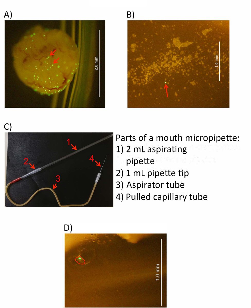
Figure 1: IBC harvesting from bladder cells. (A) An infected and inverted blad...
Access restricted. Please log in or start a trial to view this content.
Materials
| Name | Company | Catalog Number | Comments |
|---|---|---|---|
| 1 mL Tuberculin Syringe | BD Biosciences | 302100 | |
| Aspirating pipette | BD Biosciences | 357558 | |
| Aspirator tube | Sigma-Aldrich | A5177 | |
| Digital camera for microscope | Olympus | DP71 | For image capture and harvesting of IBCs. Any other fluorescent microscope with a GFP channel will suffice |
| Glass Capillaries | Kimax | 6148K07 | |
| Iris Scissors STR SS 110MM | Braun | BC110R | |
| Light source unit for microscope | Olympus | LG-PS2 | For image capture and harvesting of IBCs. Any other fluorescent microscope with a GFP channel will suffice |
| Macro fluorescence microscope | Olympus | MVX10 | For image capture and harvesting of IBCs. Any other fluorescent microscope with a GFP channel will suffice |
| Micropipette + micropipette tips | For static bacterial culture and OD measurement | ||
| PBS (Phosphate-bufered saline) 1x | For static bacterial culture and OD measurement | ||
| Polyethylene Tubing | BD Intramedic | 427401 | |
| Sterilin 90mm standard Petri dish | Thermo | 101VR20 | Any sterile petri dish |
| Stevens, vascular and tendon scissors, curved, delicate, 110 mm | Braun | OK366R | Recommended for harvesting of bladder |
| Surgical scissors STR S/B 105MM | Braun | BC320R | |
| Tabletop centrifuge | Eppendorf | 5810R | Any refridgerated centrifuge for 15ml conicals |
