Method Article

Microscopy-Based Evaluation of Bacterial Attachment to Plant Root Surfaces

February 26th, 2026

In This Article

Abstract

Loading...
$$\rightleftharpoonup{xx}$$ $$\longleftharp{xx}$$, $$\longrightharp{xx}$$,

Source: Matthysse, A. G. Adherence of Bacteria to Plant Surfaces Measured in the Laboratory. J. Vis. Exp. (2018).

This video demonstrates the evaluation of bacterial attachment to plant roots using phase-contrast microscopy. Root samples from seedlings incubated with bacteria are first mounted to check for free bacteria. The samples are then washed to remove loosely attached bacteria and remounted using a suitable coverslip to prevent damage. Finally, the number of bacteria attached to the root hairs is counted under the microscope.

Protocol

Loading...
$$\rightleftharpoonup{xx}$$ $$\longleftharp{xx}$$, $$\longrightharp{xx}$$,

1. Inoculation of the Bacteria

  1. Determine the number of bacteria to be inoculated with reference to the measurement to be used to determine bacterial adhesion and the length of the incubation time.
  2. For microscopic studies involving incubation times less than 1 day, inoculate relatively large numbers of bacteria. Add an amount of culture to reach a final bacterial concentration of more than 106 bacteria per mL. For longer incubation times, decrease the bacterial inoculum size.
  3. For studies in which bacterial adhesion will be measured by viable cell counts, add an amount of culture to reach a final bacterial concentration of 103 to 106 bacteria per mL.
  4. Avoid adding so many bacteria that their metabolism changes the pH or oxygen concentration in the incubation medium. Measure pH using pH paper or an electrode. Measure oxygen concentration using an oxygen electrode.
  5. For plants grown in sand, inoculate in three possible ways.
    1. Inoculate the seed with the bacteria before planting by soaking the seeds for 1 min in a suspension in water of about 106 bacteria per mL.
    2. Inoculate the root by germinating axenic seedlings. When the seedling root is about 1 cm long, dip it or place the whole seedling in a suspension of 106 bacteria per mL in water for 1 min.
    3. Inoculate the sand by mixing the bacteria with the sand before planting to give a final concentration of about 103 bacteria per mL or by watering the seedling with a suspension of 106 bacteria per mL after planting.

2. Incubation of the Bacteria with the Plant Material

  1. For incubation in liquid media, incubate the bacteria with the plant material in sterile water or sucrose and mineral salts or plant tissue culture medium (such as a 1:10 dilution of Murashige and Skoog basal salt mixture or MS salts).
    1. Use a container to which the bacteria do not adhere. Try to keep the plant surface covered continuously, either by submersion or by gentle agitation. Vigorous agitation may prevent adhesion or even remove bacteria from the plant surface.
    2. Observe the plant material in the light microscope after varying time intervals to determine when to stop the incubation and make measurements. A time course of adhesion is often valuable with samples taken every 1 to 4 h or every day, depending on the speed of the interaction.
  2. To incubate in sand, apply the same considerations described for incubation in liquid medium in step 2.1.

3. Measurement of Adhesion Using Microscopy

  1. Make microscopic measurements with Nomarski or phase-contrast optics for easy viewing of the bacteria on surfaces. However, any bright-field microscope with magnification of 20X or higher can be used.
    1. Use microscopic observation to determine if the bacteria are randomly distributed or located in specific sites. Also, check whether they are bound singly or in clusters. Look for the presence of microcolonies suggesting bacterial growth or entrapment after adhesion. Check whether the bacteria seem to form a biofilm on the surface.
      NOTE: A biofilm is a large number of bacteria bound to the surface and surrounded by an extracellular matrix. The structure may be smooth and uniform or have a more complicated architecture.
    2. Use bacteria tagged with a fluorescent marker. If other bacteria are present and the bacteria of interest are identified by a fluorescent tag such as green fluorescent protein (GFP), use fluorescence microscopy to determine the presence of the tagged bacteria in clusters of other bacteria. For GFP, use a filter with 490 nm excitation and 520 nm emission.
      1. Check that the fluorescent tag is not adversely affecting the bacteria by observing the adherence to axenic material of an equal mixture of wild-type bacteria and tagged bacteria of the same strain using both Nomarski and fluorescence optics. If the fluorescent and dark bacteria are randomly mixed and present in equal numbers, then the tag did not interfere with the assay.
  2. Determine the number of attached bacteria.
    ​NOTE: It is very difficult to determine the number of attached bacteria in the microscope. When the binding is to irregular plant surfaces, it is generally not possible to obtain a quantitative measurement. Scanning electron microscopy (not discussed in this article) can be used to make such measurements.
    1. When the bacteria are bound to a smooth surface, such as a root hair, count the number of bacteria bound to the edge of the root hair per mm of root hair length. Take care to use root hairs of roughly the same size and shape in comparing measurements.
    2. To determine the size of objects in the microscope, use a commercial slide with measured markings on it. Observe and photograph this slide at the same settings as used for the experimental material and use the resulting images to determine the size of objects in the photomicrographs.
  3. Prepare the sample for microscopy.
    1. Wash the sample. Move the sample to a drop of water or incubation medium on a microscope slide and observe it directly.
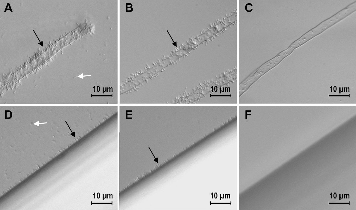
      NOTE: This has the advantage that if there was no bacterial growth or actual bacterial death, there are unlikely to be many free bacteria. Take the absence of free bacteria as a warning sign that there may have been bacterial death or bacterial binding to the container in which the incubation was carried out. The effect of washing the sample is shown in Figure 1.
    2. Wash the sample gently in water or incubation medium by placing it in a vial of liquid and inverting the vial gently. Then place the sample on the microscope slide in fresh liquid for observation.
    3. Mount the sample in liquid using an ordinary cover slip and a microscope slide.
      1. If the sample is thick and so would make a bulge under the cover slip, use a press-apply cover slip. These cover slips have a ring of rubber or plastic around the edge of the cover slip. Place the liquid and sample in the well in the cover slip and then place the slide on top and press down gently to seal the cover slip to the slide. Invert and examine.
      2. Alternatively, use an algae counting slide and cover slip in a similar manner. Note that slides with this depth cannot generally be examined with an objective lens of greater than 20X magnification.

Access restricted. Please log in or start a trial to view this content.

Results
Loading...
$$\rightleftharpoonup{xx}$$ $$\longleftharp{xx}$$, $$\longrightharp{xx}$$,
24465_Figure_1.jpg

Figure 1: Steps in the preparation of a sample for determining the number of bound bacteria. Agrobacterium tumefaciens binding to tomato root hairs (A, B, and C) and to nylon threads (D, E, and F). In samples mounted in water without...

Access restricted. Please log in or start a trial to view this content.

Materials

List of materials used in this article
NameCompanyCatalog NumberComments
Light microscopeanyN/Aphase contrast or Nomarski optics are helpful, for studies using fluorescent markers a fluorescence microscope is required.
SeedsanyN/Amake a note of the seed lot number and the cultivar
BleachanyN/A 
Bath sonicatorany scientific supply companyN/A 
Triton X-100Sigma-AldrichT9284 
Nutrient agarDifco2001-01-08 
SoytoneDifco24360 
Sandsea sand Fisher ScientificS25 
SandSigma-AldrichS9887 
ContainersStuewe & Sons, Inc.Ray-Leach cone-tainersmany different sizes are available to suit the type of plant you wish to grow
Parafilmany scientific supply companyN/A 
MS saltsSigma-AldrichM5524 
Sedgwick-rafter counting cellHauser ScientificHS3800 
Probe-clip press-seal incubatin chamberSigma-AldrichZ359483 
RifampicinSigma-AldrichR3501 

Reprints and Permissions

Request permission to reuse the text or figures of this JoVE article

Request Permission

Tags

Bacterial AttachmentPlant Root SurfacesPhase Contrast MicroscopyRoot Hair AnalysisBacterial WashingSample MountingCoverslip TechniqueMicroscopy EvaluationRoot Sample PreparationBacterial Counting

Related Articles