This video demonstrates a pellicle formation assay using Bacillus subtilis to evaluate small-molecule inhibitors that disrupt matrix synthesis and biofilm development, providing a model to screen compounds that selectively inhibit surface-associated growth without affecting free-floating cells.
A subscription to JoVE is required to view this content. Sign in or start your free trial.
Method Article
Evaluating Inhibitor Effects on Pellicle Formation in Bacillus subtilis
370 views
⸱
February 26th, 2026
In This Article
Abstract
Protocol
1. Assessing the Effect of Small Molecule Inhibitors on Pellicle and Biofilm Colony Formation
- Prepare a 2x solution of defined biofilm-inducing MSgg (monosodium glutamate-glycerol) medium without the calcium chloride and iron(III) chloride hexahydrate. After filter sterilization, add the calcium chloride. The medium is ready to use directly, or it can be stored at 4 °C in the dark.
- Prepare the 1x MSgg dilution on the day of the experiment.
- Dilute the 2x MSgg medium to 1x with sterile distilled water (pellicles) or sterile 3% hot (80 °C) agar (biofilms) and add iron(III) chloride hexahydrate to a final concentration of 50 μM (pellicles) or 250 µM (biofilms). Add antibiotics or small-molecule inhibitors to the desired concentration and mix well. For example, to obtain a final concentration of 0.5 mM D-leucine in 30 ml to establish pellicles or biofilms, add 196.6 µl of a 76.3 mM (10 mg/ml) D-leucine stock solution.
NOTE: The final 1x MSgg composition is described in Table 1. Compared to the original recipe, the medium contained 50 µg/ml threonine, and the iron concentration to grow biofilm colonies on solid MSgg medium was increased 2.5x to optimize the wrinkled colony morphology.
- Dilute the 2x MSgg medium to 1x with sterile distilled water (pellicles) or sterile 3% hot (80 °C) agar (biofilms) and add iron(III) chloride hexahydrate to a final concentration of 50 μM (pellicles) or 250 µM (biofilms). Add antibiotics or small-molecule inhibitors to the desired concentration and mix well. For example, to obtain a final concentration of 0.5 mM D-leucine in 30 ml to establish pellicles or biofilms, add 196.6 µl of a 76.3 mM (10 mg/ml) D-leucine stock solution.
- After solidification of the agar, dry the solid MSgg plates in a biological hood for 30-45 min prior to the inoculation.
- To select specific inhibitors that interfere with the mechanisms of pellicle formation (Figure 1), rule out that the concentrations used affect planktonic and static growth.
- Determine planktonic growth (increase in optical density over time in liquid culture) in a simple growth curve by measuring the optical density at 600 nm every hour until the stationary growth phase.
- To confirm that the measured culture turbidity represents live cell counts, determine the number of colony forming units (CFU) of cells in the planktonic growth phase from a shaking culture at several time points.
- To assess the effect of small molecule inhibitors on static pellicle growth, harvest cells at the end of a 3-day incubation at 23 °C from a 24-well cell-culture well, inoculated under the same conditions as described in sections 1.7-1.9, and determine the CFU. For this control, use a pellicle-deficient strain that lacks the operons encoding the extracellular matrix components (i.e., B. subtilis ΔepsH, ΔtasA).
NOTE: This strain is capable of growing under static conditions, but in contrast to a pellicle-forming wild type, it is deficient in the ability to float to the liquid-air interface, where growth is favored due to increased oxygen levels. Thus, this extracellular matrix- and pellicle-deficient strain is a recommended reference strain to assess growth under static conditions.
NOTE: For the specific example of the non-canonical D-amino acid D-leucine described below, an effect on planktonic and static growth at concentrations that interfered with pellicle formation was ruled out. The methods to determine planktonic and static growth are described in detail.
- Streak out B. subtilis from a -80 °C stock (LB culture of 109 cells/ml frozen in 20% glycerol) to isolate single colonies on an LB-1.5% agar plate with a sterile tip or applicator stick.
- Grow overnight at 30 °C.
- CRITICAL STEP: For a robust pellicle inhibition by the non-canonical D-amino acids such as D-leucine, grow a single colony picked from the LB-1.5% agar plate in 3 ml LB broth at 37 °C for 4 hr in a shaking incubator (shaking speed 200 rpm). Replace the LB broth with biofilm-inducing MSgg medium prior to inoculation by centrifuging 1.5 ml starter culture for 4 min at 6,000 x g, carefully removing the supernatant, and resuspending the pellet in 1.5 ml MSgg medium. The rest of the culture can be discarded.Important: To ensure the robustness of the system, the optical density at 600 nm (OD600) of the washed starter culture should be between 0.6 and 1.
- During the growth of the starter culture, prepare a 12-well cell-culture multidish plate containing 3 ml of MSgg medium without or with a concentration range of the small molecule inhibitor (e.g., 0.3, 0.5, 1 mM D-leucine). To rule out edge effects, distribute the location of the different concentrations across the multidish plate. Alternatively, use 24-well cell-culture multidish plates containing 1.5 ml of MSgg medium.
- Inoculate the wells of the 12-well cell-culture multidish plate with 3 µl of the washed starter culture (1:1,000 dilution).
NOTE: A lower dilution ratio, i.e., 1:500 can be used. This decreases the development time of the pellicles. - Grow the pellicles at 23 °C under static conditions for three days. Do not move the pellicles during this time, as it can affect the final surface morphology of the pellicle.
- Acquire pictures with a binocular and homogeneous exposure of lightning. Alternatively, take a picture of the pellicles with a high-resolution camera. To avoid artefacts caused by inconsistent light angles and shadows, take top-down pictures with the camera fixed on a tripod and use a soft and large light source at 45° from both sides.
NOTE: An alternative method to study B. subtilis multicellularity is the biofilm colony assay on solid, biofilm-inducing MSgg medium. Like pellicles, this assay allows the study of spatiotemporal processes. Once the active range of small molecule inhibitors is determined, their effect on biofilm colony formation can be studied. - To grow biofilm colonies, symmetrically spot 1.5 µl of the unwashed pre-culture (Step 1.7) on the dried MSgg 1.5% agar plate with the help of a template — 4 drops per Petri dish of 8.5 cm diameter. Let the drops absorb to the plate before moving them.
NOTE: The template helps to get an equal distribution of the biofilm colonies within the area where the cells are grown. To prepare the template, draw the total area of growth at original scales, divide it to equal sectors and mark the center. For a round Petri dish of 8.5 cm diameter, this assigns 14 cm2 to one biofilm colony. - Incubate the plates at 30 °C for three days. During this time, biofilm colonies develop and form a three-dimensional, wrinkled structure.
- Take pictures as in step 1.11.
Table 1. Final 1x MSgg composition used in this study.
| 1.925 mM | Potassium phosphate monobasic |
| 3.075 mM | Potassium phosphate dibasic |
| 100 mM | 3-(N-morpholino)propanesulfonic acid, pH 7.1 |
| 2 mM | Magnesium chloride hexahydrate |
| 700 μM | Calcium chloride anhydrous |
| 50 μMa | Iron(III) chloride hexahydrate |
| 125 μMb | |
| 1 μM | Zinc chloride anhydrous |
| 2 μM | Thiamine hydrochloride |
| 50 μg/ml | Tryptophan |
| 50 μg/ml | Phenylalanine |
| 50 μg/ml | Threonine |
| 0.5% (v/v) | Glycerol anhydrous |
| 0.5% (w/v) | L-glutamic acid monosodium salt hydrate |
| afor pellicle assay; bfor biofilm assay | |
Access restricted. Please log in or start a trial to view this content.
Results
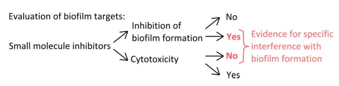
Figure 1. Conceptual overview for the identification of a robust experimental setup to assess t...
Access restricted. Please log in or start a trial to view this content.
Materials
| Name | Company | Catalog Number | Comments |
|---|---|---|---|
| Luria Broth, Lennox | Difco | 240230 | |
| Potassium phosphate monobasic | Sigma, 136.09 g/mol | P0662-500G | |
| Potassium phosphate dibasic | Fisher Scientific, 174.18 g/mol | BP363-1 | |
| 3-(N-morpholino)propanesulfonic acid | Fisher Scientific, 209.27 g/mol | BP308-500 | |
| Magnesium chloride hexahydrate | Merck, 203.30 g/mol | 1.05833.0250 | |
| Calcium chloride anhydrous | J.T. Baker, 110.98 g/mol | 1311-01 | |
| Manganese(II) chloride tetrahydrate | Sigma, 197.91 g/mol | 31422-250G-R | |
| Iron(III) chloride hexahydrate | Sigma, 270.30 g/mo) | F2877-500G | |
| Zinc chloride anhydrous | Acros Organics, 136.29 g/mol | 424592500 | |
| Thiamine hydrochloride | Sigma, 337.27 g/mol | T1270-100G | |
| L-tryptophan | Fisher Scientific, 204.1 g/mol | BP395-100 | |
| L-phenylalanine | Sigma, 165.19 g/mol | P5482-100G | |
| L-threonine | Sigma, 119.12 g/mol | T8625-100G | |
| Glycerol anhydrous | Bio-Lab Itd | 712022300 | |
| L-glutamic acid monosodium salts hydrate | Sigma, 169.11 g/mol | G1626-1KG | |
| D-leucine | Sigma, 169.11 g/mol | 855448-10G | |
| Ethanol anhydrous | Gadot | 830000054 | |
| Shaker 37 °C | New Brunswick Scientific Innowa42 | NA | |
| Centrifuge | Eppendorf table top centrifuge 5424 | NA | |
| Incubator 30 °C | Binder | NA | |
| Incubator 23 °C | Binder | NA |
