This video demonstrates the method of establishing a foodborne bacterial intestinal infection model in zebrafish larvae by feeding them paramecia infected with fluorescent E. coli.
A subscription to JoVE is required to view this content. Sign in or start your free trial.
Method Article
Modeling Intestinal Infection in Zebrafish Larvae Using Foodborne Bacteria
353 views
⸱
February 26th, 2026
In This Article
Abstract
Protocol
- Food-borne Infection of Zebrafish
- Incubate bacteria with Paramecia.
- Prepare a co-culture of Paramecium caudatum and Escherichia coli MG1655 the night prior to infection. Combine 8 mL of E3 media, 1 mL of an ongoing paramecia culture, and 1 mL of an E. coli MG1655 culture (OD600 = 1.0) resuspended in 1x E3 in T25 tissue culture flasks. Incubate the flasks at room temperature (RT) overnight. For each treatment condition, prepare two flasks of paramecia.
- Inoculate bacterial growth media (LB (Lysogeny Broth): 1 g/L tryptone, 0.5 g/L yeast extract, 1 g/L NaCl) with the infectious strain of bacteria by picking an individual bacterial colony from a plate using a sterile inoculation loop. Incubate the liquid culture at 37 °C and leave shaking at 110 rotations per minute (rpm) overnight.
NOTE: Personal protective equipment (a laboratory coat and gloves) should be worn, and biosafety level 2 facilities should be used when handling infectious agents. - On the next day, measure the OD600 of the overnight culture. Calculate the volume of culture required to achieve an OD600 of 1 when resuspended in 11 mL of media.
- Harvest the volume of bacteria via centrifugation at 6,000 x g for 5 min, one volume for each flask of paramecia. Discard the supernatant and resuspend the bacterial pellet in 1 mL of E3 media.
- Optionally, pre-stain the bacteria with a fluorescent dye.
- Add 1 µL of FM 4-64FX bacterial stain (5 mg/mL stock solution). Cover tube with foil to protect from photobleaching and incubate rotating end-over-end at RT (Room temperature) for 15 min.
- Remove excess dye by washing with 1x E3: Pellet bacteria via centrifugation at 6,000 x g for 1.5 min, then resuspend the pellet in 1 mL of E3 media. Repeat the wash step two times.
- Harvest stained bacteria via cntrifugation at 6,000 x g for 5 min. Discard the supernatant and resuspend the bacterial pellet in 1 mL of E3 media.
- Add 1 mL of the bacterial suspension to each of the two flasks of fresh paramecia. Incubate at RT for 2 h.
NOTE: If working with stained bacteria, incubate in the dark at RT for 2 h
- Incubate bacteria with Paramecia.
- Wash bacteria/paramecia co-culture
- Combine the contents of both flasks of paramecia/bacteria co-culture into a 50 mL conical tube. Centrifuge samples at 300 x g at 15 °C for 10 min. Make sure that the centrifuge is pre-cooled prior to this step.
- Remove approximately 10 mL of the E3 supernatant using a serological pipette and add approximately 10 mL of fresh 1x E3 to the conical tube.
NOTE: During all wash steps, it is essential to be very quick when removing the supernatant, as the paramecia will begin to swim out of the pellet. Spin and remove supernatant from one tube at a time to ensure quick enough handling at this step, and avoid loss of paramecia in the supernatant. - Spin samples via centrifugation at 300 x g at 15 °C for 5 min. Remove approximately 10 mL of the E3 supernatant using a serological pipette, and add approximately 10 mL of fresh 1x E3 to the conical tube. Repeat this step twice.
- Centrifuge samples at 300 x g at 15 °C for 5 min. Remove approximately 10 mL of the E3 supernatant, taking care not to disrupt the pellet.
- Resuspend pellet into the remaining 10 mL of E3 media and transfer 500 µL of the suspension into a new 1.5 mL microcentrifuge tube. Pellet the 500 µL of paramecia by centrifuging at 300 x g for 5 min to count the number of paramecia.
- Remove 400 µL of the E3 supernatant from the 500 µL sample. Add 20 µL of 36.5% formaldehyde solution to the remaining 100 µL of paramecia and gently resuspend, and incubate for 5 min at 22 °C.
NOTE: This step kills the paramecia to allow for counting. - Measure actual total volume using the pipette and record. Dilute the paramecia suspension 1:1 v/v with 0.4% trypan blue solution.
- Use a cell counter or hemocytometer to count the number of dead paramecia/mL.
NOTE: Because of the prior fixation step, most paramecia will be dead at this point, but this number reflect the number of live paramecia for the co-incubation experiment. The authors have not found significant paramecia death due to bacterial co-incubation, so this can be disregarded as a factor here.
- Co-incubate Paramecia and zebrafish larvae
- Calculate the concentration of paramecia in the 50 mL conical tube
- Calculate the volume of washed paramecia required for a concentration of 2 x 10⁵ paramecia/mL in a final volume of 3 mL of E3.
NOTE: The concentration of paramecia can be adjusted based on the desired bacterial dosage, which is subject to optimization. - Anesthetize zebrafish by adding tricaine in 100 mM Tris pH 8.0 to a final concentration of 100 mg/L. Transfer 10 zebrafish into each well of a 6-well plate into a total volume of 3 mL of fresh E3 containing the appropriate concentration of paramecia. Ensure that larvae are transferred in a minimal amount of liquid to ensure they recover from anesthesia in the recipient well.
- Incubate at 30 °C for 2 h in a diurnal incubator under daylight conditions, to ensure optimal lighting conditions for preying.
- Wash zebrafish at least 5 times by transferring fish into a new well containing 3 mL of fresh E3 containing 100 mg/L tricaine each time.
NOTE: Do not attempt to omit the tricaine during the washing step. Transferring mobile larvae without anesthesia increases the risk of damage and distress to the animal. - Optionally, prepare zebrafish for imaging by embedding zebrafish in 3 mL of 1% low-melt agarose in a black-walled 6-well plate: Low-melt agarose is made up in 1xE3 and heated in a microwave. Once molten, add tricaine to a final concentration of 160 mg/mL. Position fish under a stereomicroscope, using a clipped gel loading tip, making sure that the head is on the left and the tail is on the right (Figure 1). Wait for 5 minutes for the agarose to set, then overlay the embedded fish with 1x E3 containing 160 mg/mL tricaine for imaging.
Access restricted. Please log in or start a trial to view this content.
Results
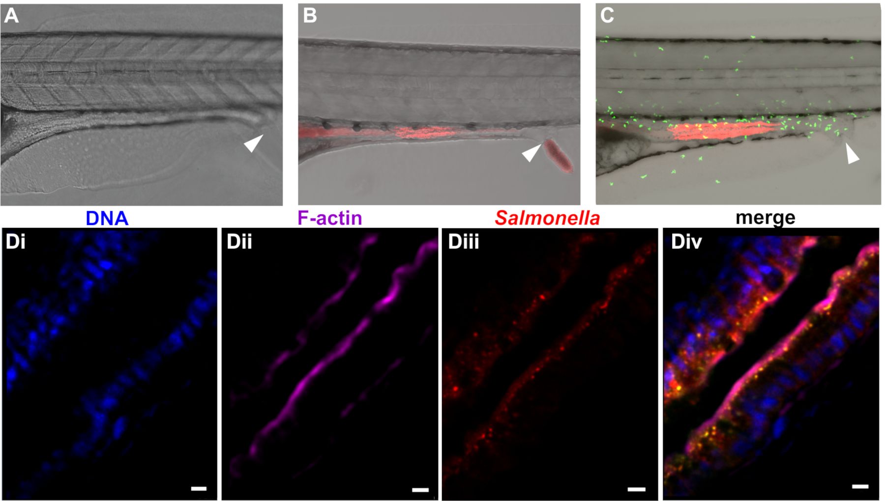
Figure 1: Colonization of zebrafish with bacteria. Zebrafish at 5 dpf were left uninfected (A) or colonized with mCherry expressing (B) E. coli or (C) Salmonella enterica Inf...
Access restricted. Please log in or start a trial to view this content.
Materials
| Name | Company | Catalog Number | Comments |
|---|---|---|---|
| Paramecium caudatum, live | Carolina | 131554 | no not store growing cultures below room temperature |
| 0.4% Trypan Blue Solution | Sigma | T8154-20ML | liquid, sterile-filtered, suitable for cell culture; prepared in 0.81% sodium chloride and 0.06% potassium phosphate, dibasic |
| Dimethyl sulfoxide (DMSO) | Sigma | 276855-100ML | store in a solvent safety cabinet |
| Escherichia coli, MG1655 | ATCC | ATCC 700926 | can be replaced by any other non-pathogenic E. coli strain |
| FM 4-64FX stain | Thermo Fisher | F34653 | aliquot and store frozen |
| Formaldehyde | Sigma | F8775-4X25ML | |
| LB Broth | Sigma | L3397-1KG | |
| Phosphate buffered saline tablets | Thermo Fisher | 18912014 | |
| Tetracycline | Sigma | 87128-25G | toxic, irritant |
| Tricaine (Ethyl 3-aminobenzoate methanesulfonate) | Sigma | E10521-10G | |
| Triton X-100 | Sigma | X100-100ML | |
| Trypan Blue Solution, 0.4% | Sigma | 93595-50ML | |
| UltraPure Low Melting Point Agarose | Thermo Fisher | 16520050 | |
| Hemocytometer or cell counter | any | ||
| Stereomicroscope | any | ||
| Table-top centrifuge | |||
| Microwave | |||
| Rotator wheel | |||
| Heated shaking incubator | |||
| Aquatics facilities | |||
| Breeding tanks |
