This video demonstrates the step-by-step procedure for preparing mouse retinal cryosections to visualize adeno-associated virus permeability across disrupted retinal barriers.
A subscription to JoVE is required to view this content. Sign in or start your free trial.
Method Article
Preparation of Mouse Retinal Cryosections to Assess Retinal Permeability to Adeno-Associated Virus
102 views
⸱
August 31st, 2026
In This Article
Abstract
Protocol
All procedures involving animal samples have been reviewed and approved by the appropriate animal ethical review committee.
- For retinal cryosections (Figure 1), dissect enucleated eyes to remove the lens and cornea, and immersion fix in 4% paraformaldehyde for 1 hr.
- Cryoprotect the previously fixed eyes in 10% sucrose for 1 hr at room temperature, 20% sucrose for another hr at room temperature. Cryoprotect in 30% sucrose solution, overnight at 4 °C. Freeze and embed eyecups in embedding resin such as Cryo-matrix. Make 10 µm cryosections and mount on slides specially treated for frozen tissue sections and which improve tissue adherence during staining.
- Permeabilize sections with 0.1% Triton X-100 in phosphate-buffered saline (PBS) pH 7.4 for 5 min. Wash 2x in PBS and block for 1 hr at room temperature in PBS with 1% bovine serum albumin (BSA), 0.1% Tween 20.
Access restricted. Please log in or start a trial to view this content.
Results
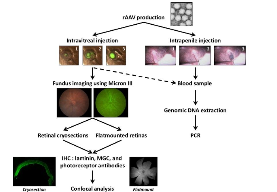
Figure 1: Schematic representation of the general protocol. After adeno-associated virus (AAV) production, particles are injected into the vitreous or into the penile vein to anesthetize adult mice. Fundus images with a Micron III camera are...
Access restricted. Please log in or start a trial to view this content.
Materials
| Name | Company | Catalog Number | Comments |
|---|---|---|---|
| Cryomatrix embedding resin | Thermo Scientific | 6769006 | |
| Superfrost plus adhesion slides | Thermo Scientific | 10143352 | |
| Binocular magnifier SZ76 | ADVILAB | ADV-76B2 | Zoom 0.66 x 5 x LEDs with stand epi and dia / Retinas dissection |
| Spring scissors straight - 8.5 cm | Bionic France S.a.r.l | 15003-08 | Retinal dissection |
