This video demonstrates the temporal enumeration of spontaneous phage induction in lysogenic Pseudomonas aeruginosa by quantifying viable bacterial cells (CFUs) and infective phage particles (PFUs) over time. By sampling at defined intervals during growth and analyzing both colony and plaque formation, the method reveals when spontaneous induction is minimal. This enables the identification of optimal time points for downstream transcriptomic analysis with minimal lytic interference.
A subscription to JoVE is required to view this content. Sign in or start your free trial.
Method Article
Measuring Spontaneous Phage Induction in Lysogenic Bacteria Over Time
114 views
⸱
August 31st, 2026
In This Article
Abstract
Protocol
1. Temporal direct enumeration of spontaneous induction (Figure 1)
- Set up overnight starter cultures of both the lysogen (e.g., P. aeruginosa PAO1 lysogen harboring LES (Liverpool Epidemic Strain (LES) bacteriophages) phages) and indicator host (PAO1-RifR) (rifampicin-resistant Pseudomonas aeruginosa PAO1) by inoculating a single colony in 5 mL of LB (Luria–Bertani broth), and incubate at 37 °C with shaking at 180 rpm (18–24 h).
- Set up fresh lysogen and indicator host cultures by inoculating the overnight cultures in 100 mL of LB at a ratio of 1:100, and incubate at 37 °C with shaking (180 rpm).
- Monitor the lysogen growth by measuring the OD600 and viable count using the Miles Misra technique. To do this, collect a 1 mL sample from each lysogen culture every hour from the point of inoculation for 8 h.
- Serially dilute the sample immediately after collection by adding 100 µL of the sample into 900 µL of the respective medium. Vortex well at the maximum speed, discard the tip at each dilution, and continue the dilution series from 10−1 to 10−9.
- Spot 10 µL of the required dilutions in triplicate onto an LB agar plate, allow to dry, and incubate at 37 °C for 18–24 h.
To calculate the number of viable bacterial cells, find a dilution with easily countable colonies. Count the number of colonies in each spot, and then use the following formula:

NOTE: As the lysogen culture grows, active phage particles will be produced by spontaneous induction. The production of infective phages means that the transcriptome of the lysogen population is now contaminated with lytic replication cycle-associated gene expression from the lytic phage replication signal and the corresponding host cell response. It is, thus, important to identify the growth stage at which the proportion of lysogenic cells to free infective phage particles is highest in order to limit as much background transcription noise (generated by the lytic phage transcriptome) as possible in the data set.
- To enumerate the infective phage particles in each temporal sample, inoculate 5 mL of sterile 0.4% bacteriological agar in LB (top agar) with 100 µL of mid-exponential phase indicator host (OD600: 0.4–0.5; in this case, PAO1-RifR) in the presence of an appropriate selective agent (50 µg·mL−1 rifampicin in this case, as the MIC of the PAO1 host is only 16 µg·mL−1)
- Spot 10 µL of the same serial dilution (see step 1.2.2) onto the inoculated top agar layer, and allow to dry before incubating at 37 °C for 18–24 h.
To calculate the infective phage particles, find a dilution with easily countable plaques. Count the number of plaques in each spot.

- Find the time/condition for which the spontaneous induction per CFU (colony forming unit) is minimal for the further experimental steps.
Access restricted. Please log in or start a trial to view this content.
Results
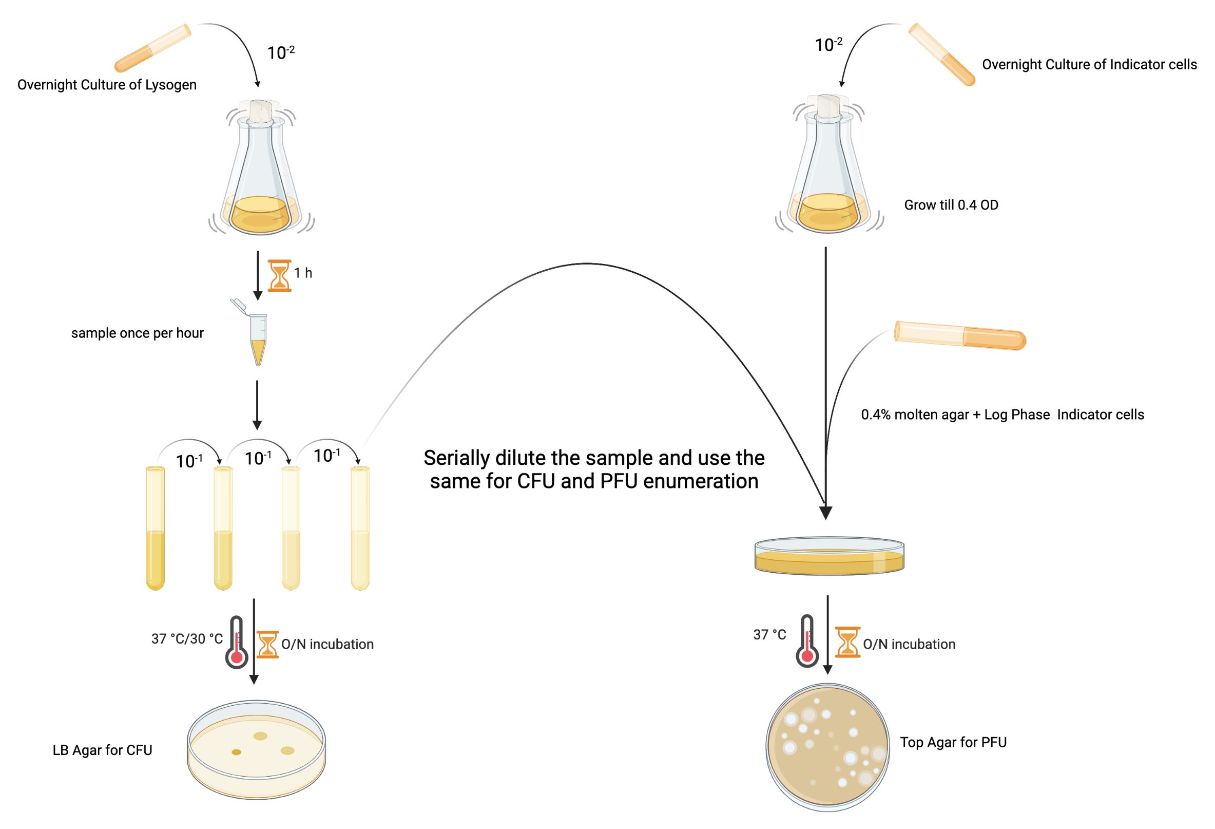
Figure 1: The experimental design for enumerating the PFU and CFU of a lysogen from the same sample.
Access restricted. Please log in or start a trial to view this content.
Materials
| Name | Company | Catalog Number | Comments |
|---|---|---|---|
| PAO1 | 6 | ||
| LESB58 | 6 | ||
| LES phages | Induced and purified from LESB58 using Norfloxacin. | ||
| Lysogeny Broth (LB) | Merck | 1.10285.500 | |
| LB Agar | Merck | 1.10283.500 | |
| Agar Agar | Fisher | A/1080/53 | |
| Top Agar | 0.4 g Agar Agar+2.5 g LB Broth in 100 mL water; autoclave and use. | - | |
| Rifampicin | Sigma (Stock: 50 mg/mL in Methanol- Mix well and use 0.22µm filter to sterilize and store it in -20°C until use) | R3501 |
