This video shows a luciferase-based antiviral assay in human embryonic kidney cells infected with recombinant measles virus. It shows that serially diluted test compounds inhibit viral replication, leading to lower luminescence in a dose-dependent manner while maintaining host cell viability.
Method Article
Luciferase-Based Assay to Evaluate Antiviral Compounds in Human Embryonic Kidney Cells
91 views
⸱
September 30th, 2026
In This Article
Abstract
Protocol
1. Preparation of Cell Cultures Infected by measles virus (MV) expressing firefly luciferase (rMV2/Luc)
- Grow HEK-293T cells at 37 °C and 5% CO2 in Dulbecco's modified Eagle's medium (DMEM) with stabilized L-glutamine and supplemented with 10% fetal calf serum (FCS), streptomycin (100 μg/ml) and penicillin (100 IU/ml). Cells are passaged by trypsinization every 4-5 days and diluted 1:10 into a fresh flask. Cells should be in the log phase for experiments.
- On the day of the experiment, recover cells by trypsinization and perform cell counting. Pellet cells by centrifugation, and resuspend them at 3 x 10⁵ cells/ml in culture medium. Consider that 12.5 ml of cell suspension is required for each screening plate.
- Again, resuspend cells at 3 x 10⁵ cells/ml in culture medium. Consider that 12.5 ml of cell suspension is required for each screening plate.
- Optional: supplement culture medium with uridine at 20 μg/ml.
NOTE: When screening chemical libraries for viral replication inhibitors, it seems that compounds targeting early steps of the pyrimidine biosynthesis pathway are frequently isolated. The addition of uridine to the culture medium is used to filter out such antiviral molecules. Indeed, this efficiently restores viral replication when upstream enzymes of the pyrimidine biosynthesis pathway are blocked. - Save 1:10 of the cell suspension for control wells with non-infected cells, and proceed to infection with the remaining volume.
- Thaw the appropriate volume of rMV2/Luc stock solution. MV stock solutions are usually at 106-108 infectious particles per ml. Culture must be infected with 0.1 infectious particles of rMV2/Luc per target cell, which corresponds to 0.1 multiplicity of infection (MOI).
NOTE: rMV2/Luc is derived from MV vaccine (Schwarz strain) and is manipulated in a BSL2 environment. - Add the virus to cell suspension and mix gently. Transfer infected cells to a trough, and dispense 100 μl of infected cell suspension in columns 2 to 11 of D2 plates containing spiked chemical compounds. Cell suspension within the trough is regularly mixed gently.
- In columns 1 and 12, dispense 100 μl of non-infected cells into each well. Infected cells are dispensed in the other wells.
- Incubate cells for 24 hr at 37 °C and 5% CO2.
2. Secondary Screen for Toxicity and Broad-spectrum Antiviral Activity
- Make 1:2 serial dilutions for each hit compound in dimethylsulfoxide (DMSO), starting from 500 µM to 4 µM (8 dilutions). Fill white, bar-coded tissue culture plates with 50 µl of culture medium. Add 1 µl of each compound dilution in culture wells. Repeat twice to have 3 culture plates with duplicate serial dilutions for each hit compound.
- Prepare 37.5 ml of a HEK-293T cell suspension at 6 x 10⁵ cells/ml in culture medium as described above (in 1.1 and 1.2). Dispense 2x 12.5 ml in Falcon tubes and infect cells with either rMV2/Luc at MOI = 0.1 or recombinant chikungunya virus (CHIKV) expressing Renilla luciferase (CHIKV/Ren) at MOI = 0.2. Mix by inverting.
NOTE: CHIKV/Ren is derived from a wild-type strain of CHIKV and therefore should be manipulated in a BSL3 environment. Plates can be moved from BSL3 to BSL1 once Renilla substrate has been added to culture wells (see below). - Dispense 50 µl of uninfected, rMV2/Luc-infected, or CHIKV/Ren-infected cells in culture plates containing serial dilutions of hit compounds. Incubate 24 hr at 37 °C.
- Add 50 µl of firefly luciferase substrate to determine firefly luciferase activity in rMV2/Luc-infected wells. Add 50 µl of Renilla luciferase substrate to determine Renilla luciferase activity in CHIKV/Ren-infected wells. Finally, add 50 µl of luciferase-based viability assay reagent to uninfected cells to determine cellular viability.
- Plot data (Figure 1) and determine hit compound concentrations that inhibit MV and CHIKV replication by 50%. Disregard compound showing some toxicity in this assay.
Results
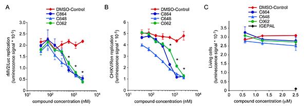
Figure 1. Dose-response antiviral activity and toxicity of compounds C864, C648 and C062. (A) HEK-293T cells were infected with rMV2/Luc strain of MV expressing luciferase (MOI=0.1), and incubated with increasing doses of C864, C648, C062 or DMSO alone. After 24 hr, luciferase expression was determined. * indicates that observed luciferase inhibitions were statistically significant with all three compounds (p-values < 0.05). (B) HEK-293T cells were infected with CHIKV/Ren strain of CHIKV expressing Renilla luciferase (MOI=0.2), and incubated with increasing doses of C864, C648, C062 or DMSO alone. After 24 hr, Renilla luciferase expression was determined. * indicates statistically significant inhibitions with all three compounds (p-values < 0.05). (C) HEK-293T cells were incubated with increasing doses of C864, C648, C062 or DMSO alone. As a control for toxicity, cell cultures were supplemented with 2.5 μl of a 0.5% IGEPAL solution. After 24 hr, the number of living cells was determined using the luciferase-based viability assay.
Materials
| Name | Company | Catalog Number | Comments |
|---|---|---|---|
| Freedom EVO platform | TECAN | Robotic platform | |
| 96-well polystyrene cell culture microplates, white | Greiner Bio One | 655083 | |
| CellTiter-Glo luminescent cell viability assay | Promega | G7570 | Luciferase-based viability assay |
| Bright-Glo luciferase assay system | Promega | E2610 | Reagent containing firefly luciferase substrate |
| Renilla-Glo luciferase assay system | Promega | E2710 | Reagent containing Renilla luciferase substrate |
| Britelite plus reporter gene assay system | Perkin-Elmer | 6016761 | Reagent containing firefly luciferase substrate. Can be used as an alternative to Brigh-Glo reagent to determine luciferase activity. |
