This video demonstrates single-cell micro-aspiration to isolate and expand a single giant virus population from a mixed viral population using amoebae as host cells.
Method Article
Isolation of a Single Giant Virus from a Mixed Viral Population Using Amoebae
83 views
⸱
September 30th, 2026
In This Article
Abstract
Protocol
1. Amoeba Culture
- Use Vermamoeba vermiformis (strain CDC19) as a cell support.
- Add 30 mL of protease-peptone-yeast extract-glucose medium (PYG) (Table 1) and 3 mL of the amoebae at a concentration of 1 x 10⁶ cells/mL in a 75 cm2 cell culture flask.
- Maintain the culture at 28 °C.
- After 48 h, quantify the amoebae using counting slides.
- To rinse, harvest the cells at a concentration of 1 x 10⁶ cells/mL and pellet the amoebae by centrifugation at 720 x g for 10 min. Remove the supernatant and resuspend the pellet in the appropriate volume of starvation medium to obtain 1 x 10⁶ cells/mL (Table 1).
2. End-point dilution
- Perform a serial dilution (10-1 to 10-11) of the viral sample in starvation medium (Table 1).
- Inoculate 2 mL of 1 x 10⁶ Vermamoeba vermiformis in each Petri dish with 100 µL of the mixture inoculum.
- Place the Petri dishes into a sealable plastic bag at 30 °C.
- Begin observing the Petri dishes with inverted optical microscopy at 6 h post-infection and check cell morphology every 4 to 8 h.
- At the appearance of the cytopathic effect characterized by rounding cells, begin the single-cell micro-aspiration process.
3. Single Cell Micro-aspiration
- Prepare the host
NOTE: This preparation is made for the release of infected single cells to a fresh cell support.- Treat the amoebae in the culture with an antimicrobial agent containing 10 µg/mL of vancomycin, 10 µg/mL of imipenem, 20 µg/mL of ciprofloxacin, 20 µg/mL of doxycycline, and 20 µg/mL of voriconazole. This mixture is used to avoid bacterial and fungal contamination.
NOTE: The procedure takes place on a bench outside the microbiological safety station. Add 2 mL of amoebae concentrated at 1 x 10⁶ cells/mL each into 15 Petri dishes. For amoeba adherence, incubate the culture at 30 °C for 30 min.
- Treat the amoebae in the culture with an antimicrobial agent containing 10 µg/mL of vancomycin, 10 µg/mL of imipenem, 20 µg/mL of ciprofloxacin, 20 µg/mL of doxycycline, and 20 µg/mL of voriconazole. This mixture is used to avoid bacterial and fungal contamination.
- Select the Petri dish used for the micro-aspiration from the limit dilution according to the following criteria: 1) absence of any visible contamination by fungal and bacterial agents, 2) evidence of cytopathic effect of amoebae due to the viruses, and 3) prelysis and rounding phase of the amoebae (to avoid aspiration of viral particles).
- Set up a workstation with the following materials (Figure 1A,B):Micromanipulator, which allows microcapillary positioning;Manual control pressure device, used to aspirate and release the cells into the microcapillary;Inverted microscope;Plug and play motor modules;Camera;Computer module to visualize manipulation and take pictures.
- Choose a microcapillary (Figure 1C).
NOTE: The size of the cells, the deformation and adhesion of their membranes to the surfaces, and the cellular motility can impact the smooth progress of the micro-aspiration. The microcapillary diameter can be precisely chosen and adapted to specific cell types depending on their sizes and methods of aspiration. A microcapillary of 20 µm inner diameter was used to aspirate a rounding amoeba (diameter ~10 µm). This allows the upkeep of an internal position and an easy release of the cell. - Mount the system.
- Fix the operating angle of the gripping system on the motorized module at 45°.
- Perform a double installation, first on the gripping system, and then on the microcapillary.
- Focus on the cells after running a few drops of oil through the microcapillary.
NOTE: The mineral oil with biological compatibility is supplied by the device. - Complete mounting following manufacturer's recommendations.
- Clone cells (Figure 2A,B)
- Place the Petri dish containing 2 mL of infected amoebae under the microscope.
- Focus first on the cells, and then on the microcapillary immersed in the culture.
- Pick a rounded single cell and bring the microcapillary closer to the micromanipulator.
- Exert soft aspiration with manual pressure control on the cell, taking it inside the microcapillary. Remove the single cell from the first sample and release it in the cellular support, then incubate it at 30 °C.
- Conduct daily observations with an inverted optical microscope to observe the appearance of the cells and to monitor the emergence of the cytopathic effect.
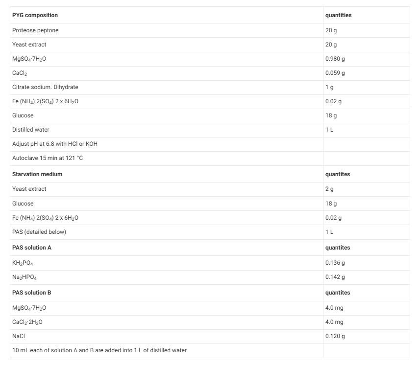
Table 1: Composition of culture media
Results
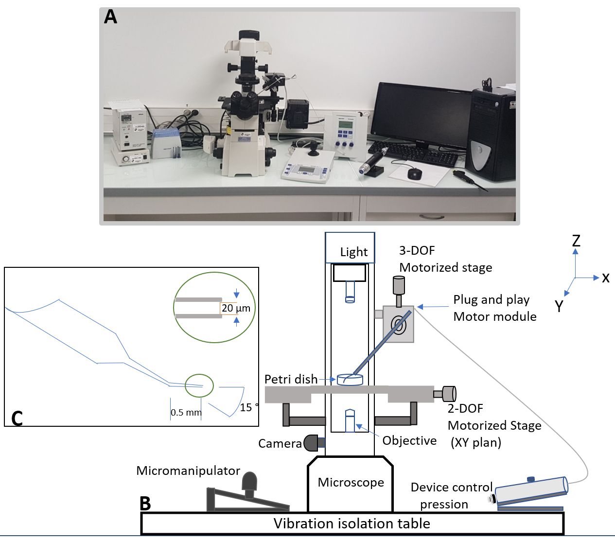
Figure 1: Materials for micromanipulation. (A) Actual setup of the workstation. (B) Schematic illustration of the workstation’s components. (C) Schematic illustration of the microcapillary.
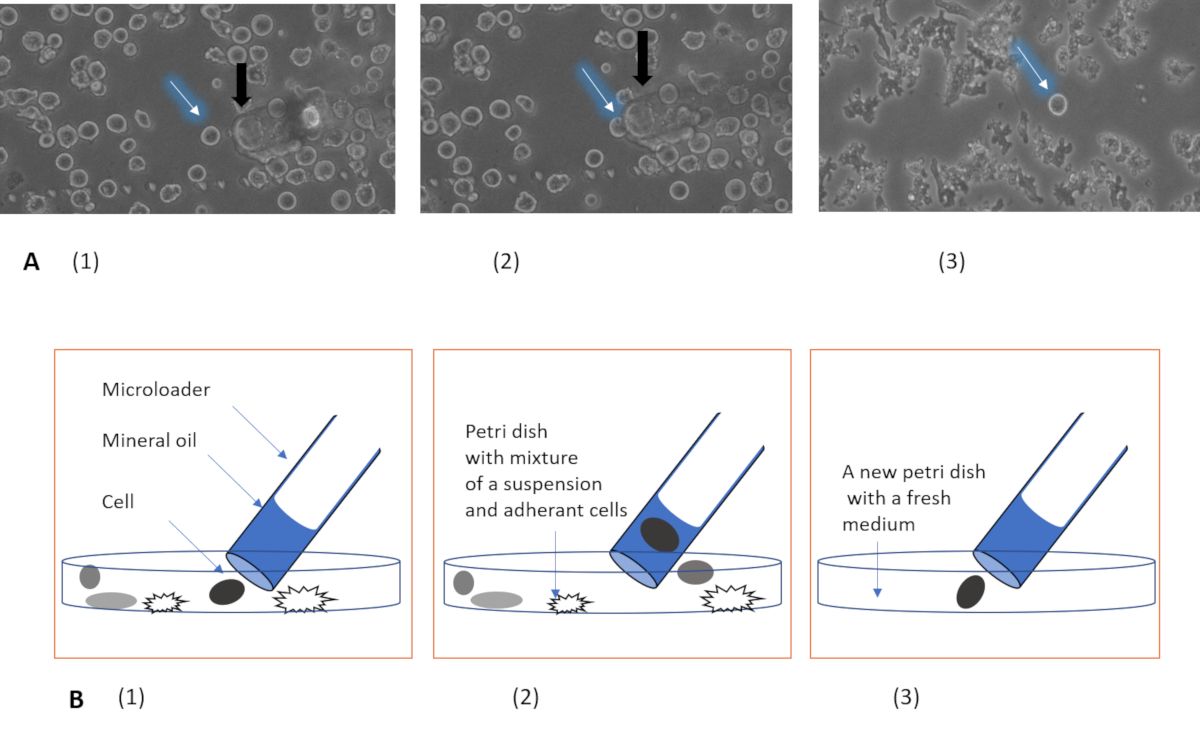
Figure 2: Micro-aspiration steps. (A) Single-cell isolation procedure (zoom x40). (B) Schematic illustration of the different steps of single-cell aspiration: 1) Localization of the cell. 2) Aspiration of the cell. 3) Release step. The black arrows show the microcapillary and the white ones show the single cell.
Materials
| Name | Company | Catalog Number | Comments |
|---|---|---|---|
| Corning cell culture flasks 150 cm² | Sigma-aldrich | CLS430825 | Culture |
| Corning cell culture flasks 25 cm² | Sigma-aldrich | CLS430639 | Culture |
| Corning cell culture flasks 75 cm² | Sigma-aldrich | CLS430641 | Culture |
| DFC 425C camera | LEICA | Unkown | Observation/Monitoring |
| Eclipse TE2000-S Inverted Microscope | Nikon | Unkown | Observation/Monitoring |
| Microcapillary 20 µm | Eppendorf | 5175 107.004 | Microaspiration and release of cells |
| Micromanipulator InjectMan NI2 | Eppendorf | 631-0210 | Microcapillary positioning |
| Petri dish 35 mm | Ibidi | 81158 | Culture/observation |
