This video demonstrates the process of infecting kidney epithelial cells with genetically engineered viruses for their production and harvesting to study viral replication.
Method Article
Production and Harvesting of Therapeutic Virus-Producing Epithelial Cells
65 views
⸱
September 30th, 2026
In This Article
Abstract
Protocol
- Determining Replicative and Cytotoxic Capacities of Viral Vectors Encoding Immunomodulators
Compare replication kinetics of the generated recombinant virus and the unmodified vector with one-step or multi-step growth curves (GCs). For one-step GCs, viral progeny are assessed following simultaneous infection of all cells (theoretically, 100% infection). Multi-step GCs start at a low percentage of infected cells to follow several rounds of virus replication.
- For analysis of measles virus replication kinetics, seed 1 x 10⁵ Vero cells in 1 mL of DMEM (dulbecco’s modified eagle medium) with 10% FBS (fetal bovine serum) per well on 12-well plates one day prior to infection. Plate at least two wells for each timepoint of interest as technical replicates.
- Infect the cells with the virus at low MOI (multiplicity of infection) for multi-step or at high MOI for one-step GCs (0.03 and 3 for MV or measles virus) replication on Vero cells, respectively. Replace medium with 300 µL of serum-free medium containing the respective amount of virus and incubate at 37 °C and 5% CO2 for at least 2 h. Remove inoculum, add 1 mL of DMEM with 10% FBS, and continue incubation.
- At relevant timepoints such as 12, 24, 36, 48, 72, and 96 h after infection, harvest viral progeny by directly scraping cells into the medium. Transfer contents from each well to an individual tube, snap-freeze in liquid nitrogen, and store at -80 °C until titration.
NOTE: Replicate samples may be pooled at this step for analysis of average titers. - Collect samples from all timepoints. Thaw simultaneously at 37 °C for assessment of viral progeny. Vortex and pellet cell debris by centrifugation for 5 min at 2,500 x g and 4 °C.
- Calculate average titers of each construct and time point as described above. Plot titers over time. Compare growth curves of vectors with and without inserted transgenes, with different transgenes, or with transgenes inserted at different positions (see Figure 1).
Results
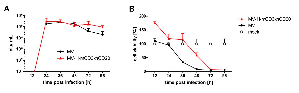
Figure 1: Replication kinetics and lytic activity of recombinant measles viruses. (A) One-step growth curves were generated after infection of Vero cells with unmodified oncolytic measles virus (MV) or measles virus encoding a bispecific T cell engager (MV-H-mCD3xhCD20) at an MOI of 1. Titers of viral progeny were evaluated at designated timepoints after infection, indicating similar virus replication kinetics. Means and standard deviations of eight samples (four technical replicates each of two biological replicates per time point) are shown. (B) Cell viability was assessed on MC38 murine colorectal carcinoma cells stably expressing human carcinoembryonic antigen and the MV receptor CD46 (MC38-CEA-CD46) after inoculation with medium only (mock) or indicated viruses at an MOI of 1. XTT cell viability assay was performed at the indicated timepoints. Reduction in cell viability was observed at earlier timepoints for the unmodified vector. Values of over 100%, as calculated for MV-H-mCD3xhCD20 at the 12 h timepoint, are frequently observed shortly after infection, which may be due to cellular stress or cell-derived factors present in the virus suspension. Mean values plus standard deviations of three technical replicates are shown for each time point.
Materials
| Name | Company | Catalog Number | Comments |
|---|---|---|---|
| Dulbecco’s Modified Eagle Medium (DMEM) | Invitrogen, Darmstadt, Germany | 31966-021 | Used for seeding and infection of Vero cells |
| Fetal Bovine Serum (FBS) | Biosera, Boussens, France | FB-1280/500 | For supplementation of DMEM |
| Vero cells | ATCC, Manassas, VA, USA | CCL81 | Host cells for measles virus propagation and assay |
| 12-well plates | Neolab, Heidelberg, Germany | 353043 | For seeding Vero cells (1 × 10⁵ per well) |
| Opti-MEM (serum-free medium) | Gibco Life Technologies, Darmstadt, Germany | 31985070 | For virus infection step (serum-free incubation) |
| Phosphate-Buffered Saline (PBS) | Gibco Life Technologies, Darmstadt, Germany | 14190-094 | For washing and medium preparation |
| Cell lifter | Corning, Reynosa, Mexico | 3008 | Used for scraping infected cells at harvest |
| 50 mL tubes | nerbe plus, Winsen/Luhe, Germany | 02-572-3001 | For sample collection and centrifugation |
| Liquid nitrogen | — | — | For snap-freezing harvested viral samples |
| Centrifuge (capable of 2,500 × g) | — | — | For pelleting cell debris |
| XTT Cell Viability Kit (Colorimetric Cell Viability Kit III) | PromoKine, Heidelberg, Germany | PK-CA20-300-1000 | Includes XTT reagent; for cytotoxicity measurement |
| 96-well plates, flat-bottom | Neolab, Heidelberg, Germany | 353072 | For absorbance measurement at 450 nm |
| Spectrophotometer / Microplate reader | — | — | For measuring optical absorbance at 450 nm (reference 630 nm) |
| Carbon dioxide incubator | — | — | 37 °C, 5% CO₂ for all cell incubations |
