This video demonstrates a cell-based assay to evaluate the target-specific immunomodulatory activity of a virally encoded bispecific T-cell engager (BTE). The BTE links tumor cells expressing a target receptor with T cells, triggering tumor lysis and enzyme release. A colorimetric readout, based on substrate conversion, indicates T-cell activation. Higher color intensity in the test sample confirms BTE-mediated, receptor-specific immune engagement.
Method Article
Evaluation of Immunomodulatory Activity of a Virally Encoded Bispecific T-Cell Engager
80 views
⸱
September 30th, 2026
In This Article
Abstract
Protocol
- Assess immunological functionality of isolated immunomodulators.
- The following describes in detail how to evaluate bispecific T-cell engager (BTE)-induced, cell-mediated cytotoxicity by lactate dehydrogenase (LDH) release assay.
- Decide on conditions to be tested during the experiment (see Figure 1 as an example). Timepoints (hours to days), concentrations of the immunomodulator (pg/mL to µg/mL), and effector to target cell (E:T) ratios (between 1:50 and 50:1, for example) can be varied. Always prepare samples in triplicate.
- Include samples without BTE protein and without effector cells, controls for spontaneous LDH release of the single cell types (Tsp and Esp), a target cell maximum lysis control (Tmax) with detergent added before readout, a medium-only control, and a volume control for Tmax.
NOTE: Required numbers of target cells and incubation times can vary. Perform initial test runs without effector cells and immunomodulators to identify optimal parameters. Include Tsp and Tmax samples and corresponding controls to assess different cell numbers and timepoints. - Isolate immune effector cells (e.g., by density gradient centrifugation of human blood to obtain peripheral blood mononuclear cells (PBMCs) or negative selection of murine T cells from mouse splenocytes).
- Seed target cells (e.g., 5 x 10³ per well) in a U-bottom 96-well plate.
- Add the isolated protein at desired concentrations to the respective samples. Add immune effector cells at desired ratios. Add medium to a total volume of 100 µL per well.
- Incubate for the determined time frame, typically between 4 and 48 h (24 h for unstimulated PBMCs and 48 h for freshly isolated murine T cells; shorter incubation times can apply for prestimulated immune cells), at 37 °C and 5% CO2.
- 45 minutes before collecting the samples, add 10 µL of 10x lysis solution to wells containing Tmax samples and the corresponding medium controls. Continue incubation.
- Spin down cells at 250 x g for 4 min. Transfer 50 µL of each supernatant to a flat-bottom 96-well plate. Do not transfer cells to avoid cell-bound LDH.
- Prepare the substrate solution according to the manufacturer's instructions. Add 50 µL to each well and incubate at room temperature [RT] in the dark for 30 min or until Tmax samples turn deep red.
- Add 50 µL of stop solution to each well. Remove air bubbles by centrifugation for 1 min at 4,000 x g, and manually with a hollow needle. Measure optical absorbance at 490 nm.
- Calculate % specific lysis values for each sample.
- Calculate averages for replicates of medium controls with and without lysis solution. Subtract obtained values from experimental samples and from Tmax and spontaneous release controls, respectively. Use these background-corrected values for further calculations.
- Calculate averages for background-corrected Tmax, Tsp, and Esp controls. Using these average values, the following equation yields the percentage of specific lysis for each sample:

- Plot % specific lysis values vs. protein concentration or E:T ratio and compare to the non-targeting control samples.
- The following describes in detail how to evaluate bispecific T-cell engager (BTE)-induced, cell-mediated cytotoxicity by lactate dehydrogenase (LDH) release assay.
Results
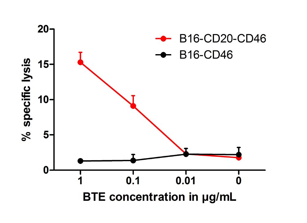
Figure 1: Cytotoxic activity of measles virus-encoded BTEs. Target tumor cells (5 x 10³ B16-CD20-CD46 cells per well) were incubated with murine T cells at a ratio of 1:50. BTEs (Bispecific T-cell engagers) previously purified from the supernatant of MV(Measles virus)-H-mCD3xhCD20-infected cells were added at indicated concentrations. Relative lysis of target cells was assessed by LDH release assay after 48 h. Cells lacking the BTE target antigen (B16-CD46) served as a reference to evaluate antigen-specific cytotoxicity. Means plus standard deviations of three technical replicates per sample are shown. Target antigen-expressing cells were specifically lysed in a BTE concentration-dependent manner. Purity of the BTE product and target antigen expression levels influence cell killing. In the present example, 15% specific cell killing was achieved at a relatively high BTE concentration of 1 µg/mL. This is a typical value for such an experimental setup with long co-incubation times and suboptimal T cell culture conditions. In other settings, up to 60% specific killing was achieved using BTEs purified from MV-infected supernatants, reaching a plateau at BTE concentrations of 100 ng/mL and higher. This indicates that, counter-intuitively, the limit of this assay is less than 100% specific killing, which can be explained by different growth kinetics in Tmax controls compared to co-culture samples.
Materials
| Name | Company | Catalog Number | Comments |
|---|---|---|---|
| Dulbecco's Modified Eagle's Medium (DMEM) | Invitrogen, Darmstadt, Germany | 31966-021 | |
| Fetal bovine serum (FBS) | Biosera, Boussens, France | FB-1280/500 | |
| Kanamycin | Sigma-Aldrich, Taufkirchen, Germany | K0129 | |
| B16-CD46/ B16-CD20-CD46 | J. Heidbuechel, DKFZ Heidelberg | available upon request | |
| Colorimetric Cell Viability Kit III (XTT) | PromoKine, Heidelberg, Germany | PK-CA20-300-1000 | includes XTT reagent |
| Dulbecco's Phosphate-Buffered Saline (PBS) | Gibco Life Technologies, Darmstadt, Germany | 14190-094 | |
| QIAquick Ni-NTA Spin Columns | QIAGEN, Hilden, Germany | 31014 | |
| 96-well plates, U-bottom | TPP, Trasadingen, Switzerland | 92097 | |
| 96-well plates, flat bottom | Neolab, Heidelberg, Germany | 353072 |
