This video demonstrates the enrichment of giant viruses from an environmental water sample using amoeba-based co-culture to enable downstream detection and analysis.
Method Article
Amoeba-Based Enrichment of Giant Viruses from an Environmental Water Sample
11 views
⸱
September 30th, 2026
In This Article
Abstract
Protocol
1. Sample Collection
- Collect 70 samples from different environments and regions. In this case, use the following: 5 dirty water samples from the village of Saint Pierre de Meyzoargues (France), 15 samples from the lake in Parc Borély in Marseille (France); 15 sea water samples with sediment from the rocky inlets at Samena in Marseille (France), 25 river water samples from the Alps (France), and finally, 10 samples from sewage in La Ciotat (France).
- Vortex samples for homogenization before inoculating, for one minute at room temperature without any treatment.
2. Isolation Procedure
- Host Preparation for Blind Culture Procedures and Sample Inoculation
- Use Vermamoeba vermiformis (strain CDC19) as a cell support for the co-culture.
- Maintain the amoebae at 28 °C, in a 75 cm2cell culture flask with 30 ml of protease-peptone-yeast extract-glucose medium (PYG). Please check PYG composition in Table 1.
NOTE: After 48 hr, amoebae are quantified by a normal count using counting slides (e.g., Kovaslides). - Harvest cells at a concentration of 5 x 10⁵ to 106 cells/ml, and pellet amoebae by centrifugation at 720 x g for 10 min.
- Remove the supernatant by aspiration, and re-suspend the amoebae pellet in 30 ml of sterile Page's amoebal saline (PAS) Table 1. Repeat this rinsing procedure.
- Re-suspend amoebae in 30 ml of starvation medium with an antibiotic and antifungal mixture at a concentration of approximately 106 amoeba/ml. The starvation medium is a survival medium for the amoeba, enabling it to stay alive without encystment or multiplication. Check the composition in Table 1.
NOTE: The antimicrobial agent mix contained 10 µg/ml vancomycin, 10 µg/ml imipenem, 20 µg/ml ciprofloxacin, 20 µg/ml doxycycline, and 20 µg/ml voriconazole. This mix served to reduce the bacterial and fungal contamination that can decrease or alter viral multiplication. - Directly inoculate 25 µl of each sample onto the 200 µl amoeba monolayer in a 96-well plate flat bottom favoring the amoeba adherence and incubate at 30 °C in a humid environment (a humid environment consists of placing a wet compress inside the bag containing the plate in order to avoid evaporation). This is the primo-culture. Leave two wells of amoebae without any sample inoculation; this acts as the negative control. Incubate this primo-culture for three days.
- Three days after the first inoculation, sub-culture the primo-culture plate on fresh amoebae as described above without any microscopic observation. Incubate the new plate for three days under the same conditions as the primo-culture.
- Perform the final culture after these three days of incubation the same way as for the primo- and sub-culture. Incubate the final culture for two days. Then proceed to detection.
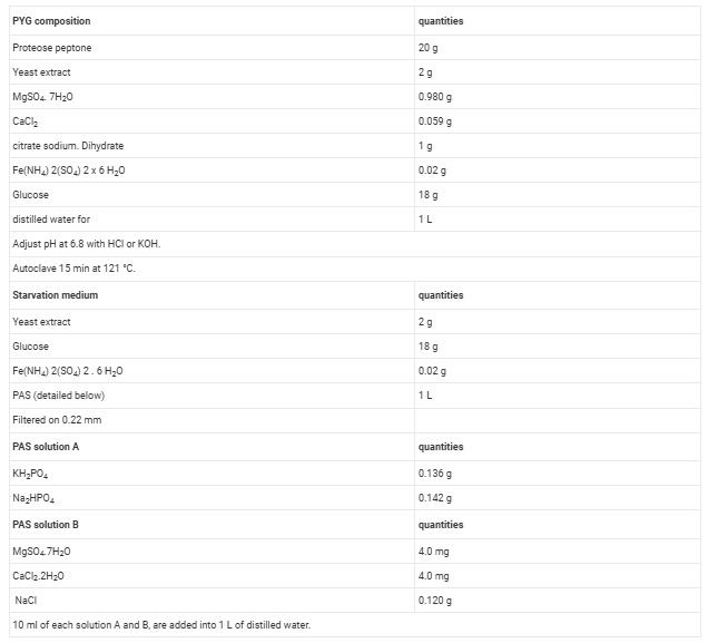
Table 1. Solution recipes
Materials
| Name | Company | Catalog Number | Comments |
|---|---|---|---|
| Protease-peptone-yeast extract-glucose (PYG) medium, Page's amoebal saline (PAS), Starvation medium | In house laboratory production | Marseille URMITE | x |
| Amoeba strain CDC-19 | ATCC | France | 50237 |
| Plates | Cellstar | France | 655180 |
| Flasks | SARSTEDT | Germany | 833911 |
| Ultracentrifuge Sorval WX 80 | Thermo scientific | France | 9102448 |
