This video demonstrates the fluorescence focus assay used to quantify infectious Hepatitis C virus in cell culture. It demonstrates the serial dilution of viral supernatant, the infection of hepatoma cells, and the detection of viral replication sites using antibody-based fluorescence microscopy.
Protocol
1. Measuring Virus Titer
Plate naive Huh-7.5.1 cells at approximately 3 x 10³ cells/well using a 96-well plate. The next day, perform 10-fold serial dilutions of cell-free culture supernatant harvested from Hepatitis C virus (HCV) RNA-transfected cells using growth media and inoculate in triplicate onto Huh-7.5.1 cells.
Fix cells at 72 hr post-infection using methanol (for 30 min at -20 ºC) and immunostain for HCV NS5A protein.
Wash the cell with PBS three times and block with IFA blocking buffer.
Use rabbit polyclonal anti-NS5A primarily antibody or mouse monoclonal anti-dsRNA antibody J2 at a dilution of 1:200 (1 μg/ml) and incubate for 5 hr to overnight at 4 ºC.
Wash the cells with PBS three times after the primary antibody.
Add goat anti-rabbit IgG-488 polyclonal secondary antibody or goat anti-mouse IgG-594 polyclonal secondary antibody at a 1:1,000 dilution (1 μg/ml) and incubate for 1 hr at room temperature.
Wash cells with PBS three times and view using a fluorescent microscope
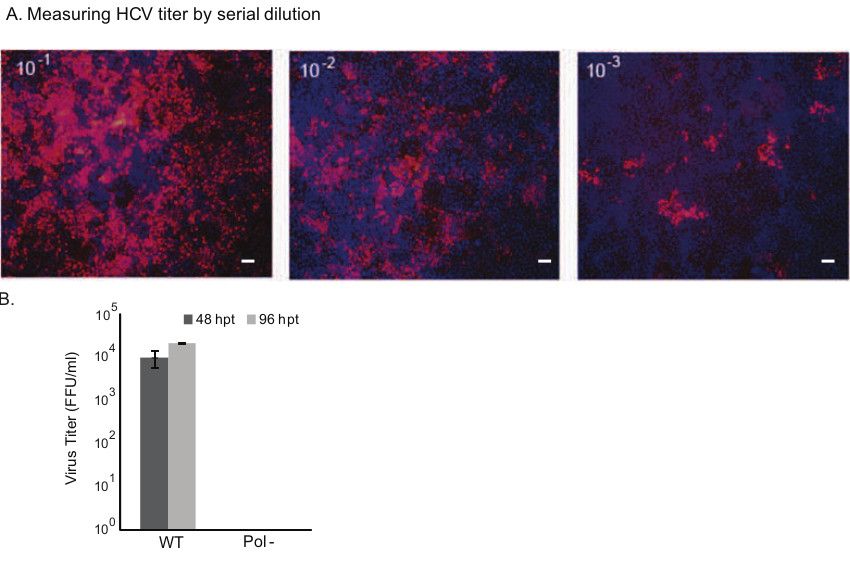
Use the highest dilution to count for the NS5A positive foci and calculate the average number of focus-forming unit (FFU) per milliliter. See Figure 1 for HCV titer.
Results
Figure 1. Examining the infectivity of wild-type HCV and measuring virus titer. A) Naive Huh-7.5.1 cells were used for infection studies. The cell-free supernatant collected at 48 and 96 hr post-transfection (hpt) of HCV RNA were subjected to 10-fold serial dilution and added to the cells in a 96-well plate in triplicate. 72 hr post-infection, the cells were fixed and immunostained for HCV NS5A protein. The cells with NS5A positive staining (red) are infected with virus. For assessing Foci Forming Unit (FFU), the positive foci at the highest dilution were counted. Representative panels of images are shown (Scale bar 100 μm). B) Mean values and standard deviations of viral titer in FFU per milliliter are shown in the graph. The polymerase null mutant did not produce infectious particles. WT: wild-type; Pol-: Polymerase null.
Materials
List of materials used in this article
Name
Company
Catalog Number
Comments
Dulbecco’s modified Eagle’s medium (DMEM)
Fisher Scientific
10-017-CV
Huh-7.5.1
The Scripps Research Institute
The cell line was kindly provided by Dr. Francis Chisari to Dr. Arumugaswami under executed MTA between The Scripps Research Institute and Cedars-Sinai Medical Center