This video demonstrates the recovery of infectious Japanese encephalitis virus from synthetic RNA transcribed from a bacterial artificial chromosome clone. It shows how cells are electroporated with the synthetic RNA, allowing the viral genome to direct the production of new virions. The resulting plaques formed in infected cell monolayers confirm the successful rescue of infectious virus.
Method Article
Rescue of Infectious Virions from Cells Transfected with Bacterial Artificial Chromosome-Derived Synthetic RNA
102 views
⸱
September 30th, 2026
In This Article
Abstract
Protocol
1. Determine RNA Infectivity and Virus Yield
- Cultivate baby hamster kidney-21 (BHK-21) cells in 150 mm culture dishes at a density of 3 × 10⁶ cells/dish for 24 hr at 37 °C with 5% CO2.
NOTE: Maintain the BHK-21 cells in alpha minimum essential medium supplemented with 10% fetal bovine serum, 2 mM glutamine, vitamins, and penicillin/streptomycin. - Rinse the cell monolayer with 10 ml of cold Solution A (137 mM NaCl, 2.7 mM KCl, 8.1 mM Na2HPO4, and 1.5 mM KH2PO4). Detach the cells from the dishes by treatment with 4 ml of trypsin-ethylenediaminetetraacetic acid (EDTA) (0.25%), and collect them by centrifugation at 270 × g in a desktop centrifuge for 2 min.
- Resuspend the cell pellet with 50 ml of cold Solution A in a 50 ml conical tube and centrifuge the cell suspension at 270 × g for 2 min. Repeat this wash procedure three times; after the last wash, resuspend the cell pellet at a density of 2 × 10⁷ cells/ml in Solution A.
- Mix a 400 µl aliquot of cell suspension with 2 µg of synthetic RNA in a 2-mm gap cuvette, and promptly electroporate the mixture with an electroporator under optimal electroporation conditions: 980 V, 99 µsec pulse length, and 5 pulses (Figure 1C).
- Leave the electroporated cells at room temperature (RT) for 10 min and transfer them to a 1.7 ml microtube containing 600 µl of complete culture medium.
- Prepare a 10-fold serial dilution of the electroporated cells in 1 ml of complete culture medium and plate a 100 µl aliquot of each dilution on the monolayers of unelectroporated BHK-21 cells (5 × 10⁵) in a 6-well plate.
- After 4-6 hr of incubation, overlay the cells with 0.5% agarose in minimal essential medium containing 10% fetal bovine serum. Incubate the plates for 4 days at 37 °C with 5% CO2.
- Visualize the infectious centers (plaques) by fixation with 7% formaldehyde and staining with 1% crystal violet in 5% ethanol (Figure 2A).
- Optional: Examine RNA-electroporated cells at 18-20 hr post-transfection for Japanese encephalitis virus (JEV) protein expression by immunofluorescence assays (Figure 2B), and harvest the supernatants from the RNA-electroporated cells at 22 and 40 hr post-transfection for virus titration by plaque assays (Figure 2C).
Results
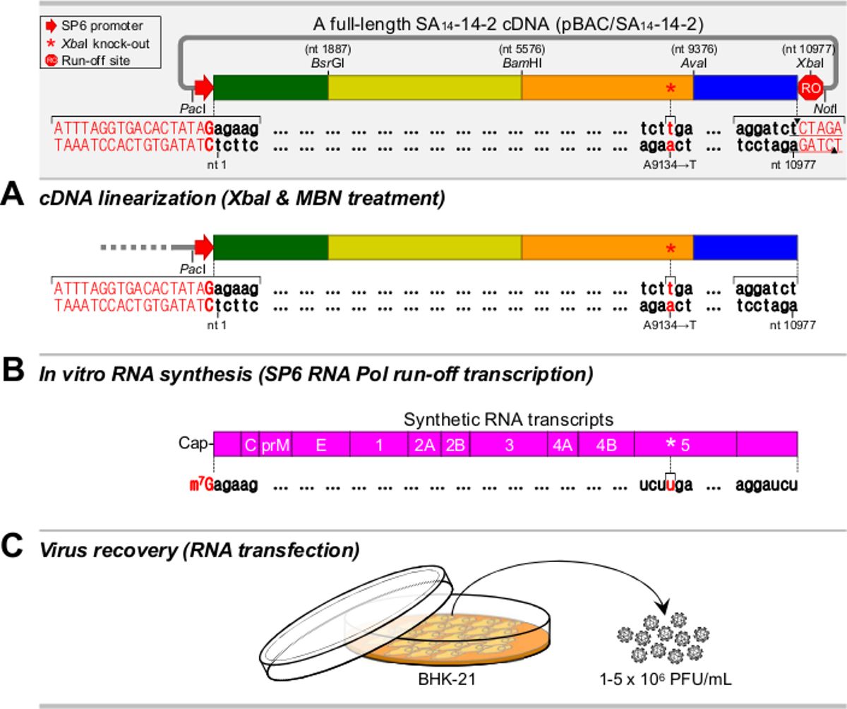
Figure 1. Overview of the recovery of infectious viruses from a full-length JEV SA14-14-2 complementary DNA (cDNA) assembled in a bacterial artificial chromosome (BAC). (A) Linearization of the cDNA template. The full-length JEV BAC is cut with XbaI and treated with mung bean nuclease (MBN). (B) Synthesis of the RNA transcripts. The linearized cDNA is transcribed by SP6 RNA polymerase in the presence of the m7G(5')ppp(5')A cap analog. (C) Recovery of the synthetic JEVs. The in vitro transcribed RNAs are transfected into BHK-21 cells by electroporation, which generates a high titer of synthetic virus.
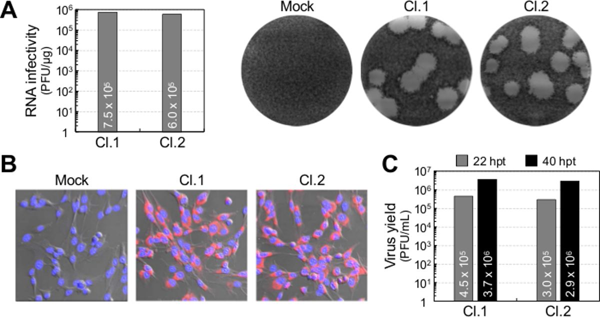
Figure 2. Specific infectivity of the synthetic RNAs transcribed from a full-length JEV BAC and the recovery of synthetic virus. BHK-21 cells are mock-electroporated (Mock) or electroporated with the RNA transcripts derived from each of the two independent clones of the full-length JEV BAC (Cl.1 and Cl.2). (A) RNA infectivity. The cells are overlaid with agarose and stained with crystal violet at 4 days post-transfection. RNA infectivity is determined by infectious center assays to estimate the amount of infectious RNA electroporated into the cells (left panel). Also, representative images of infectious centers are shown (right panel). (B) Protein expression. The cells are cultured in 4-well chamber slides. Viral protein expression in RNA-electroporated cells at 20 hr post-transfection (hpt) is analyzed by immunofluorescence assays using a primary anti-nonstructural protein 1 (NS1) rabbit antiserum and a secondary Cy3-conjugated goat anti-rabbit immunoglobulin G (IgG) (red). The nuclei are counterstained with 4',6-diamidino-2-phenylindole (blue). The immunofluorescence images are overlaid on their corresponding differential interference contrast images. (C) Virus yield. The cells are cultured in 150 mm culture dishes. The production of infectious virions accumulated in the culture supernatants of RNA-electroporated cells at 22 and 40 hours post-transfection (hpt) is examined by plaque assays.
Materials
| Name | Company | Catalog Number | Comments |
|---|---|---|---|
| Alpha Minimal Essential Medium | Life Technologies (Gibco) | 12561-049 | |
| Agarose | Lonza | 50004 | |
| Conical Tube (50 mL) | VWR | 21008-242 | |
| Crystal Violet | Sigma-Aldrich | C0775 | |
| Culture Dish (150 mm) | TPP | 93150 | |
| Cuvette (2-mm Gap) | Harvard Apparatus | 450125 | |
| Fetal Bovine Serum | Life Technologies (Gibco) | 16000-044 | |
| Formaldehyde | Sigma-Aldrich | F1635 | Toxic and carcinogenic |
| Glutamine | Life Technologies (Gibco) | 25030-081 | |
| Minimal Essential Medium | Life Technologies (Gibco) | 61100-061 | |
| Penicillin/Streptomycin | Life Technologies (Gibco) | 15070-063 | |
| Potassium Chloride | Sigma-Aldrich | P3911 | |
| Potassium Phosphate Monobasic | Sigma-Aldrich | P9791 | |
| Six-Well Plate | TPP | 92006 | |
| Sodium Chloride | Sigma-Aldrich | S3014 | |
| Sodium Phosphate Dibasic | Sigma-Aldrich | S3264 | |
| Trypsin-EDTA (0.25%) | Life Technologies (Gibco) | 25200-056 | |
| Vitamins | Sigma-Aldrich | M6895 | |
| CO₂ Incubator | Thermo Scientific | Heracell 150i | |
| Desktop Centrifuge | Thermo Scientific | ST16R | |
| Electroporator | Harvard Apparatus | ECM 830 |
