All procedures involving animal models have been reviewed by the local institutional animal care committee and the JoVE veterinary review board.
1. Isolation of Metastatic Colonies
- At day 5 post injection, remove embryos from the incubator and inspect the embryo CAMs for metastatic colony distribution. Identify the embryos with uniform colony distribution in which compact (or overly invasive) colonies are present.
NOTE: The process of culturing chicken embryos is not sterile, therefore, all the screening steps must be performed under highly clean conditions to avoid future tissue culture contamination. Contamination is quite rare and can be easily avoided by wearing gloves and a mask and using only sterile tools during cell injection and colony isolation procedures. It is recommended to inspect embryos one by one to decrease their exposure to the ambient laboratory temperature and to prevent contamination.
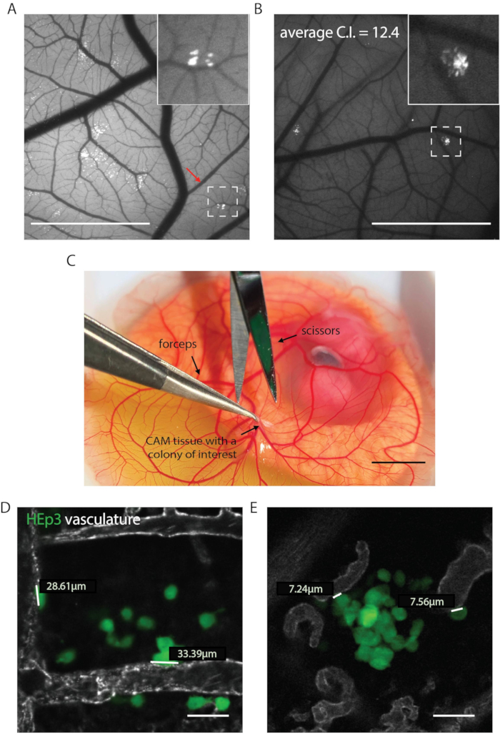
- Locate the metastatic colony of interest. A compact colony can be described as one with most of the cancer cells located within a limited area in the CAM tissue (cells appear "clumped together"). An invasive colony can be described as a colony where cancer cells "scatter" in the CAM tissue (Figure 1A, B).
NOTE: "Compactness" of the metastatic colony can be explained either by the inhibition of cancer cell invasion, inhibition of cancer cell proliferation, or both. Attention should be paid to all scenarios, and contact colonies should be isolated (Figure 1A, B). A simple fluorescent stereomicroscope can be used to discriminate between compact and invasive metastatic colony phenotypes.
- Under the dissection microscope, gently pull the CAM tissue that contains the metastatic colony of interest upwards using fine forceps (Figure 1C).
- Cut off the CAM tissue that contains the metastatic colony using surgical scissors.
- Transfer the CAM tissue that contains the metastatic colony into an empty, sterile 1.5 mL tube (on ice) and close the tube lid.
NOTE: Isolated colonies can be kept on ice for up to 3 h without loss of viability.
- Repeat the excision procedure until all the colonies of interest are collected into separate tubes. To avoid animal suffering, do not excise more than 2–3 colonies from one embryo. Euthanize embryos by freezing at -20 °C (or another approved method) immediately after the colony excision.
- Gently mince the CAM tissue in a microcentrifuge tube using a sterile 18G needle. Use a separate needle for each colony.
- Add 100 µL of 1x collagenase solution and incubate for 30 min at 37 °C.
- Spin down the cells and CAM tissue at 300 x g for 5 min at ambient temperature.
- Aspirate the collagenase solution and resuspend cells in complete media used for the cell line of interest.
NOTE: Normally, CAM tissue is not completely dissociated after collagenase treatment, and cancer cells will first proliferate within the pieces of CAM tissue and only later migrate onto the tissue culture dish.
- Spin the cells and CAM tissue again at 300 x g for 5 min at ambient temperature.
- Resuspend cells and CAM tissue pieces in 1 mL of complete media plus selection factor (if any), then transfer into a single 12-well tissue culture dish well.
NOTE: Chicken CAM fibroblasts may persist in tissue culture for multiple passages inhibiting clonal expansion. The ability to expand the metastatic colonies in the presence of a selection factor (i.e., if a vector that was used to render cancer cells fluorescent also encodes a mammalian antibiotic resistance gene) may greatly speed up clonal expansion. A kill curve experiment should be performed before the screening to ensure that the proper concentration of antibiotics is used.
- For the next 1–3 weeks, monitor cancer cells daily for growth and contamination.
- When cells reach 70–80% of confluency, transfer the cells into a larger volume culture dish.
NOTE: It is recommended to expand cells till at least two cryogenic vials of each clone can be frozen. Maintaining large amounts of tissue culture can be laborious and unnecessary.
- Proceed to sequencing or next round of selection as soon as enough cancer cell numbers are reached. Generally, 1 x 10⁶ of cancer cells are sufficient for modern high-throughput sequencing techniques.
- Proceed to the clone reinjection and imaging and quantification of colony compactness or cancer cell-blood vessel contact (Figure 2A). Alternatively, proceed to high throughput sequencing or repeat the colony selection.
NOTE: To decrease the number of false positives at least two rounds of selection are recommended. Two approaches have been successfully employed. 1) Expand each clone and reinject individually to confirm the colony phenotype. 2) Mix all the expanded clones in a 1:1 ratio each, reinject as a mixture, and repeat the selection cycle.