Method Article

In Vitro Phagocytosis Assay: A Live Imaging Based Method to Study Real-time Astrocyte Mediated Synaptosome Phagocytosis

July 8th, 2025

In This Article

Abstract

Loading...
$$\rightleftharpoonup{xx}$$ $$\longleftharp{xx}$$, $$\longrightharp{xx}$$,

Source: Byun, Y. G., Chung, W. S. A Novel In Vitro Live-imaging Assay of Astrocyte-mediated Phagocytosis Using pH Indicator-conjugated Synaptosomes. J. Vis. Exp. (2018)

In this video, we evaluate the phagocytic capacity of purified astrocytes in vitro by live imaging assay. The assay serves as an effective screening platform to identify chemical compounds that can enhance or inhibit the phagocytic capacity of astrocytes.
Protocol

Loading...
$$\rightleftharpoonup{xx}$$ $$\longleftharp{xx}$$, $$\longrightharp{xx}$$,

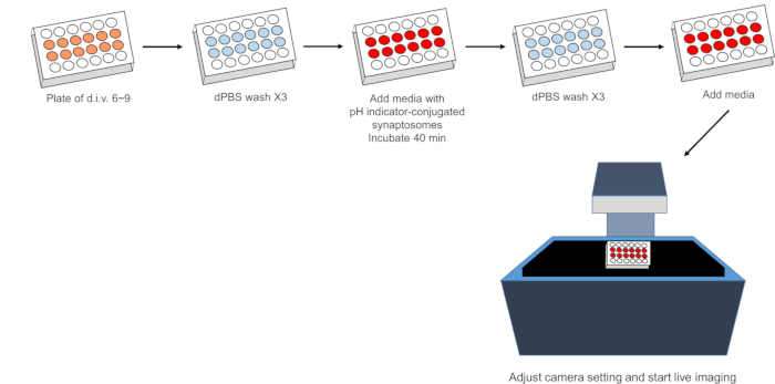
1. Phagocytosis Live-imaging Assay (Figure 1)

  1. Prepare a 24-well plate (or any plate/dish that is prepared for live-imaging) in which astrocytes are confluent (generally, 7 to 10 days of incubation after the purification is ideal for stabilizing the cells).
  2. Remove the media in each well and wash the wells with 1 mL of Dulbecco’s phosphate buffer saline (DPBS), three times. To minimize air exposure, wash the wells quickly.
  3. Add 300 µL of immunopanned astrocyte basic media (IP-ABM) with 5 µL of pH indicator-conjugated synaptosomes and additional factors such as immunopanned astrocyte-conditioned media (IP-ACM) that can modulate glial cell phagocytosis.
  4. Incubate the plate in a CO2 incubator for 40 min. This step allows the pH indicator-conjugated synaptosomes to settle to the bottom of the plates.
  5. Remove the media with unbound pH indicator-conjugated synaptosomes and wash each well with 1 mL of DPBS, three times.
    CAUTION: Do not wash harshly. To minimize air exposure, wash the wells quickly.
  6. Add 500 µL of IP-ABM with additional factors to test into the wells.
  7. Take the plate to a live-imaging instrument and select positions. Adjust focus, exposure time, brightness, and LED power.
    NOTE: The settings for exposure time, brightness, and LED power are variable depending on fluorescence intensity and the purpose of the experiment. In the live-imaging assay with pH indicator-conjugated synaptosomes, we usually set those values as follows: exposure: ~150–200 ms, brightness: 15, and LED power: 4–6.
  8. Set the image format, time interval, and the total number of cycles for live imaging. Set the time interval to 1 or 2 h depending on how many positions are selected. 2-hour intervals are recommended when more than 150 positions are selected for live-imaging.
  9. Start the live-imaging experiments.

Access restricted. Please log in or start a trial to view this content.

Results

Loading...
$$\rightleftharpoonup{xx}$$ $$\longleftharp{xx}$$, $$\longrightharp{xx}$$,

Flowchart of live imaging process for synaptosomes with pH indicator, using dPBS washes on plate.
Figure 1. Schematic of phagocytosis live-imaging assay.

Access restricted. Please log in or start a trial to view this content.

Disclosures

Loading...
$$\rightleftharpoonup{xx}$$ $$\longleftharp{xx}$$, $$\longrightharp{xx}$$,
No conflicts of interest declared.

Materials

List of materials used in this article
NameCompanyCatalog NumberComments
pHrodo red, succinimidyl ester Molecula probes P36600For pH indicator conjugation
Fetal bovine serum (FBS) Gibco16000-044
DMEMGibco11960-044
dPBSWelgene LB001-02
<strong>Phagocytosis live imaging assay</strong>
Juli stage&nbsp;NanoEntek
Time Series Analyzer V3 plugins&nbsp;https://imagej.nih.gov/ij/plugins/ time-series.html

Reprints and Permissions

Request permission to reuse the text or figures of this JoVE article

Request Permission

Tags

Astrocyte PhagocytosisSynaptosome IsolationpH Indicator AssayFluorescence MicroscopyAstrocyte CultureSynaptosome BindingPhagosome FormationReal time Imaging

Related Articles