1. Preparation of Reagent
- Prepare low-fluorescence DMEM medium for assays using a microplate fluorescence reader. Mix 25 mM glucose, 0.4 mM glycine, 0.4 mM arginine, 0.2 mM cysteine, 4.0 mM glutamine, 0.2 mM histidine, 0.8 mM isoleucine, 0.8 mM leucine, 0.8 mM lysine, 0.2 mM methionine, 0.4 mM phenylalanine, 0.4 mM serine, 0.8 mM threonine, 0.078 mM tryptophan, 0.4 mM tyrosine, 0.8 mM valine, 1.8 mM CaCl2, 0.81 mM MgSO4, 5.33 mM KCl, 44.0 mM NaHCO3, 110 mM NaCl, 0.9 mM NaH2PO4. Adjust the pH of the solution using HCl or NaOH to pH 7.4. Sterilize the medium through filtration.
NOTE: This medium contains components of the standard DMEM medium with high glucose except that Fe(NO3)3, vitamins, and Phenol Red are omitted. Phenol Red, riboflavin, and pyridoxal in regular DMEM culture medium significantly interfere with the detection of fluorescence signal. Culture medium without those components is crucial for successful live cell GFP imaging. The medium is stable for 12 months when stored refrigerated.
2. Real-time Degradation Assay of NLS-luciferase-GFP Using Fluorescence Microplate Reader
- Seed approximately 1 x 10⁴ HeLa cells into black 96-well tissue culture plates with transparent bottom. After O/N culturing, a confluence of 50-70% is reached at the time of transfection.
NOTE: 60 µl of medium containing completely suspended HeLa cells are seeded directly into each well. Do not add additional medium or rock plate back and forth after seeding, otherwise, cells may be distributed unevenly.
- Transfect HeLa cells with 0.05-0.1 µg of NLS-luciferase-GFP/pRK5 plasmid into each well using transfection reagent according to manufacturer's instruction. Make a master transfection mix containing DNA and transfection reagent and aliquot it for each well. Three wells with cells are not transfected with DNA, which serves as control for background fluorescence signal for each treatment condition.
NOTE: An alternative way to reduce transfection variation is to transfect cells before seeding. However, a large portion of cells transfected with misfolded proteins fail to attach or show reduced viability using this method. It is likely that some cells may not stand the stress caused by trypsin digestion when expressing toxic misfolded proteins. As a result, the overall fluorescent signal is significantly reduced.
- 20-24 hr after transfection, examine the live cells under an inverted fluorescent microscope for GFP expression with excitation wavelength 450-490 nm.
- Remove the medium by vacuum aspiration. Add approximately 200 µl 1x PBS to each well and then aspirate it to remove residual amount of DMEM medium.
- Add 60 µl of low-fluorescence DMEM medium with 5% FBS and 50 µg/ml CHX. To examine proteasomal degradation, include additional proteasome inhibitor MG132 (10 µM) in one set of samples. Set triplicate of wells for each treatment/condition.
NOTE: MG132 treatment is included as controls for proteasomal degradation because it is crucial to rule out other factors that can cause drop of fluorescence signal, including cell death and fluorescence quenching.
- Measure the fluorescence signal of GFP immediately on a fluorescent plate reader after adding CHX.
NOTE: The software measurement settings are shown in Table 1.
- Read the plate every hour for up to 8-10 hr. After reading, return the plate back to cell culture incubator.
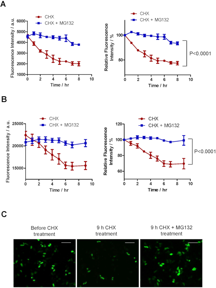
- Export the data as spreadsheet file. Use mean value of multi-reads for each well as fluorescence intensity. Normalize values of each data point to the mean values of Time 0 in the respective groups. Plot the normalized fluorescence intensity over time as shown in Figure 1A and 1B, and Figure 2, right panels.
- Perform statistical analysis of degradation rates between two conditions using two-way ANOVA with repeated measures.
NOTE: In this analysis, "fluorescence intensity" is the dependent variable whereas "treatment conditions" and "time" are the two factors. Instead of comparing data at individual time points, two-way ANOVA with repeated measures analyzes the difference between the two treatment groups over the entire time course.
Table 1: Measurement settings on fluorescence microplate reader.
| Plate definition file | GRE96fb |
| Measurement mode | Fluorescence Intensity Bottom |
| Wavelength | Excitation wavelength | 485 nm |
| Excitation bandwidth | 9 nm |
| Emission wavelength | 535 nm |
| Emission bandwidth | 20 nm |
| Number of flashes | 5 |
| Gain | Manual 200 |
| Integration time | 20 µs |
| Multiple reads per well | Type | Circle (filled) |
| Size | 5×5 |
| Border | 400 µm |