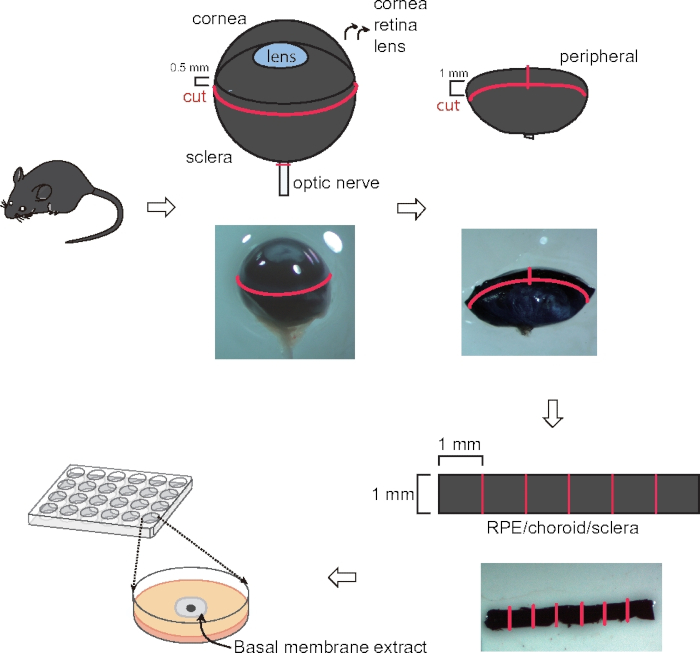
All procedures involving animal models have been reviewed by the local institutional animal care committee and the JoVE veterinary review board.
1. Preparation
- Add 5 mL of Penicillin/Streptomycin (10000 U/mL) and 5 mL and 10 mL of commercially available supplements to 500 mL of complete classic medium with serum. Aliquot 50 mL of the medium initially.
NOTE: Do not return any medium back to the stock to avoid contamination.
- Put an aliquot of the complete classic medium on ice.
- Use 70% ethanol to clean the dissecting microscope, forceps, and scissors.
- Prepare two cell culture dishes (10 cm), one on the dissection microscope, and one on ice; put 10 mL of complete classic medium in each dish.
2. Experimental steps (Figure 1)
- Sacrifice C57BL/6J mice around postnatal (P) 20 using 75-100 mg/kg ketamine and 7.5 -10 mg/kg xylazine injected intraperitoneally. Keep the eyes in complete classic medium on ice before dissection.
- Remove the connective tissue (muscle and fatty tissue) and the optic nerve on the eye.
- Use a micro-scissor to circumferentially cut 0.5 mm posterior to the corneal limbus. Remove the cornea/iris complex, vitreous, and the lens.
- Make a 1 mm incision perpendicular to the cut edge towards the optic nerve and cut a circumferential band of 1 mm width. Separate the central and peripheral regions of the complex. Use forceps to peel off the retina from RPE/choroid/sclera complex.
- Keep the peripheral choroid band in complete classic medium on ice; isolate the other eye and repeat the process to cut a second band.
- Cut the circular band into 6 ~equal square pieces (~1 mm x 1 mm).
NOTE: Never touch any edge.
- Thaw the basal membrane extract (BME) per the manufacturer's instruction. Add 30 μL/well of BME into the center of each well of a 24-well tissue culture plate. Make sure the droplet of BME forms a convex dome at the bottom of the plate without touching the edges.
NOTE: Thaw the BME overnight in a refrigerator. BME should be on the ice any time after thawing.
- Place the tissue in the middle of the BME.
NOTE: Do not flatten the choroid explant; generally, let the tissue expand within the BME. The orientation of the tissue (scleral side up or down) does not impact the experimental outcome.
- Incubate the plate at 37 °C for 10 min to let the gel solidify.
- Add 500 μL of the complete classic medium/well.
- Change the classic medium every other day (500 μL). Choroid sprouting can be observed after 3 days with a microscope.
NOTE: For growth factor treatment, starve the tissue for 4 h. Dilute a trial compound in a growth factor-reduced medium (1:200 boost instead of 1:50).
3. SWIFT-Choroid computerized quantification method (Figure 2)
NOTE: A computerized method to measure the area covered by growing vessels was used. A macro plugin to ImageJ software is needed prior to quantification.
- Open the choroid sprouting image with ImageJ and check "Image |Type| 8-bit" with grayscale.
- Go to "Image | Adjust | Brightness/Contrast (Ctrl/shift/ C)" and optimize the contrast.
- Use the magic wand function to outline and remove from the image the choroid tissue which are present in the center of the sprouts (using shortcut key "F1") (Figure 2A, B).
NOTE: Set the tolerance rate of the magic wand to 20-30%.
- Remove the background of the image with the free selection tools (Figure 2C). Go to "Image | Adjust | Threshold (Ctrl/shift/T)". Use the threshold function to define the microvascular sprouts against the background and periphery (Figure 2D).
- Click "F2" and a summary will appear. Save an image of the selected area by clicking "Save". Save it in the same folder as the original image for future reference.
- After a group of samples is measured, copy the recorded for data analysis.
NOTE: It is also possible to measure the area (µm²) by "Analyze | Set Scale" using images with scale bars.