Method Article

High-Resolution Melting PCR to Determine Sequence Variations in Mutant DNA Sequences

July 8th, 2025

In This Article

Abstract

Loading...
$$\rightleftharpoonup{xx}$$ $$\longleftharp{xx}$$, $$\longrightharp{xx}$$,

Source: Kisserli, A., et al. High-resolution Melting PCR for Complement Receptor 1 Length Polymorphism Genotyping: An Innovative Tool for Alzheimer's Disease Gene Susceptibility Assessment. J. Vis. Exp. (2017).

In this video, we describe a high-resolution melting PCR technique to determine the length polymorphism of DNA fragments of varying sequence lengths, based on melting temperature analysis.

Protocol

Loading...
$$\rightleftharpoonup{xx}$$ $$\longleftharp{xx}$$, $$\longrightharp{xx}$$,

1. HRM-PCR Protocol

  1. Thaw the DNA samples. Dilute the DNA samples in 1.5 mL tubes with water to adjust them to a concentration of 10 ng/µL
    NOTE: The total volume of diluted DNA should be between 2 µL and 10 µL.
  2. Thaw the primer solutions. Dilute the primer solutions in 1.5-mL tubes with water to adjust them to the same concentration of 6 µM.
    NOTE: The primer sequences and reaction conditions are provided in Table 1.
  3. Thaw the HRM-PCR kit solutions and mix carefully by vortexing to ensure the recovery of all contents. Briefly spin the three vials containing the enzymatic mixture with DNA binding dye, MgCl2, and water in a microcentrifuge before opening them. Store them at room temperature.
  4. In a 1.5 mL tube at room temperature, prepare the PCR mix for one 20 µL reaction by adding the following components in the order listed below:
    1. 10 µL of enzymatic mixture with DNA binding dye;
    2. 2 µL of 25 mM MgCl2;
    3. 1 µL of primer 1, 6 µM (final concentration: 300 nM);
    4. 1 µL of primer 2, 6 µM (final concentration: 300 nM); and
    5. 5 µL of water.
      NOTE: To prepare the PCR mix for more than one reaction, multiply the volumes above by the number of reactions to be run, plus one additional reaction.
  5. Mix carefully by vortexing.
  6. Pipette 19 µL of PCR mix, prepared above, into each well of a white multiwell plate.
  7. Add 1 µL of concentration-adjusted DNA template.
    NOTE: For control reactions, always run a negative control with the samples. To prepare a negative control, replace the template DNA with water.
  8. Seal the white multiwell plate with sealing foil.
  9. Place the white multiwell plate in the centrifuge and balance it with a suitable counterweight (i.e., another multiwell plate). Centrifuge for 1 min at 1,500 x g in a standard swing-bucket centrifuge containing a rotor for multiwell plates with suitable adaptors.
  10. Load the white multiwell plate into the HRM-PCR instrument.
  11. Start the HRM-PCR program with the following PCR conditions:
    Denaturation: 95 °C for 10 min; 1 cycle.
    Amplification: 95 °C for 10 s, 62 °C for 15s, and 72 °C for 20s; 47 cycles.
    Melting curve: 95 °C; ramp rate: 0.02 °C/s; 25 acquisitions per °C; 1 cycle.
    Cooling: 40 °C for 30 s; ramp rate 2.2 °C/s; 1 cycle.

HRM-PCR conditions table with CR1 target, primer sequences, MgCl2 concentration, and melting curve.

Table 1: Primers and parameters used in the high-resolution melting analysis.

2. HRM Analysis to Determine the CR1 Length Polymorphism

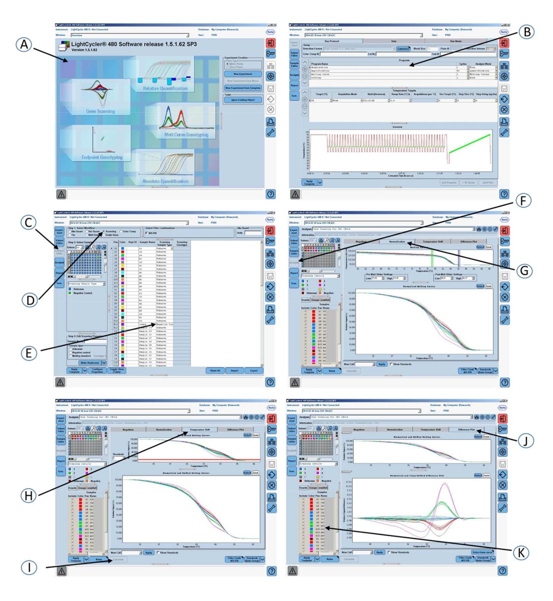
NOTE: The methodology described (Figure 1) is specific to our software (See the Table of Materials), although other software packages may be used.

  1. Open a gene scanning software to perform the CR1 length polymorphism scanning analysis.
  2. Open the experiment containing the amplification program and the melting curve program.
  3. Click Sample editor in the Module bar and then select the Scanning workflow.
  4. Define the properties of the samples (i.e., name; unknown or negative control).
  5. Click Analysis in the Module bar.
  6. In the Create New Analysis list, select Gene Scanning.
  7. Click the Normalization tab to normalize the melting curves.
  8. Click the Temperature shift tab to reset the temperature axis (x-axis) of the melting curves.
    NOTE: The lower graph shows melting curves that are both normalized and temperature-shifted.
  9. Click the Calculate button to analyze the results and determine the grouping.
  10. Click the Difference plot tab in the charts area to view the Normalized and Shifted Melting Curves and the Normalized and Temperature Shifted Difference Plot.

Access restricted. Please log in or start a trial to view this content.

Results

Loading...
$$\rightleftharpoonup{xx}$$ $$\longleftharp{xx}$$, $$\longrightharp{xx}$$,

Real-time PCR analysis setup, LightCycler software interface, data visualization, quantification curves.
Figure 1: Screenshots of the graphical interface of the software used in step 2 of the protocol. (A) Open the gene scanning software. (B) Amplification program and melting curve program. (C) Click on Sample edit...

Access restricted. Please log in or start a trial to view this content.

Disclosures

Loading...
$$\rightleftharpoonup{xx}$$ $$\longleftharp{xx}$$, $$\longrightharp{xx}$$,
No conflicts of interest declared.

Materials

List of materials used in this article
NameCompanyCatalog NumberComments
Lab coatprotection
SensiCareIce powder-free Nitrile Exam glovesMedline Industries, Inc, Mundelein, IL 60060, USA486802sample protection
Eppendorf Reference 2 pipette, 0.5-10µLEppendorf France SAS, F-78360 Montesson, France4920000024sample pipetting
Eppendorf Reference 2 pipette, 20-100µLEppendorf France SAS, F-78360 Montesson, France4920000059sample pipetting
Eppendorf Reference 2 pipette, 100-1000µLEppendorf France SAS, F-78360 Montesson, France4920000083sample pipetting
TipOne 10µL Graduated, filter tipStarlab GmbH, D-22926 Ahrenburg, GermanyS1121-3810sample pipetting
TipOne 1-100µL bevelled, filter tip Starlab GmbH, D-22926 Ahrenburg, GermanyS1120-1840sample pipetting
ART 1000E Barrier TipThermo Fischer Scientific , F-67403 Illkirch, France2079Esample pipetting
Eppendorf Safe-Lock Tubes, 1.5 mL, Eppendorf QualityEppendorf France SAS, F-78360 Montesson, France30120086mix
Vortex-Genie 2Scientific Industries, Inc, Bohemia, NY 111716, USASI-0236mix
Mikro 200 centrifugeHettich Zentrifugen, D-78532, Germany0002020-02-00centrifugation
Multipette E3Eppendorf France SAS, F-78360 Montesson, France4987000010distribution
Light Cycler 480 multiwell plate 96, whiteRoche Diagnostics GmbH, D-68305 Mannheim, Germany4729692001reaction place
Light Cycler 480 sealing foilRoche Diagnostics GmbH, D-68305 Mannheim, Germany4429757001coverage
LightCycler 480 Instrument II, 96-wellRoche Diagnostics GmbH, D-68305 Mannheim, Germany05015278001high resolution melting polymerase chain reaction
Heraeus Megafuge 11R centrifugeThermo Fischer Scientific , F-67403 Illkirch, France75004412centrifugation
LightCycler 480 High Resolution Melting MasterRoche Diagnostics GmbH, D-68305 Mannheim, Germany04909631001reaction reagents
CN3 primer: 5'ggccttagacttctcctgc 3'Eurogentec Biologics Division, B4102 Seraing, Belgiumreaction reagent
CN3re primer: 5'gttgacaaattggcggcttcg 3'Eurogentec Biologics Division, B4102 Seraing, Belgiumreaction reagents
light cycler 480 SW 1.5.1 softwareRoche Diagnostics GmbH, D-68305 Mannheim, Germanysoftware used for HRM-PCR CR1 polymorphism data analysis

Reprints and Permissions

Request permission to reuse the text or figures of this JoVE article

Request Permission

Tags

DNA PolymorphismMelting Curve AnalysisPCR ProtocolDNA Fragment AnalysisFluorescent Reporter DyeCR1 GenotypingMolecular Biology

Related Articles