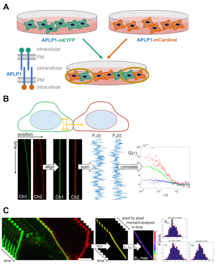
1. Sample Preparation: Cell-Cell Mixing Assay
NOTE: The following protocol describes the mixing procedure for adherent cells. It may be modified for cells cultured in suspension.
- Seed an appropriate number of cells on a 6-well plate, e.g., 800,000 HEK 293T cells (counted with a Neubauer counting chamber), a day before transfection. The number can be modified depending on the time between seeding and transfection and adjusted for other cell types. To perform a basic experiment (i.e., proteins of interest and negative control), prepare at least 4 wells. Culture cells at 37 °C, 5% CO2 in Dulbecco's Modified Eagle Medium (DMEM) medium, supplemented with fetal bovine serum (10%) and L-glutamine (1%).
- Transfect cells according to the manufacturer's instructions (see Table of Materials).
- To perform a basic experiment, transfect, in separate wells, plasmids for the protein of interest fused to a 'green' (e.g., monomeric enhanced green fluorescent protein (mEGFP), or yellow fluorescent protein (mEYFP)) or 'red' (e.g., mCherry, or mCardinal) fluorescent protein.
NOTE: In this protocol, we focus on APLP1-mEYFP and APLP1-mCardinal, and the corresponding negative control, e.g., myristoylated-palmitoylated-mEYFP (myr-palm-mEYFP) and -mCardinal (myr-palm-mCardinal). Generally, 200 ng - 1 µg of plasmid DNA is sufficient. High transfection efficiency increases the chance to find 'red' and 'green' cells in contact. Modify the amount of plasmid and transfection reagent to optimize transfection efficiency. Critical: Cell confluency should be around 70% when transfecting the cells. If cells are over-confluent, the transfection efficiency will decrease. If cells are not confluent enough, transfection and mixing may induce stress and prevent many cells from proper attachment after mixing.
- Perform cell mixing ~4 ± 2 h after transfection.
- Remove the growth medium and wash each well gently with 1 mL PBS supplemented with Mg2+ and Ca2+. Then, remove the PBS. (Critical) Drop PBS on the well edge to prevent detachment of cells during washing.
- Add ~50 µL trypsin ethylenediaminetetraacetic acid (EDTA) solution drop-wise to each well to facilitate the detachment of cells. Incubate at 37 °C for 2 min. Afterward, slowly shake the 6-well plate laterally to detach the cells.
NOTE: Extended incubation times may be required for some cell types.
- Add 950 µL of growth medium to each well and resuspend cells by pipetting a few times up and down, thereby detaching all cells from the well bottom. (Critical) Ensure that cells are resuspended properly and detached from each other by visually checking for the absence of large cell aggregates after resuspension. Otherwise, many 'red'-'red' or 'green'-'green' contacts will be obtained after mixing.
- Transfer the cell solution of one well (protein of interest or negative control) to the corresponding well, i.e., 'red' (e.g., APLP1-mCardinal transfected) to 'green' (e.g., APLP1-mEYFP transfected) cells. Mix by gently pipetting a few times up and down. Then, seed the mixed cells on 35-mm glass bottom dishes (1 mL of mixed cell solution per dish, plus 1 mL of growth medium) and culture seeded cells for another day at 37 °C, 5% CO2.
2. Sample Preparation: Positive Control for Cross-Correlation Experiments and Homo-Dimer Construct for Brightness Analysis
- Seed 600,000 HEK 293T cells, counted with a cell counting chamber, on 35-mm glass bottom dishes one day before transfection. Culture the cells at 37 °C, 5% CO2 in a complete DMEM medium (see step 1.1) for another day.
- Transfect cells with ~250 ng of plasmid DNA according to manufacturer instructions. For the positive cross-correlation control, use a plasmid encoding a membrane-anchored fluorescent protein hetero-dimer, e.g., myr-palm-mCherry-mEGFP or myr-palm-mCardinal-mEYFP corresponding to the FPs of the protein of interest. For brightness calibration, use plasmids encoding both a membrane-anchored FP monomer and homo-dimer corresponding to the FPs fused to the protein of interest, e.g., myr-palm-mEYFP and myr-palm-mEYFP-mEYFP to calibrate the brightness analysis of APLP1-mEYFP.
- Culture cells at 37 °C, 5% CO2 in complete DMEM medium (see step 1.1) for another day.
3. Confocal Laser Scanning Microscopy: Setup and Focal Volume Calibration
NOTE: The following protocol is written for experiments performed with mEGFP/mEYFP and mCherry/mCardinal on the laser scanning confocal microscope used in this study. The optical setup, the software settings (laser lines, dichroic mirrors, filters), and the choice of calibration dyes may be modified for other FPs and microscope setups.
- Turn on the microscope and lasers at least an hour before the experiment to ensure laser stability and equilibration of temperature.
- Prepare 100-200 µL of appropriate water-soluble fluorescent dye solutions (see the Table of Materials for examples) in water or PBS to calibrate the focal volume, with concentrations in the 10-50 nM range.
- Place the dye solutions on a clean 35-mm glass bottom dish #1.5, i.e., having a thickness of 0.16-0.19 mm.
NOTE: Ideally, use dishes with high-performance cover glass having a low thickness tolerance, e.g., 0.170 ± 0.005 mm, allowing an optimal collar ring correction (step 3.6). It is important to use the same type of dish as used later for the following experiments.
- Place the dish containing the dye solution directly on the objective (preferably, water immersion, with NA 1.2) to ensure focusing into the solution. Alternatively, place the dish on the sample holder and focus into the sample (e.g., 10-20 µm above the bottom of the dish).
NOTE: We do not recommend using oil objectives due to the poor signal obtained when focusing deep into aqueous samples.
- Set up the excitation and emission path, e.g., choose the 488 nm laser, a 488/561 nm dichroic mirror, detection window 499-552 nm, and a pinhole size of 1 Airy unit (AU). Make sure that the pinhole size is the same as the one that will be used in cross-correlation measurements.
- Adjust the pinhole position (pinhole adjustment) and the objective collar ring to maximize the count rate. To this aim, turn the collar ring until the maximum count rate is detected.
NOTE: The collar ring correction accounts for the specific thickness of the cover glass used. Maximizing the count rate, i.e., collecting as many photons per molecule as possible, is crucial to maximizing the signal-to-noise ratio (SNR) of the measurements.
- Perform a series of point FCS measurements (e.g., 6 measurements at different locations, each consisting of 15 repetitions of 10 s, i.e., 2.5 min total time, sampled with 1 µs dwell time or less) at the same laser power as used in cross-correlation measurements (typically ~1%, i.e., ~1-2 µW).
- Fit a three-dimensional diffusion model including a triplet contribution (Equation 1) to the data.

NOTE: Typically, the obtained diffusion times are around 30 µs and the structure factor is around 4-8.
- Calculate the waist w0 from the measured average diffusion time and published values for the diffusion coefficient of the used dye at room temperature according to Equation 2.

Typical values are 200-250 nm.
- Repeat the calibration routine (steps 3.4-3.9) with a different fluorescent dye for a second detection channel if needed (e.g., 561 nm excitation and detection between 570 nm and 695 nm). Keep the pinhole position and size as it was set for the first detection channel.
- Calculate the molecular brightness (Equation 3) from the calibration measurements, and store the obtained values.

NOTE: Typical values for the used setup are ~8-10 kHz/molecule (MOL) for 1.8 µW 488 nm excitation power. Lower than usual values might indicate dirt on the objective, misalignment of the setup, or a reduced laser output. Check and store laser output powers at the objective regularly using a power meter. For comparison of different setups, molecular brightness normalized by the excitation laser power is the most meaningful parameter to assess microscope performance.
4. Scanning Fluorescence Cross-Correlation Spectroscopy: Acquisition
NOTE: The following protocol is written for experiments performed with mEGFP/mEYFP ('green') and mCherry/mCardinal ('red') on the laser scanning confocal microscope used in this study. The optical setup and the software settings (laser lines, dichroic mirrors, filters) may be different for other FPs or microscope setups.
- Set up the optical path, e.g., 488 nm and 561 nm excitation and a 488/561 nm dichroic mirror, pinhole on 1 AU for 488 nm excitation. To avoid spectral cross-talk, select two separate tracks to excite and detect mEGFP/mEYFP (488 nm excitation, green channel) and mCherry/ mCardinal (561 nm excitation, red channel) sequentially and select switch tracks every line. For the detection, use appropriate filters for both channels, e.g., 499-552 nm in the green channel and 570-695 nm in the red channel.
- If alternated excitation is not possible, use appropriate filter settings for the red channel to minimize spectral cross-talk (i.e. detect mCherry/mCardinal fluorescence not below 600 nm). This may reduce the amount of photons detected in the red channel and thus reduce the SNR.
- Place the dish containing the mixed cells on the sample holder. Wait at least 10 min to ensure temperature equilibration and to reduce focus drift.
- Focus on the cells using the transmission light in the Locate menu.
- Search for a pair of 'red' and a 'green' cells in contact with each other. For the positive cross-correlation or homo-dimer brightness control (see section 2), search for an isolated cell emitting fluorescence in both channels or the respective homo-dimer signal at the PM.
NOTE: (Critical) Minimize sample exposure while searching for cells to avoid pre-bleaching, which may reduce the cross-correlation. Therefore, scan at the fastest scan speed and with low laser powers. To avoid detector saturation while imaging strongly expressing cells, search in integration mode. However, to minimize exposure, scanning at lower laser powers is possible in photon counting mode.
- Select a scan path perpendicular to cell-cell contact (or to PM of a single cell for the positive cross-correlation or homo-dimer brightness control) using the Crop button as depicted in Figures 1B and 2A.
NOTE: Some older microscopes do not allow arbitrary scan directions. In this case, cell-cell contacts with an orientation perpendicular to the scan direction have to be located.
- Zoom to achieve a pixel size of 50-200 nm and select Line in Scan Mode. Set Frame Size to 128 × 1 pixels.
NOTE: Typical pixel size is 160 nm, corresponding to a scan length of around 20 µm.
- Set Scan speed to the maximum allowed value, e.g., 472.73 µs per line.
NOTE: For an alternate excitation scheme, this corresponds to 954.45 µs scan time, i.e. ~1000 scans/s on the setup used. The scan speed may be adjusted depending on the diffusion coefficient of the protein of interest. For membrane-anchored proteins, typical diffusion times are around 10-20 ms. The scan time should be at least ten times smaller than the diffusion times. Lower scan speeds may induce stronger photobleaching and require lower illumination powers. Alternatively, one can impose a pause, e.g., 5 ms, in between each scan for very slowly diffusing complexes using Interval in the Time Series submenu.
- Choose the appropriate laser powers, e.g., ~1-2 µW for 488 nm and ~5-10 µW for 561 nm excitation.
NOTE: Higher laser powers improve SNR, but increase photobleaching. Therefore, laser powers should be chosen such that photobleaching is less than 50% of the initial count rate.
- Set Cycles to 100,000-500,000.
NOTE: The number of scans, i.e., duration of the measurement, may vary: Longer measurement times will improve SNR and may be more appropriate for slowly diffusing molecules, however, the motion of the cells and photobleaching limit the maximal measurement time. Data presented here were routinely acquired for ~3-6 min, i.e., 200,000-400,000 line scans.
- Set detectors to Photon counting mode. Press Start Experiment to start the acquisition. Repeat steps 4.5-4.11 to measure another cell.
NOTE: It is recommended to measure 10-15 cells per sample at different expression levels. (Critical) Avoid detector saturation at high expression levels. The maximum count rate should not exceed ~1 MHz.
- If brightness analysis is carried out to determine oligomeric states, perform homo-dimer brightness calibration measurements according to modified steps 4.1-4.11: Measure each fluorescent protein homo-dimer separately (in isolated cells, prepared using protocol section 2) and perform measurements only in one spectral channel.