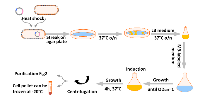
1. Production of 15N, 13C-Tau (Figure 1)
- ATransform pET15b-Tau recombinant T7 expression plasmid into BL21(DE3) competent Escherichia coli bacterial cells.
NOTE: the cDNA coding for the longest (441 amino acid residues) Tau isoform is cloned between NcoI and XhoI restriction sites in the pET15b plasmid.
- Mix gently 50 µl of competent BL21(DE3) cells, forming 1-5 x 10⁷ colonies per µg of plasmid DNA, with 100 ng of plasmid DNA in a 1.5 ml plastic tube.
NOTE: Codon-usage optimized bacterial strains for eukaryotic cDNA expression are not essential to produce human Tau.
- Place the cell mixture on ice for 30 min and then heat shock for 10 sec at 42 °C. Place the tube back on the ice for 5 min and add 1 ml of room temperature LB (Luria-Bertani) medium. Incubate the bacterial suspension at 37 °C for 30 min under gentle agitation.
- Spread using an inoculation loop 100 µl of cell suspension evenly onto an agar plate of LB medium containing 100 µg/ml of ampicillin antibiotic.
- Incubate the selection plate for 15 hr at 37 °C.
- Keep the selection plate at 4 °C until proceeding to the culture step, for a maximum of 2 weeks approximately.
NOTE: a glycerol stock of bacterial culture (50% glycerol), stored at -80 °C, can be prepared to start the culture at a later stage.
- Add 1 ml 1 M MgSO4, 1 ml 100 mM CaCl2, 10 ml 100x MEM vitamin complement, 1 ml 100 mg/ml ampicillin to 1 L of autoclaved M9 salts (6 g Na2HPO4, 3 g KH2PO4, 0.5 g NaCl).
NOTE: A white precipitate will form upon the addition of the CaCl2 solution to the M9 salts that quickly dissipates.
- Solubilize 300 mg of 15N, 13C-complete medium, 1 g of 15NH4Cl, and 2 g of 13C6-glucose in 10 ml of M9 medium. Filter-sterilize the isotope solution using a 0.2 µm filter, directly into the M9 medium.
- Suspend using an inoculation loop one colony of pET15b-Tau transformed bacteria from the selection plate in 20 ml of LB medium supplemented with 100 µg/ml of ampicillin.
- Incubate the inoculated medium at 37 °C for about 6 hr.
- Measure optical density at 600 nm (OD600) on 1 ml of a ten-fold dilution of bacterial culture in a plastic spectrometer cuvette.
NOTE: Turbidity of the bacterial culture corresponding to OD600 of 3.0-4.0 indicates that the saturation growth phase is reached.
- Add 20 ml of the saturated LB culture to 1 L of M9 growth medium supplemented with ampicillin (100 µg/ml final concentration), in a 2 L Erlenmeyer plastic baffled culture flask.
- Place the culture flask in a programmable incubator set to 10 °C and 50 rpm. Program the incubator to switch to 200 RPM and 37 °C early in the morning of the next day.
- Measure OD600 on 1 ml of bacterial culture in a plastic spectrometer cuvette. Add 400 µl of 1 M IPTG (isopropyl β-D-1-thiogalactopyranoside) stock solution (kept at -20 °C) when OD600 reaches a value of about 1.0 to induce the expression of recombinant Tau protein.
- Continue the incubation at 37 °C for a further 3 hr. Collect the bacterial cells by centrifugation at 5,000 x g for 20 min.
- Freeze the bacterial pellet at -20 °C. Keep frozen until the purification step, for an extended period if needed.
2. Purification of 15N, 13C-Tau (Figure 2)
- Autoclave cation-exchange (CEX) purification buffers at 121 °C under 15 psi for 20 min. Store buffers at 4 °C.
- Thaw the bacterial cell pellet and resuspend thoroughly in 45 ml of extraction CEX A buffer (50 mM NaPi buffer pH 6.5, 1 mM EDTA) freshly supplemented with protease inhibitor cocktail 1x (1 tablet) and DNAseI (2,000 units).
- Disrupt the bacterial cells using a high-pressure homogenizer at 20,000 psi. 3-4 passes are necessary. Centrifuge at 20,000 x g for 40 min to remove insoluble material.
- Heat the bacterial cell extract for 15 min at 75 °C using a water bath.
NOTE: a white precipitate is observed after a few minutes.
- Centrifuge at 15,000 x g for 20 min and keep the supernatant containing the heat-stable Tau protein.
- Store at -20 °C until the following purification step if needed.
- Perform cation-exchange chromatography on a strong CEX resin packed as a 5 ml bed column using a fast protein liquid chromatography (FPLC) system (Figure 3 A).
- Set flow rate to 2.5 ml/min.
- Equilibrate the column in CEX A buffer
- Load the 60-70 ml heated extract containing Tau using a sample pump, or alternatively pump A, depending on the system. Collect the flow-through for analysis to verify that Tau protein is efficiently binding to the resin (see 2.8).
- Wash the resin with CEX A buffer until the absorbance at 280 nm is back to the baseline value.
- Elute Tau from the column using a three-step NaCl gradient obtained by a gradual increase of CEX B buffer (CEX A buffer with 1 M NaCl). Program the FPLC as follows: the first step of the gradient to 25% CEX B buffer in 10 column volumes (CV) to reach 250 mM NaCl, the second step to 50% CEX B buffer in 5 CV to reach 500 mM NaCl, and the third step to 100% CEX B buffer in 2 CV to reach 1 M NaCl. Collect 1.5 ml fractions during the elution steps.
- Analyze 10 µl of the fractions collected during the elution step by SDS-PAGE (12% SDS-acrylamide gel) and Coomassie staining (Figure 3 A). Check the loading step on the column as well by analyzing 10 µl of the flow-through.
- Choose the fractions containing Tau and pool these fractions for the next step.
- Perform a buffer exchange on Tau-containing pooled fractions (Figure 3 B).
- Equilibrate a desalting column of 53 ml G25 resin-packed bed (26 x 10 cm) in 50 mM ammonium bicarbonate (volatile buffer) using an FPLC system.
- Set the flow rate to 5 ml/min. Inject the Tau sample on the column via a 5 ml injection loop. Collect fractions corresponding to the absorption peak at 280 nm.
- Repeat the injection 3-4 times, depending on the volume of the initial CEX pool.
- Calculate the amount of purified Tau protein by using the peak area of the chromatogram at 280 nm (1 mg of Tau corresponds to 140 mAU*ml).
NOTE: The extinction coefficient of Tau protein at 280 nm is 7,550 M-1cm-1. Tau does not contain any Trp residues.
- Pool all Tau fractions.
- Aliquot the sample into tubes containing the equivalent of 1 to 5 mg of Tau. Choose these tubes so that the volume of solution is small compared to the volume of the tube (for example 5 ml of solution in a 50 ml tube).
- Punch holes in the tube caps using a needle. Freeze Tau samples at -80 °C.
- Lyophilize Tau samples. Lyophilized Tau protein can be kept at -20 °C for long periods of time.
3. In Vitro Phosphorylation of 15N-Tau
- Dissolve 5 mg of lyophilized Tau in 500 µl phosphorylation buffer (50 mM Hepes·KOH, pH 8.0, 12.5 mM MgCl2, 1 mM EDTA, 50 mM NaCl).
- Add 2.5 mM ATP (25 µl of 100 mM stock solution kept at -20 °C), 1 mM DTT (1 µl of 1 M stock solution kept at -20 °C), 1 mM EGTA (2 µl of a 0.5 M stock solution), 1x protease inhibitor cocktail (25 µl of a 40x stock obtained by dissolving 1 tablet in 1 ml phosphorylation buffer) and 1 µM activated His-ERK2 (250 µl in conservation buffer 10 mM Hepes, pH 7.3, 1 mM DTT, 5 mM MgCl2, 100 mM NaCl and 10% glycerol, stored at -80 °C) in a total sample volume of 1 ml.
NOTE: The activated His-ERK2 can be prepared in-house by phosphorylation with the MEK kinase.
- Incubate 3 hr at 37 °C.
- Heat the sample at 75 °C for 15 min to inactivate ERK kinase.
- Centrifuge at 20,000 x g for 15 min. Collect and keep the supernatant.
- Desalt the protein sample into 50 mM ammonium bicarbonate using a column of 3.45 ml G25 resin-packed bed (1.3 x 2.6 cm), which is suitable for a 1 ml sample.
- Run a 12% SDS-PAGE with 2.5 µl of the protein sample to check both its integrity and efficient phosphorylation (Figure 4).
- Lyophilize the phosphorylated Tau sample. Store the powder at -20 °C.
4. Acquisition of NMR Spectra (Figure 5)
- Solubilize 4 mg of lyophilized 15N, 13C ERK-phosphorylated-Tau in 400 µl NMR buffer (50 mM NaPi or 50 mM deuterated Tris-d11.Cl, pH 6.5, 30 mM NaCl, 2.5 mM EDTA, and 1 mM DTT).
- Add 5% D2O for field locking of the NMR spectrometer and 1 mM TMSP (3-(trimethylsilyl)propionic-2,2,3,3-d4 acid sodium salt)) as internal NMR signal reference. Add 10 µl of a 40x stock solution of a complete protease inhibitor cocktail.
- Transfer the sample to a 5 mm NMR tube using an electronic syringe with a long needle or Pasteur pipette. Close the NMR tube using the plunger. Remove any air bubbles trapped between the plunger and the liquid by plunger movements.
- Place the NMR tube in a spinner. Adjust its vertical position in the spinner with the appropriate gauge for the NMR probe head used, such that most of the sample solution will be inside the NMR coil.
- Start the airflow: click lift in the magnet control system window. Carefully place the spinner with the tube in the airflow at the top of the magnet bore. Stop the airflow (click lift) and let the tube descend into place inside the probe head in the magnet.
- Set the temperature to 25 °C (298 K).
- Perform semi-automatic tuning and matching of the probe head to optimize power transmission. Type atmm on the command line.
- Lock the spectrometer frequency using the D2O signal of the sample recorded on the deuterium channel. Click lock in the magnet control system window.
- Start the shimming procedure to optimize the homogeneity of the magnetic field at the position of the sample. Type topshim gui on the command line to open the shim window. Click Start in the shim window. Check the residual B0 standard variation value to verify that shims are optimal (less than 2 Hz is good).
- Calibrate the p1 parameter (length of a proton radiofrequency pulse in µsec), which is necessary to obtain a 90° rotation of proton spins. Aim for the 360° pulse using a 1D spectrum of water protons (Figure 6).
- Adjust the frequency offset by setting the o1 parameter (in Hz) to the proton water frequency in the 1D spectrum (Figure 6).
- Start acquisition of a 1D proton spectrum (pulse sequence with watergate sequence for water signal suppression, for example, zggpw5) to verify signals from the sample (Figure 7). Adapt the number of scans to the relative protein concentration. Type zg on the command line to start the acquisition.