A subscription to JoVE is required to view this content. Sign in or start your free trial.

Method Article

Super-Resolution Imaging of NK Cell Immunological Synapse Formation on a Supported Lipid Bilayer

876 views

July 8th, 2025

In This Article

Abstract

Source: Zheng, P., et al. Super-resolution Imaging of the Natural Killer Cell Immunological Synapse on a Glass-supported Planar Lipid Bilayer. J. Vis. Exp. (2015).

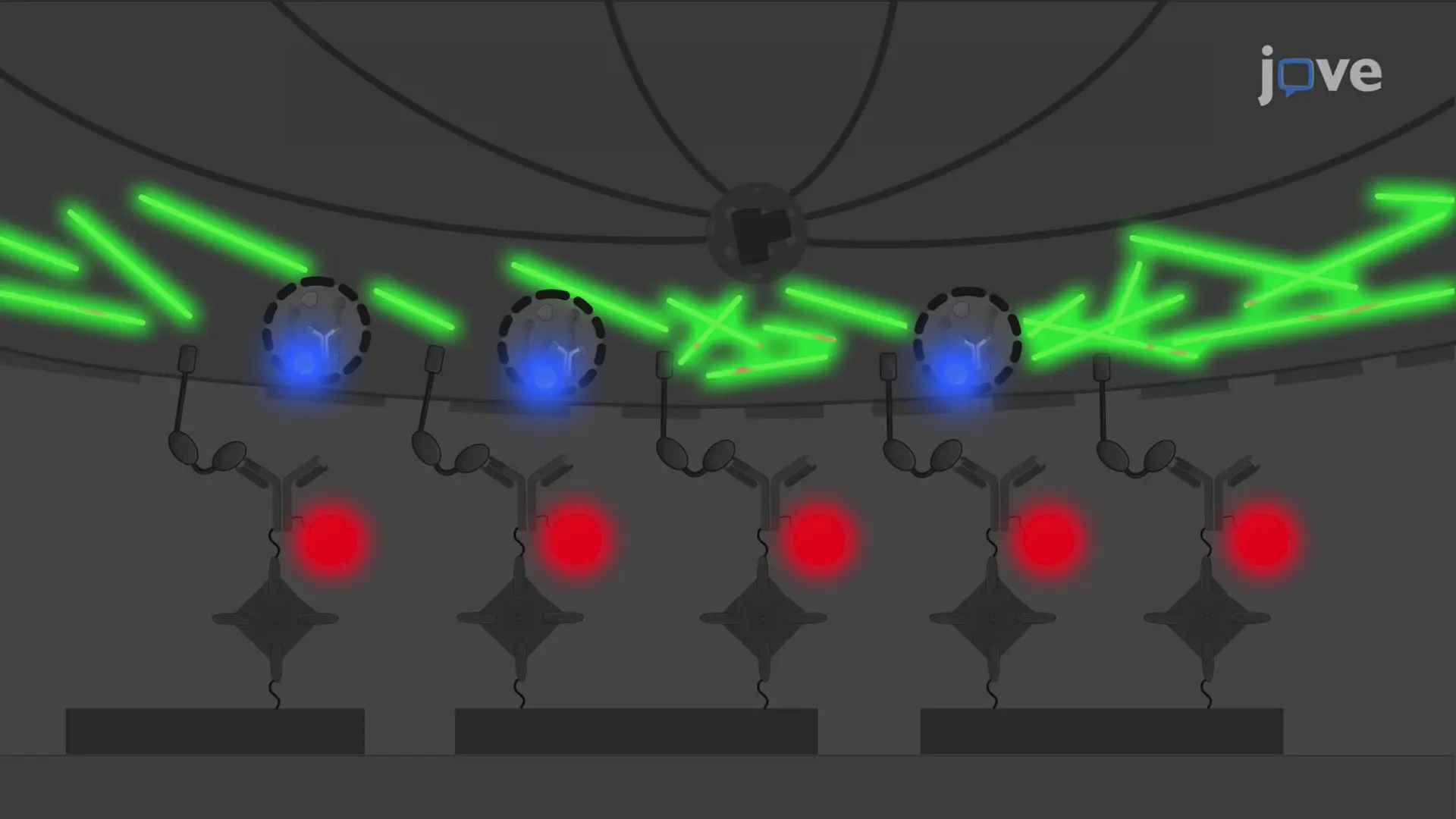
This video demonstrates a technique for visualizing the formation of the immunological synapse in natural killer (NK) cells on a supported lipid bilayer (SLB). The SLB is first labeled with a fluorophore-tagged NK cell activating receptor ligand, followed by incubation with NK cells. Subsequently, the cells are stained for polymerized actin and perforin-positive lytic granules. This approach enables the visualization of synapse formation using stimulated emission depletion (STED) microscopy.
Protocol

1. Preparation of Liposomes

  1. Calculate the amount of chloroform-suspended stock solutions of 1,2-dioleoyl-sn-glycero-3-phosphocholine (DOPC) and 1,2-dioleoyl-sn-glycero-3-phosphoethanolamine-N-cap biotinyl (Biotin-PE) to make diluted stocks at the desired final concentration. To make final concentrations of 400 μM DOPC and 80 μM Biotin-PE phospholipids at 10 ml each, start by placing 629 μl of 10 mg/ml DOPC and 88 μl 10 mg/ml Biotin-PE into separate glass chromatography tubes.
    NOTE: it is important to clean glass Hamilton syringes and glass chromatography tubes by cleaning solution (1 L 95% ethano....

Access restricted. Please log in or start a trial to view this content.

Disclosures

No conflicts of interest declared.

Materials

List of materials used in this article
NameCompanyCatalog NumberComments
18:1 (Δ9-Cis) PC (DOPC)
1,2-dioleoyl-sn-glycero-3-phosphocholine
Avanti850375CLiposome preparation
18:1 Biotinyl Cap PE
1,2-dioleoyl-sn-glycero-3-phosphoethanolamine-N-(cap biotinyl) (sodium salt)
Avanti870273CLiposome preparation
Argon gas, compressedAirgasUN1006Liposome preparation
A lyophilizerLabconcoFreezone 7740020Liposome preparation
Lyophilizer tubesLabconco7540200Liposome preparation
Chromatography ColumnsSanta Cruzsc-205558Liposome preparation
1 M Tris pH 8.0AmbionAM9856Liposome preparation
5 M NaClAmbionAM9759Liposome preparation
Octyl-β-D-glucopyranosideSigma-AldrichO1008Liposome preparation
Dialysis tubingSpectrum Labs132676Liposome preparation
96-well V-bottom polystyrene plate (untreated)Corning3896Antibody density determination
Non-functionalized silica beadsBangs LaboratoriesSS06NAntibody density determination
FACS tubesFisher Scientific14-959-2AAntibody density determination
QuantumTM MESF beadsBangs LaboratoriesVariableAntibody density determination
CaseinSigma-AldrichC0875Lipid bilayer preparation
HEPES buffered saline + 1% HSAHomemadeLipid bilayer preparation
StreptavidinLife technologies434301Lipid bilayer preparation
Fluorescently-labeled biotinylated proteinHomemadeLipid bilayer preparation
ibidi Sticky-Slide VI 0.4ibidi80608Lipid bilayer preparation
25x75 mm glass coverslipibidi10812Lipid bilayer preparation
Hydrogen peroxide (30%)Fisher ScienticBP2633Lipid bilayer preparation
Sulfuric acidSigma-Aldrich258105Lipid bilayer preparation
D-BiotinInvitrogenB20656Lipid bilayer preparation
Lens paperVWR54826-001For imaging
Type F Immersion OilLeica11 513 859For imaging
A Leica TCS STED microscopeLeicaFor imaging

Tags

Natural Killer CellsSTED MicroscopyActin PolymerizationPerforin GranulesCD16 ReceptorsFluorescent LabelingChamber Slide