All procedures involving animal models have been reviewed by the local institutional animal care committee and the JoVE veterinary review board.
1. Materials and Reagents
- Use female Anopheles stephensi mosquitoes (Sda500 strain) that feed on infected mice 3-5 days after emergence and rear as described previously.
- Use parasites Plasmodium berghei ANKA clone expressing the Green Fluorescent Protein (GFP) gene under the control of the constitutive Heat Shock Protein 70 (HSP70) promoter. This yields bright fluorescence throughout the parasite life cycle.
- Use female C57BL/6JRj mice (7 week-old).
NOTE: All the reagents used in the described protocol are listed in the Table of Materials/Equipment.
2. Radiation-attenuated Sporozoite Isolation from Mosquito Salivary Glands
- Between 18-25 days after the infectious blood meal, collect and cold-anesthetize mosquitoes into a 15 ml tube on ice as described previously. Carefully transfer them onto a Petri dish by inverting the tube. Maintain the insects on ice and expose the mosquitoes to a 12 krad dose of γ- or x-irradiation.
- Isolate attenuated sporozoites from irradiated mosquito salivary glands as previously described. Collect infected salivary glands in a small volume (around 15 μl) of 1x Dulbecco's phosphate-buffered saline (DPBS) on ice and gently crush them to release sporozoites.
NOTE: The injection of a highly concentrated parasite suspension in a small volume being critical, it is therefore essential to harvest a sufficient number of salivary glands from a high number of preselected well-infected mosquitoes, as evidenced by their robust expression of green-fluorescence.
- Filter the sporozoite suspension through a 35 µm cell strainer cap adapted to a low adhesion 1.5 ml microcentrifuge tube in order to eliminate clumps and mosquito debris.
- Count the total number of isolated sporozoites as previously described and adjust the concentration of parasite suspension with cold 1x DPBS to obtain 83,000 sporozoites/μl. Store the parasites on ice while continuing the next steps.
- Collect the equivalent number of salivary glands from uninfected mosquitoes that will be used as negative control and process the sample as described in steps 2.2 and 2.3. Dilute the sample as indicated above to obtain similar concentration of salivary gland extracts in the suspension.
NOTE: Sporozoites can be stored on ice for a maximum of 2 hr. However, ideally they are injected within an hour after crushing the salivary glands.
3. Injection of Sporozoites into the Dermal Layer of the Ear
- Anesthetize animals by intraperitoneal injection of ketamine (50 mg/kg)/xylazine (5 mg/kg) mixture. Wait 5-8 min for the mouse to be in a state of unconsciousness and apply ophthalmic ointment to the eyes to prevent corneal drying. Evaluate the depth of anaesthesia by performing a gentle toe pinch on both rear feet.
NOTE: The dose of anaesthesia lasts for less than 20 min, so the next steps must be done quickly.
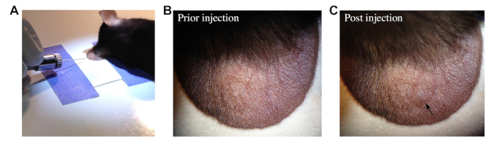
- Stabilize the ear pinna of the mouse by fixing the ventral side with clear tape previously placed under a stereomicroscope (Figure 1A). Gently press the ear to avoid damage of the vasculature.
- Load a 10 μl syringe/35 gauge bevelled needle with 0.6 µl of resuspended parasites.
- Deliver the sporozoite suspension in 4 injection sites (0.15 µl per site) by carefully inserting the needle on the dorsal side of the ear (Figure 1A), beneath the epidermis, with the bevel up, taking care to avoid any blood vessels. Allow the needle to remain in the dermis for few sec to prevent backflow of the injectant before removing it. Observe a characteristic papule at each injection site at the end of the procedure (Figure 1B and 1C).
- After injection, carefully remove the ear from the tape.
- Inject the contralateral ear with the same dose of parasites by following the steps 3.2-3.4.
- Inject 2 additional mice with either 0.6 µl of 1x DPBS or 0.6 µl of 1x DPBS + salivary gland extract from uninfected mosquitoes in order to evaluate the inflammatory response mediated by the injection alone and the deposition of mosquito material in the dermis.
- Monitor mouse recovery from anaesthesia before placing it back into the cage.
NOTE: The proper sporozoite deposition in the skin can be monitored by fluorescence microscopy (Figure 2A and 2B), the mouse being prepared as described previously.