Method Article

Using a Macrophage Conditioned Medium to Promote Neuron Extensions

July 8th, 2025

In This Article

Abstract

Loading...
$$\rightleftharpoonup{xx}$$ $$\longleftharp{xx}$$, $$\longrightharp{xx}$$,

Source: Yun, H. J., et al. Neuron-Macrophage Co-cultures to Activate Macrophages Secreting Molecular Factors with Neurite Outgrowth Activity. J. Vis. Exp. (2018).

This video demonstrates an assay for promoting the growth of neuron projections known as neurite outgrowth using the macrophage-conditioned medium. Co-culture neurons and macrophages with cAMP and harvest the macrophage-derived conditioned medium. Growing the neurons with the conditioned medium promotes neurite outgrowths. These outgrowths are fluorescently labeled and visualized using a fluorescence microscope.

Protocol

Loading...
$$\rightleftharpoonup{xx}$$ $$\longleftharp{xx}$$, $$\longrightharp{xx}$$,

1. Treatment of Db-Camp and Collection of Macrophage Conditioned Medium

NOTE: Start db-cAMP treatment 4 h after the neuron-macrophage co-cultures.

  1. Add 2 µL of 100 µM dibutyryl cyclic AMP (db-cAMP) solution to the neuron-macrophage co-cultures. Add the same volume of phosphate-buffered salin (PBS) for a control experiment.
  2. After 24 h, fill an empty well with 1 mL of macrophage culture medium in the same 6-well plate. Transfer the cell culture insert in the neuron-macrophage co-cultures to the empty well with macrophage culture medium. Keep the cells under the same condition for 72 h without changing the medium.
    NOTE: We add only 1 mL of macrophage culture medium during conditioned medium (CM) collection to make concentrated CM. During the CM collection, if needed, we added more to completely cover the macrophages within the insert.
  3. After 72 h, centrifuge the macrophage CM at 239 x g for 5 min to remove the cellular components. Pass the supernatant through a 0.2-µm filter to remove any remaining cellular debris. Store the collected CM at -70 °C until use.

2. Neurite Outgrowth Assay with Collected CM

  1. Before setting up a separate adult DRG neuron culture, pre-coat an 8-well chamber slide with poly-D-lysine and laminin. Incubate a 6-well plate with 0.01% poly-D-lysine at 37°C for 2 h or at 4°C overnight. Then, wash the plate twice with distilled water.
  2. Incubate the plate with laminin solution at a concentration of 3 µg/mL for 2 h at room temperature, and then wash the plate twice with distilled water. Dry the plate at room temperature at least for 1 h.
  3. Obtain dissociated adult DRG neurons in Neurobasal medium supplemented with B27. Plate 5 x 10⁴ cells per well onto the pre-coated 8-well chamber slide.
  4. Place the chamber slide in a 37 °C incubator for 2 h, allowing the cells to attach to the bottom. Then, replace the culture medium with the thawed CM that is preheated at 37 °C.
    NOTE: Be careful not to touch the bottom of the 8-well chamber slide when removing the culture medium and adding the collected CM.
  5. 15 h after the initial plating, remove the medium and wash the cells with PBS once. Then, add 200 µL of ice-cold 4% paraformaldehyde solution to the wells, and incubate the cells with the paraformaldehyde solution for 20 min at 4 °C
    NOTE: The DRG neuron culture for neurite outgrowth assay should be precisely restricted to 15 h. Under this condition, there is no appreciable neurite outgrowth.
  6. Wash three times with ice-cold PBS and then perform blocking with 10% normal goat serum (NGS) with 0.1% Triton-X for 30 min. Incubate the fixed cells with primary antibody (anti-Tuj-1) solution diluted with 10% NGS (at a concentration of 1 µg/mL) for 4 h at room temperature or overnight at 4 °C.
  7. Wash three times with PBS, and then incubate the cells with secondary antibody (goat-anti mouse) solution for 2 h at room temperature. Then, wash twice with PBS and mount the culture slide with a coverslip (24 × 50 mm) using a mounting solution.
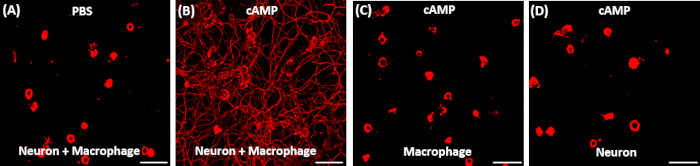
  8. Take images using a fluorescence microscope to visualize neurite outgrowth (Figure 1).
Access restricted. Please log in or start a trial to view this content.

Results

Loading...
$$\rightleftharpoonup{xx}$$ $$\longleftharp{xx}$$, $$\longrightharp{xx}$$,

Neuron-macrophage interaction via cAMP; fluorescence microscopy comparison; cellular response analysis.
Figure 1: Representative results of neurite outgrowth assay using conditioned medium obtained from various conditions. (A, B) Adult DRG neurons were acutely dissociated and cultured for exactly 15 h. 2 h after plating, the culture medium was replaced...

Access restricted. Please log in or start a trial to view this content.

Disclosures

Loading...
$$\rightleftharpoonup{xx}$$ $$\longleftharp{xx}$$, $$\longrightharp{xx}$$,
No conflicts of interest declared.

Materials

List of materials used in this article
NameCompanyCatalog NumberComments
Cell culture insert transparent PET membrane 0.4μm pore sizeCorning,Falcon353090Transparent PET membrane with 0.4-μm pore size, for 6-well plate
70-μm nylon cell strainerCorning, Falcon352350
8-well culture slideBiocoat354632with a uniform application of Poly-D-Lysine
Neurobasal mediumThermo Fisher Scientific, Gibco21103-049Containing 1% glutamax and 1% penicilin-streptomycin
B-27 supplement, serum freeThermo Fisher Scientific, Gibco17504-044extracellular solution
GlutamaxThermo Fisher Scientific, Gibco35050-061
Penicillin-streptomycinThermo Fisher Scientific, Gibco15140-122
Poly-D-lysine HydrobromideSigma-AldrichP6407-5MG
LamininThermo Fisher Scientific, Invitrogen23017-015
Adenosine 3', 5'-cyclic monophosphate, N6,O2'-dibutyryl-, sodium saltMerck Millipore Corporation, Calbiochem28745
10% Normal Goat SerumThermo Fisher Scientific16210072
Triton-X-100Daejung Chemical and Metal Co8566-4405
Anti β III tubulin (Tuj-1)Promega CorporationG7121Mouse monoclonal antibody
Goat anti-Mouse IgG (H+L) secondary antibodyThermo Fisher Scientific, InvitrogenA11005
HemacytometerMarienfeld-SuperiorN/A
Cell culture CO2 incubatorPanasonicN/A
Tabletop centrifugeSorvallN/A
Confocal microscopeOlympus America IncIX71
FBS (Fetal Bovine Serum)VWR International, HycloneSH30919.03

Reprints and Permissions

Request permission to reuse the text or figures of this JoVE article

Request Permission

Tags

Macrophage Conditioned MediumNeurite OutgrowthNeuron Macrophage Co culturecAMP TreatmentFluorescence MicroscopyCentrifugation ProtocolParaformaldehyde FixationPrimary AntibodiesSecondary AntibodiesPoly D lysine Coating

Related Articles