All procedures involving sample collection have been performed in accordance with the institute's IRB guidelines.
Prepare density centrifugation media mix solution by placing 90 ml of 10x PBS into 264 ml of ultrapure deionized water (3.93x) in a sterile plastic container. Titrate to pH 7.0-7.2 with HCl and filter sterilize through a 0.22 µm filter. Store at 4 °C.
Prepare 70% density centrifugation media by combining 18 ml of density centrifugation media mix solution with 30 ml of density centrifugation media (see materials list for details) in a 50 ml polypropylene centrifuge tube. Store at 4 °C, but bring to RT before use.
Prepare 37% density centrifugation media by combining 9.6 ml of DPBS with 10.4 ml 70% density centrifugation media in a 50 ml centrifuge tube. Store at 4 °C, but bring to RT before use.
1. Isolation of Glioma-infiltrating PBMCs
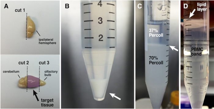
- Using a clean, single-edged razor blade, isolate the area of the brain containing the tumor by making a sagittal cut down the center of the brain to bisect the two hemispheres. Turn the ipsilateral hemisphere medial side down and make two coronal cuts at the levels of the cerebellum and the olfactory bulb to isolate the target tissue containing the tumor implant (Figure 1A). An equivalent portion of the contralateral hemisphere may also be isolated and used as a negative control.
- Place the target tissue into the respectively labeled glass Dounce tissue grinder containing 1 ml of DPBS, push the plunger all the way down, and twist 7 times to initially disrupt the tissue. Lift the plunger to allow the liquid to settle back to the bottom of the tissue grinder. Repeat this step thrice; however, only twist the plunger 4 times during the next two repeats and 3 times during the last repeat to avoid over-triturating the tissue.
- Using a P-1000 micropipette, sequentially apply three 1 ml volumes of ice-cold DPBS along the sides of the plunger to rinse into the tissue grinder.
- Resuspend the triturated brain matter by pipetting up and down and placing it into a labeled 15 ml centrifuge tube on ice. Rinse the sides of the Dounce tissue grinder with 1 additional ml of ice-cold DPBS and add to the same 15 ml centrifuge tube. Keep all tubes containing triturated brain matter on ice until all samples have been processed.
- Once all samples have been processed, spin down the triturated brain matter in the 15 ml centrifuge tubes at 740 x g (max RCF) for 20 min at 4 °C.
- Remove the supernatant with a 10 ml serological pipette and pipette gun and resuspend the pelleted brain matter in 1 ml of the previously prepared collagenase/DNase-I digestive enzymes using a P-1000. Place the 15 ml centrifuge tubes into a test tube rack and place in a 37 °C water bath for 15 min.
- Gently agitate the samples twice throughout the incubation period by flicking the tubes to facilitate tissue disaggregation.
- Add 6 ml of ice-cold DPBS using a 10 ml serological pipette to each tube to dilute the digestive enzymes. Pipette up and down to resuspend, and filter the total volumes through sterile 70 µm nylon mesh filters into new labeled 15 ml centrifuge tubes on ice.
- Spin the brain cell suspension down at 740 x g (max RCF) for 20 min at 4 °C to achieve a single-cell pellet (Figure 1B). After removing the 15 ml tubes from the centrifuge, place them on ice and increase the temperature setting on the centrifuge to about 21 °C in preparation for the next step.
- Fully remove the supernatant from each tube containing brain cells and initially resuspend the pellets in 1 ml of 70% density centrifugation media using a P-1000 micropipette. Then add 4 additional milliliters of 70% density centrifugation media to each tube using a 10 ml serological pipette. Screw the caps on securely and homogenize the cell suspension by gently inverting to tubes several times.
- Remove the 15 ml centrifuge tubes containing brain cells resuspended in 70% density centrifugation media from ice, and place in a test tube rack at RT. One at a time, carefully overlay 2 ml of 37% density centrifugation media solution onto the 5 ml of 70% density centrifugation media with a P-1000 micropipette to form a clean interface between the two density centrifugation media layers (Figure1C).
- Use a fine-tipped marker to indicate the position of the interface so it can be easily identified after centrifugation when the distinction becomes less apparent.
- Spin down the 15 ml centrifuge tubes at 740 x g (max RCF) for 20 min at RT with no break to avoid disrupting the interface.
- After centrifugation, collect the PBMCs that have accumulated at the interface between the two density centrifugation media layers (Figure 1D) by introducing a P-200 micropipette into the tube along its side, carefully bypassing the lipid layer.
- Once at the level of the PBMC band, slowly extract 200 µl from the surface of the 70% density centrifugation media layer and place it into a respectively labeled polypropylene FACS tube on ice. Repeat once for a total volume of 400 µl per sample.
- Add 3 ml of flow buffer to each FACS tube containing 400 µl of PBMCs to sufficiently dilute the density centrifugation media, then spin the cells down at 660 x g (max RCF) for 20 min at 4 °C.