$$\rightleftharpoonup{xx}$$
$$\longleftharp{xx}$$,
$$\longrightharp{xx}$$,
1. Mouse embryonic stem cell culture
- Prepare 0.1% gelatin coated cell culture dishes or plates.
- Add 2 mL of sterilized 0.1% gelatin (0.1% w/v in water) to 60 mm cell culture dishes. Rock gently to ensure even coating of the cell culture dishes.
- Put the dishes into a 5% CO2 incubator at 37 °C and allow coating for 1 h.
- Remove the 0.1% gelatin solution before seeding the cells.
NOTE: After removing the gelatin, there is no need to dry or wash the coated dishes.
- Mouse embryonic stem cells (A2lox and 129) culture
- Incubate mESCs (A2lox and 129) cells in the 0.1% gelatin coated 60 mm cell culture dishes in mESC growth medium at 37 °C in a 5% CO2 incubator, respectively. The mESC growth medium consists of 85% knock-out Dulbecco's modified Eagle medium/nutrient mixture F-12 (DMEM/F12), 15% Knock-out serum replacement (KSR), 0.1 mM β-mercaptoethanol (2ME), 2 mM GlutaMAX, 1% non-essential amino acid (NEAA), 1% penicillin/streptomycin (P/S), 1000 U/mL leukemia inhibitory factor (LIF), 10 nM CHIR-99021 (GSK-3 inhibitor) and 0.33 nM PD0325901 (MEK inhibitor).
CAUTION: β-mercaptoethanol is flammable and has inhalation toxicity. Keep away from fire sources and wear a mask to avoid inhalation when use.
- Change the mESC growth medium daily for better growth of A2lox and 129.
- When the cells reach 80% confluence, remove the medium and add 1 mL of 0.1% trypsin to the dish. Gently rock for 30 s to ensure even cover of trypsin on all cells.
- Leave the cells for about 1 min to trypsinize and then remove the trypsin using a 1 mL pipette.
- Add 2 mL of mESC growth medium to the dish, pipette up and down several times to make a single cell suspension.
- Count the density of the cells in the suspension as accurately as possible using a hemocytometer.
- Divide the cells into 7 groups and induce differentiation using different protocols shown in Table 1.
2. Differentiation from mESCs to NPCs
- Prepare 0.1% gelatin coated cell culture plates or coverslips.
- Before use, prepare 0.1% gelatin coated 6-well plates or coverslips as in step 1.1.
- Differentiation (Table 1)
- Add 1.5 x 10⁶ mESCs into a non-adhesive bacterial dish in 10 mL of basal differentiation medium I to allow for embryoid body formation at 37 °C in a 5% CO2 incubator.
- After 2 days, transfer cell aggregates into 15 mL centrifuge tubes and let them settle by gravity.
- Remove the supernatant and add 10 mL of fresh basal differentiation medium I to resuspend the embryoid bodies. Replant them into a new non-adhesive bacterial dish and allow differentiation for another 2 days.
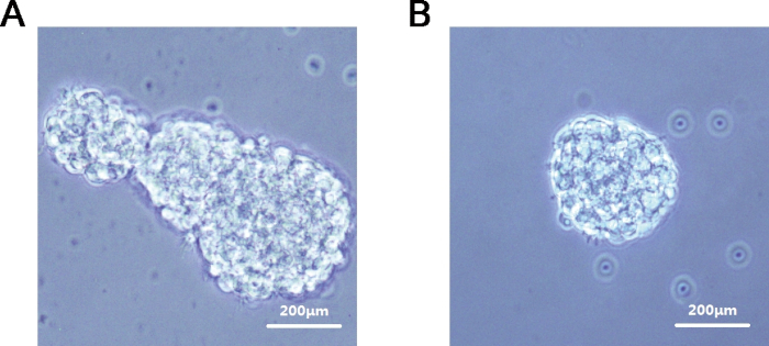
- Check the formation of embryoid bodies under the microscope (Figure 1A).
- Collect embryoid bodies as described in steps 2.2.2-2.2.3. Seed about 50 embryoid bodies in 2 mL of basal differentiation medium I per well onto 0.1% gelatin-coated 6-well plates.
- Prepare 1 mM all-trans retinoic acid (RA) stock (in DMSO) and store away from light in a -80 °C freezer after sub-packaging.
NOTE: RA is unstable, and attention should be paid to keeping it away from light and reducing air contact during preparation of RA stock.
- For RA induction, add 2 µL of RA stock into each well to make a final concentration of 1 µM.
- Place the plate into the 5% CO2 incubator at 37 °C and differentiate for another 4 days.
- Change the entire 2 mL of basal differentiation medium I (with 1 µM RA) every 2 days.
Table 1: Details of the protocol used in differentiation.
| Protocol | Media |
| 4-day Embryoid Bodies formation + 4-day RA induction | Basal differentiation medium:
DMEM/F12 +15%FBS + 1%NEAA+0.1mM 2ME+ 1%P/S |