Method Article

Generating Neurons by Reprogramming Human Dermal Fibroblast Cells

July 8th, 2025

In This Article

Abstract

Loading...
$$\rightleftharpoonup{xx}$$ $$\longleftharp{xx}$$, $$\longrightharp{xx}$$,

Source: Shrigley, S., et al. Simple Generation of a High Yield Culture of Induced Neurons from Human Adult Skin Fibroblasts. J. Vis. Exp. (2018)

The video demonstrates the generation of neurons from adult human dermal fibroblasts (aHDFs). The aHDFs are reprogrammed using a lentiviral vector carrying genes for neuronal transcription factors and short hairpin RNAs (shRNAs). The shRNAs inhibit the expression of a neuronal transcriptional repressor, and the neuronal transcription factors activate genes to differentiate aHDFs into neurons. These reprogrammed aHDFs are cultured in an early and a late neuronal conversion medium to generate mature neurons.

Protocol

Loading...
$$\rightleftharpoonup{xx}$$ $$\longleftharp{xx}$$, $$\longrightharp{xx}$$,

1. Preparation of Skin Fibroblasts for Reprogramming

  1. Using an automated cell thawing system or a 37 °C water bath, thaw the adult human dermal fibroblasts (aHDFs) and plate 200,000 per T75 flask (count with an automated cell counter) in 10 mL of fibroblast medium (see Table 1) at 37 °C in 5% CO2.
  2. Perform a complete medium change with fibroblast medium on the next day.
  3. Change the fibroblast medium every 3–4 days until the cells reach 95% confluency.
    NOTE: One confluent flask will contain approximately 1,000,000 cells.

2. Plating for Reprogramming (Day −1)

NOTE: It is recommended to use a gelatin coating for short term experiments (up to 30 days); alternatively, for long term experiments it is recommended to start on a poly-L-ornithine, fibronectin and laminin (PFL) coating.

  1. 60 min before plating the aHDFs for reprogramming, coat a 24-well plate with 0.1% gelatin (250 µL/well) and incubate at 37 °C.
  2. Aspirate the fibroblast medium on the aHDFs. Wash once with Dulbecco's phosphate buffer saline (DPBS). Dissociate the cells with 0.05% trypsin (1.5 mL per T75 flask) at 37 °C for 3–5 min.
  3. Add fibroblast medium to neutralize the trypsin (3 mL per flush per T75 flask) and collect the detached cells in a 15 mL tube by flushing out the cells in the flask twice.
  4. Spin down the cells at 400 x g for 5 min. Discard supernatant and resuspend the cell pellet in 1 mL of fibroblast medium.
  5. Count the cells using an automated cell counter (to ensure a good quality conversion check that the cell viability is above 90% with trypan blue staining).
  6. For a complete 24-well plate, prepare a suspension of 1,320,000 cells in 13.2 mL of fibroblast medium to achieve a suspension of 100,000 cells/mL of medium (or 55,000 cells/well in 550 µL of fibroblast medium multiplied by the number of wells needed).
  7. Aspirate the gelatin from the plate and wash twice with DPBS. Add 500 µL of the cell suspension to each well and incubate overnight at 37 °C in 5% CO2.

3. Viral Transduction (Day 0)

NOTE: Working with lentiviral particles requires category 2 equipment and the use of an agent to neutralize the virus. Wearing double pairs of gloves is also strongly recommended.

  1. Warm up 13.2 mL of fibroblast medium to 37 °C.
  2. Thaw a lentiviral vector containing the transcription factors achaete-scute family bHLH transcription factor 1 (Ascl1) and Brn2 with two short hairpin RNAs (shRNA) targeting RE1-silencing transcription factor (REST) at room temperature.
  3. Add the necessary volume of lentivirus to infect the aHDFs at multiplicity of infection (MOI) of 20 to the medium without any transduction enhancers.

    Virus volume calculation formula, equation for cell infection using MOI and virus titer, diagram.
  4. Replace the medium in the 24-well plate with fibroblast medium containing the lentiviral vector (500 µL/well) and incubate overnight at 37 °C in 5% CO2.
  5. The next day, replace the medium in the wells with fresh fibroblast medium without the lentiviral vector.
    NOTE: The medium is considered infectious for 7 days and as such, adequate protection and handling procedures should be used during the first week following viral transduction.

4. Maintenance of the Converting Cells

NOTE: Once conversion begins cells are susceptible to lifting; take care to tip the plate up and use a 1,000 µL pipette when removing media to avoid cells detaching.

  1. On day 3, remove the fibroblast medium and add 500 µL of early neuronal conversion medium (see Table 1).
  2. Two to three times a week, take out 225 µL of old medium from the well and add-in 250 µL of fresh early neuronal conversion medium.
  3. On day 18, remove all of the medium from each well and replace with 500 µL of late neuronal conversion medium (see Table 1).
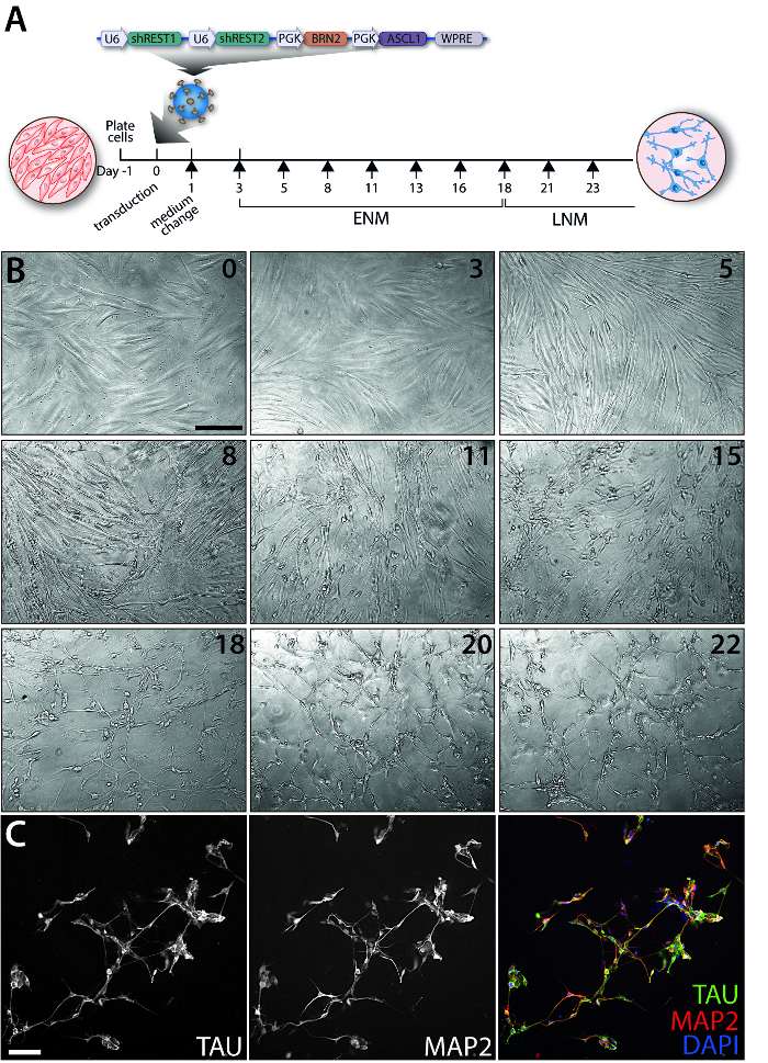
  4. Continue to change half of the medium as above with late neuronal conversion medium every 2–3 days until day 25 or experiment endpoint (Figure 1A).

Table 1. Composition of the different media used. Full description of the composition for media needed in this protocol including fibroblast medium, early neuronal conversion medium, late neuronal conversion medium.

Stock ConcentrationWorking Concentration
Fibroblast medium
Basal mediumN/AN/A
Penicillin/Streptomycin10,000 U/mL100 mg/mL
FBSN/A10%
Early neuronal conversion medium (ENM)
Neural differentiation medium N/AN/A
Penicillin/Streptomycin10,000 U/mL100 mg/mL
CHIR9902110 mM2 µM
SB-43154220 mM10 µM
Noggin100 µg/mL0.5 µg/mL
LDN-193118910 mM0.5 µM
VPA1 M1 mM
LM-22A420 mM2 µM
GDNF20 µg/mL2 ng/mL
NT310 µg/mL10 ng/µL
db-cAMP50 mM0.5 mM
Late neuronal conversion medium (LNM)
Neural differentiation medium N/AN/A
Penicillin/Streptomycin10,000 U/mL100 mg/mL
LM-22A420 mM2 µM
GDNF20 µg/mL2 ng/mL
NT310 µg/mL10 ng/µL
db-cAMP50 mM0.5 mM

Access restricted. Please log in or start a trial to view this content.

Results

Loading...
$$\rightleftharpoonup{xx}$$ $$\longleftharp{xx}$$, $$\longrightharp{xx}$$,

Gene editing process in neuron differentiation; microscopy time-lapse; TAU, MAP2, DAPI staining.
Figure 1: Evolution of the iN conversion over time. (A) Timeline of the experiment and map of the construct packaged in a lentivirus used to reprogram the adult human dermal fibroblasts. Each black arrow represents a medium change. (

Access restricted. Please log in or start a trial to view this content.

Disclosures

Loading...
$$\rightleftharpoonup{xx}$$ $$\longleftharp{xx}$$, $$\longrightharp{xx}$$,
No conflicts of interest declared.

Materials

List of materials used in this article
NameCompanyCatalog NumberComments
Cell Lines
Adult human dermal fibroblasts[C2 passage #7] Donor was a 67 year old female. Cells obtained from the Parkinson’s Disease Research and Huntington’s disease clinics at the John van Geest Centre for Brain Repair (Cambridge, UK).
Reagents for Fibroblast Culture, Transduction and Conversion
Dulbecco's phosphate-buffered saline (DPBS) [-CaCl2, -MgCl2]Gibco14190094
Trypsin-EDTA [0.5%]Gibco15400-054Dilute to 0.05% in DPBS.
Virkon (agent used to neutralize virus)Viroderm7511Dilute to 1% solution with warm water.
Milli-Q WaterMillipore
Basal medium - Dulbecco’s Modified Eagle Medium (DMEM) + GlutaMaxGibco61965059
Penicillin/Streptomycin [10,000 U/ mL]Gibco15140-122
Fetal Bovine Serum (FBS)Gibco10270-106
CryoMACS® dimethyl sulfoxide (DMSO) 10Miltenyi170-076-303
Neural differentiation medium - NDiff 227Takara-ClontechY40002
LM-22A4Tocris4607Dilute 10 mg in 1450 µL DMSO.
Stock concentration: 20 mM.
Glial cell line-derived neurotrophic factor (GDNF) [recombinant human]R&D systems212-GD-010Dilute 10 ug in 500 µL 0,1% BSA in DPBS. Stock concentration: 20 µg/mL.
NT3 [recombinant human]R&D systems267-N3-025Dilute 25 µg in 2,5 mL 0,1% BSA in DPBS. Stock concentration: 10 µg/mL.
db-cAMPSigma AldrichD0627Dilute 1 g in 40,7 mL Milli-Q water.
Filter and make 500 µL aliquots or stock tubes of 10 mL. Stock concentration: 50 mM.
CHIR99021Axon1386Dilute 2 µg in 429,8 µL DMSO.
Stock concentration: 10 mM.
LDN-193189Axon1509Dilute 2 mg in 360 µL DMSO.
Stock concentration: 10 mM.
Valproic acid sodium salt (VPA)Merck Millipore676380Dilute 5 g in Milli-Q water to acheive a stock concentration of 1 M. CAUTION: Avoid ingestion, contact with skin, and breathing dust formation.
Reagents for Coatings
GelatinSigma AldrichG2500Dilute to 0.1% in Milli-Q water.
Poly-L-ornithineSigma AldrichP3655Dissolve in Milli-Q water. Use at 15µg/mL.
FibronectinThermoFisher Scientific33010-0182 mL of Milli-Q water + 70 µL 0,25 M NaOH. Use at 5 µg/mL.
LamininThermoFisher Scientific23017-015Store at -80°C. Thaw on ice, keep cool and aliquot 30 µL. Use at 5 µg/mL.
Equipment
T75 flask [Nunclon Delta Surface]ThermoFisher Scientific156499
24-well plate [Nunc]ThermoFisher Scientific142485
1.5 mL polypropylene tubeSigma AldrichZ336769
15 mL falcon tubeSarstedt6,25,54,502
50 mL falcon tubeSarstedt6,25,47,254
Pippette controllerFor pipetting volumes 1-25 mL.
Sterile serological pipettes: 5, 10 and 25 mLSarstedt86.1253.001, 86.1254.001, 86.1685.001
Adjustable volume pipettors: 5, 20, 200, and 1,000 µL
Sterile pipette tipsFor pipetting volumes of 0.5 - 1,000 µL.
ThawSTAR Automated Cell Thawing SystemBioCisionBCS-601
Countess II Automated Cell CounterThermoFisher ScientificAMQAX1000
Cell counting chambers [50 slides] and trypan blue [0.4%]ThermoFisher ScientificC10228For use with Countess II Automated Cell Counter.
Laminar flow hood
Humidified 5% CO2 37 °C incubator
CentrifugeSuitable for 1,5, 15 and 50 mL tubes.
Orbital shaker
Inverted fluorescence microscopeLeicaDMI6000 B

Reprints and Permissions

Request permission to reuse the text or figures of this JoVE article

Request Permission

Tags

Neuronal ReprogrammingLentiviral VectorTranscription FactorsshRNA InhibitionEarly Neuronal ConversionLate Neuronal ConversionAdult Human Dermal FibroblastsNeuronal DifferentiationCell Culture ProtocolMature Neuron Generation

Related Articles