1. Microglia differentiation
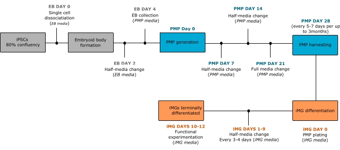
NOTE: An overview of the protocol is summarized in Figure 1.
- Induced pluripotent stem cell (iPSC) culture
NOTE: Further details describing iPSC culture techniques can be found elsewhere.
- Thawing and maintenance
- Prepare the iPSC medium, aliquot it, and store it at -20 °C for up to 6 months. Thaw the aliquots overnight at 4 °C and use them for up to 1 week. Leave the media at ambient temperature for at least 1 h before use.
- Coat the wells of a 6-well plate by adding 1 mL of 10 µg/mL of laminin 521 diluted in Dulbecco's phosphate buffered saline (DPBS) containing calcium and magnesium. Keep the plates in an incubator at 37 °C and 5% CO2 for at least 2 h or preferably overnight. Once diluted, store the laminin at 4 °C for 3 months.
- Prepare 10 mM Rho kinase inhibitor Y27632 (ROCK Inhibitor) stock solution by diluting in sterile water. Make single-use aliquots of ROCK inhibitor solution and store them at -20 °C for up to 1 year.
- Prepare 0.5 mM Ethylenediamine tetraacetic acid (EDTA) by diluting the stock solution of 0.5 M EDTA in DPBS.
- To thaw a vial of frozen iPSCs, place the vial in a water bath at 37 °C until it is mostly thawed. Immediately transfer the content of the vial to a 15 mL conical tube containing 4 mL of iPSC medium. Centrifuge at 500 × g for 1 min.
- Aspirate the supernatant and resuspend the cells by slowly adding 1 mL of iPSC medium containing 10 µM ROCK inhibitor against the wall of the tube to avoid disturbing the colonies.
- Add 1.5 mL of iPSC medium containing 10 µM ROCK inhibitor to each of the coated wells and transfer the resuspended colonies drop by drop to the wells containing the medium.
NOTE: Use 5-7 drops from step 1.1.1.6 for culture maintenance but adjust the optimal seeding density for every iPSC line.
- Evenly distribute the cells in the wells by manually shuffling the plate side-to-side and back to front. Place the cells in an incubator at 37 °C and 5% CO2. The next day, completely replace the medium by adding fresh iPSC medium without ROCK inhibitor.
- For maintenance, change the medium every day until the cells reach 80% confluency.
NOTE: The cells can be maintained without changing the medium for 2 days if the iPSC medium used allows a flexible feeding schedule. It is advised to limit this to one time per passage and only if the cells are less than 50% confluent.
- Splitting
- Aspirate the medium and wash the cells with 1 mL of DPBS (without calcium and magnesium).
- To dislodge the cells, add 1 mL of 0.5 mM EDTA and incubate for 2-3 min at room temperature until the edges of the cell colonies lift from the surface of the well. Wash the cells again with DPBS and add 1 mL of iPSC medium.
- Dissociate the cell colonies by gently scraping them using a cell lifter. Scrape each area of the well only once to avoid disturbing the colonies.
NOTE: Alternative methods to dislodge the colonies from the well can be found elsewhere.
- Using a 1 mL pipette tip, collect the cells and transfer them drop by drop at a 1:6 ratio (cells to medium) into precoated wells (as mentioned in step 1.1.1.2) containing 1.5 mL of iPSC medium.
2. iPSC differentiation to microglia-like cells (iMGs)
NOTE: The small molecules and growth factors are dissolved in sterile-filtered 0.1% bovine serum albumin in DPBS to a stock concentration 1,000x higher than the final concentration. It is recommended to differentiate iPSCs into iMGs during early passages. Routine karyotyping of iPSC lines is advised.
- Prepare standard coating solution
- Thaw the stock solution of an extracellular matrix coating reagent on ice at 4 °C overnight.
- Prechill microcentrifuge tubes and filter pipette tips at 4 °C.
- Aliquot 250 µL of the concentrated extracellular matrix coating reagent into each tube and immediately place on ice. Store the aliquots at -20 °C.
- To prepare the coating solution, thaw one of the extracellular matrix coating reagent aliquots on ice at 4 °C overnight.
- Add 50 mL of ice-cold Dulbecco's Modified Eagle Medium/Nutrient mixture F12 (DMEM-F12) optimized for growth of human embryonic and induced pluripotent stem cells (referred as to DMEM-F12) media to a prechilled conical tube and keep it on ice.
- Chill a 1 mL pipette tip by pipetting ice-cold DMEM-F12 up and down multiple times, and then immediately use the pipette tip to transfer 250 µL of the extracellular matrix coating reagent into the conical tube containing the DMEM-F12 medium.
NOTE: The standard coating solution can be stored for 2 weeks at 4 °C.
- Embryoid body (EB) formation
- Prepare EB medium that can be maintained at 4 °C for up to 4 days.
- Once iPSCs have reached 80% confluency, dissociate the colonies by washing with 1 mL of DPBS and adding 1 mL of a dissociation reagent for 2 min at 37 °C. Dislodge the colonies using a cell lifter by scraping multiple times to create a single-cell suspension. Collect the cells and transfer everything to a 15 mL conical tube containing 9 mL of DPBS.
- Centrifuge the cells at 500 × g for 1 min, remove the supernatant, and resuspend the cells in 1 mL of EB medium. Take 10 µL of cells and dilute 1:1 with Trypan blue. Count the cells with a hemacytometer, and based on the cell count, dilute the cell-stock to a final dilution of 10,000 cells per 100 µL. For plating cells, add 100 µL of the diluted cells per well into a low adherence, round-bottom, 96-well plate.
NOTE: Generally, 48 wells of the 96-well plate can be obtained from each 80% confluent well of iPSCs.
- Centrifuge the plate at 125 × g for 3 min and incubate at 37 °C and 5% CO2 for 4 days. Perform a half-medium change on day 2 by using a multichannel pipette and gently collecting 50 µL of old medium, and then adding back 50 µL of fresh EB medium.
NOTE: On day 4, iPSCs will form spherical cellular structures termed EBs as described in the results section. The EB differentiation process can be extended up to 7 days, if necessary.
- Generation of primitive macrophage precursors (PMPs)
- Prepare PMP base medium, sterile-filter, and store it at 4 °C for up to 1 month.
- Coat the wells of a 6-well plate by adding 1 mL of ice-cold Matrigel coating solution and incubate at 37 °C and 5% CO2 for at least 2 h or preferably overnight.
- On day 4 of EB differentiation, transfer the EBs to the Matrigel-coated wells by collecting the EBs with 1 mL pipette tips (using a pipette). Pipette up and down once or twice to dislodge the EBs from the well. Hold the 6-well plate at a tilted angle to allow for the EBs to settle down at the edge of the well.
NOTE: Nine or ten EBs can be plated per coated well.
- Once all the EBs have settled down, gently pipette and remove the old medium using a 1 mL pipette tip while keeping the EBs at the edge of the well. Add 3 mL of freshly prepared PMP complete medium to each well. Evenly distribute the cells in the wells by manually shuffling the plate side-to-side and back to front. Place the plate in the incubator at 37 °C and 5% CO2.
- Do not disturb the plate for 7 days to allow the EBs to attach to the bottom of the well. After that time, perform a half-medium change using PMP complete medium.
NOTE: At this point, most of the EBs should be attached to the plate. Any floating EBs can be removed.
- After 5-7 days, inspect the EBs under a light field microscope at 4x magnification to ensure that they are attached to the bottom of the wells. Change the medium as described in 1.2.3.5. On day 21, perform a complete medium change with 3 mL of PMP complete medium.
NOTE: Myeloid progenitors floating in the medium may be evident at this point. These cells should be discarded during the medium change.
- On day 28, look for round cells referred to as PMPs in the supernatant and collect the medium containing the PMPs using a 10 mL pipette and automatic pipettor. Take care not to disturb the EBs. Transfer the PMPs and the medium to a 15 mL conical tube and proceed as described in step 1.2.4.
NOTE: Usually PMPs from the same iPSC line can be collected from five wells of a 6-well plate and pooled together in a single 15 mL conical tube.
- Add 3 mL of fresh PMP complete medium for further maintenance of the EBs. As PMPs continuously emerge from EBs for more than 3 months, collect them every 4-7 days (do not allow the medium to change color to a yellow tone) as described in steps 1.2.3.7 and 1.2.3.8.
NOTE: Although PMPs can be collected for several months, they may change their phenotype over time.
- Differentiation to iMGs
- Prepare iMG base medium (Table of Materials), sterile-filter and store it at 4 °C for up to 3 weeks.
- Once the PMPs have been collected into a 15 mL conical tube, centrifuge them at 200 × g for 4 min. Aspirate the supernatant and resuspend the PMPs using 1-2 mL of iMG basal medium. Count the cells using a hemocytometer by taking a small aliquot and diluting 1:1 with Trypan blue.
NOTE: 0.5-1.5 × 10⁶ PMPs are normally obtained every week depending on the iPSC line and the age of the EB culture.
- Centrifuge the rest of the cells again at 200 × g for 4 min. Dilute the PMPs to the desired concentration such that cells are plated at a density of ~105/cm2 on cell culture-treated plates using freshly prepared iMG complete medium. Perform a half-medium change using freshly prepared iMG complete medium every 3-4 days throughout 10-12 days to allow for terminal differentiation.
NOTE: At this point, the cells should acquire microglia-like morphology. To confirm the commitment of PMPs to microglia fate, immunofluorescence analysis is performed to corroborate the expression of microglia-enriched markers such as purinergic receptor P2RY12 and transmembrane protein 119 (TMEM119).
- To preserve cell health and viability, perform all the experiments between days 10 to 12 of iMG differentiation.