Source:
Jake Griner1, Arwen Wedgeworth2, Brenton Puckett2, Kacey Idouchi1, Slivia Guglietta2, Carsten Krieg1
1Department of Pathology and Laboratory Medicine, Medical University of South Carolina.
2Department of Regenerative Medicine and Cell Biology, Medical University of South Carolina.
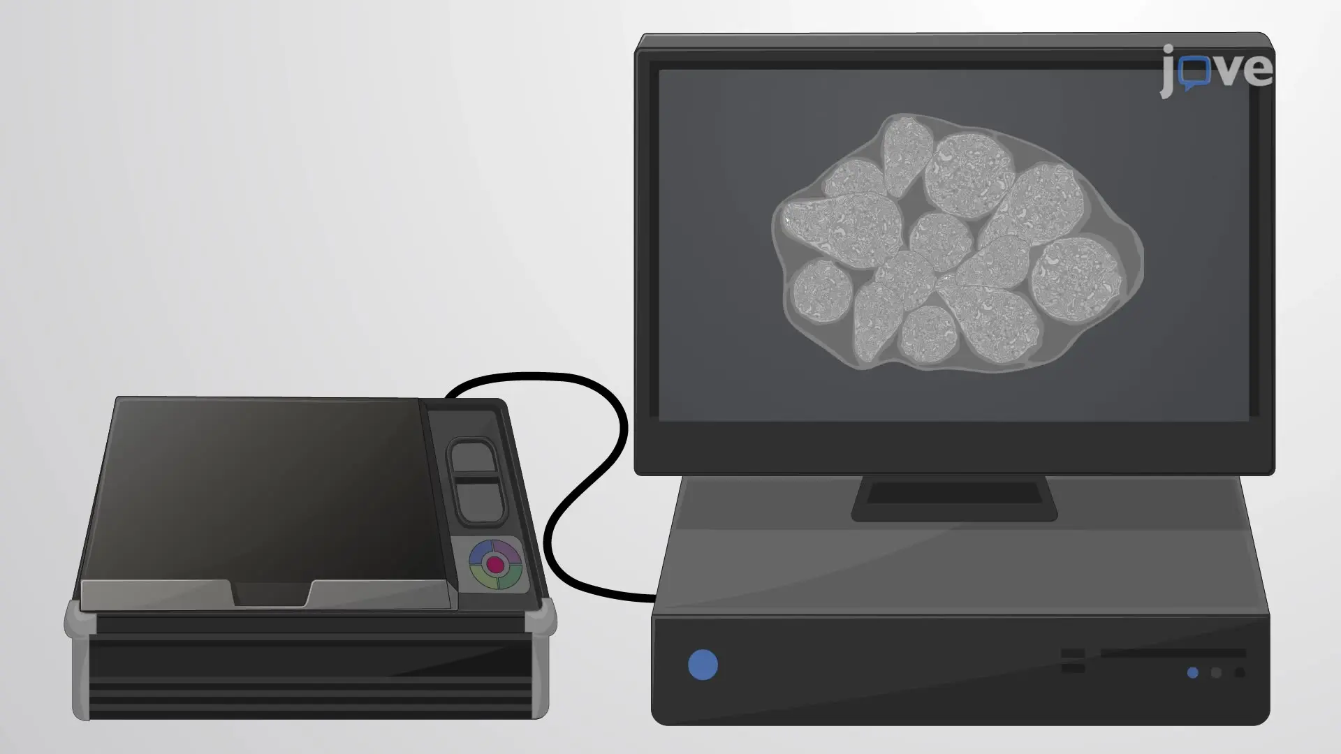
The video describes the preparation of a tissue sample for imaging mass cytometry. The tissue is first processed to unmask the antigen epitopes, permeabilize the cells, and block non-specific antibody binding sites. Then, the tissue is incubated with a metal-conjugated antibody cocktail and a nuclear stain. Finally, the treated tissue sample is imaged using an optical scanner.