$$\rightleftharpoonup{xx}$$
$$\longleftharp{xx}$$,
$$\longrightharp{xx}$$,
All procedures involving animal models have been reviewed by the local institutional animal care committee and the JoVE veterinary review board.
1. Animal Preparation and Anesthesia
- Induce anesthesia in the mouse with isoflurane/oxygen inhalation. Use 4% of isoflurane for the induction of the anesthesia and 2-3% for the maintenance at 2.5 L/min flow of oxygen. Adjust the isoflurane percentage for the maintenance of the anesthesia according to the condition of the mouse, i.e., small and weak mice require less anesthetics. Confirm adequate anesthesia e.g., by applying mild pressure to the hindlimb walking pad to check the absence of a pain withdrawal reflex.
- Control the mouse body temperature using a thermostatic heating plate at 37 °C to prevent the decrease of body temperature during anesthesia.
- Fit the mouse with the nosecone for maintenance of the anesthesia. Ensure that the animal has sufficient delivery of oxygen by checking that the nosecone does not block airways and that the animal is breathing steadily.
- During the recording, monitor whether the mouse is sufficiently anesthetized by observing the breathing rate (approximately 1 Hz in anesthesia) and the absence of a withdrawal reflex on mild pressure. Increase the isoflurane concentration manually if the anesthesia is not deep enough.
- After the measurements, leave the mouse to recover on the heating plate or in the warmth of an infrared lamp until it has regained sufficient consciousness to maintain sternal recumbency, for approximately 2-5 min. Do not leave the mouse unattended and in the company of other mice until it has fully recovered from anesthesia.
2. Measurement of CMAP in Forelimbs
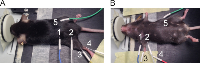
- Use the 27 G needle electrodes for forelimb CMAP measurements. See Figure 1 for recommended places of electrode positioning.
- Place electrodes on the forelimbs as follows.
- Position the mouse on the heating pad in the supine position and use adhesive tape to extend both forelimbs on the sides of the body (Figure 1B).
- Place the stimulating electrodes (1 = anode and 2 = cathode) subcutaneously on both sides of the forelimb to align with the brachial plexus nerve. Lift the skin to insert the needle perpendicularly through the skin and push approximately 5 mm of the needle under the skin without puncturing the underlying muscles.
- Place the recording electrode (3) subcutaneously on top of the biceps brachii muscle by lifting the skin. Place the reference electrode (4) on the walking pads in 3 mm depth at a 30-degree angle. Place the ground electrode (5) subcutaneously on the side of the mouse.
NOTE: Electrodes are in close proximity of each other in this setup. Prevent electrodes from touching each other as this distorts the recording.
3. Data Acquisition
- Start the stimulation by pushing the recurrent stimulus button in the controller unit and turn the intensity controller knob to increase the stimulus. Stimulate all axons using 1 pulse/s with 0.1 ms stimulus duration. Select the correct frequency and duration from dropdown menus in the software.
- To reach supramaximal stimuli (5-20 mA; in demyelinating conditions up to 60 mA), apply increasing stimuli by turning the intensity controller knob until the amplitude of the CMAP response ceases to increase. From there, further increase the stimulus by 20% to ensure that the CMAP amplitude has reached its maximal response. End the stimulation by pushing the recurrent stimulus button again.
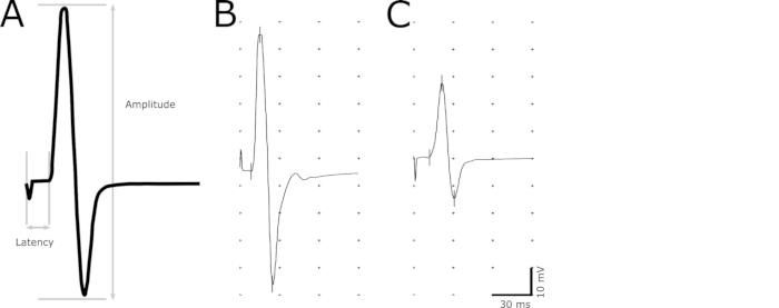
- Use the marker tool to indicate the following points in the recording: initiation of the stimulus, initiation of the response, maximum positive peak, and maximum negative peak (Figure 2).
- Determine the latency (in ms) as a delay from the initiation of the stimulus to the initiation of the response (Figure 2). Define the initiation of the response as the earliest point where the amplitude begins to increase. Use the latency to evaluate demyelination in the axons.
- Measure the amplitude (mV) from the maximum negative to the maximum positive peak (Figure 2). Use the magnitude of the amplitude to correlate the number of functional axons.