| Stereo Zoom Microscope for making the fly preparation | Olympus | SZX12 DFPLFL1.6x PF eyepieces: WHN30x-H/22 | Capable of ~150X magnification with long working distance; bespoke heavy steel table mount stand |
|---|
| Stereomicroscope in the intracellular set-up | · Olympus | Olympus SZX7; eyepieces: WHN30x-H/22 | 30X eyepieces are needed for seeing the electrode tip reflections well when driving it through the small corneal hole into the eye |
|---|
| Nikon microscope | Nikon SMZ645; eyepieces: C- W30x/7 | | |
|---|
| Anti-vibration Table | Melles Griot | With metric M6 holes on the breadboard | Our bespoke rigs have a large hole drilled through the thick breadboard that lets in the fly preparation platform pole (houses a copper heatsink with electronics) from below |
|---|
| Newport | | | |
|---|
| Micromanipulators | Narishige | Narishige NMN-21 | In our intracellular set-ups, different micromanipulator systems are used for driving the shap recording electrodes into the fly eye. All the listed manipulators are succesfully providing long-lasting stable recordings from Drosophila photoreceptors and LMCs. |
|---|
| Huxley Bertram | Huxley xyz-axis with fine manual control | | |
|---|
| Sensapex | Sensapex triple axis | | |
|---|
| Märzhäuser | Märzhäuser DC-3K with additional x-axis piezo stepper and MS 314 controller | | |
|---|
| Magnetic Stands | Any magnetic base with on/off switch will do | | For example, to manage cables inside the Faraday cage |
|---|
| Electrode Holders | Harvard Apparatus | ESP/W-F10N | |
|---|
| Silver Wire | World Precision Instruments | AGW1510 | 0.3 - 0.5 mm diameter; needs to be chloridized for the electrode holders |
|---|
| Fiber Optic Light Source | Many different, including Olympus | | |
|---|
| Fiber Optic Bundles | UltraFine Technology | | To deliver the LED light stimulus to the Cardan arm system. We use both liquid and quartz light guides (range from UV to IR) |
|---|
| Thorn Labs | | | |
|---|
| Fly Cathing Tube | | P80-50P 50ml Cent. Tube PP., Pack of 100 Pcs | Cut the conical bottom off from 50 ml Plastic Centrifuge Tube and glue a 1 ml pipette tip on it. |
|---|
| Digital Acquisition System | National Instruments | | |
|---|
| Single-electrode current/voltage- clamp microelectrode amplifier | npi SEC-10LX | http://www.npielectronic.de/ products/amplifiers/sec-single- electrode-clamp/sec-10lx.html | Outstanding performer! |
|---|
| Head-stage | Standard (+/- 150 nA) | For npi SEC-10LX | |
|---|
| LED light sources and drivers | 2-channel OptoLED (Cairn Research Ltd., UK) | | Many of our stimulus systems are in-house built |
|---|
| Self-designed and constructed | | | |
|---|
| Acquisition and Analyses Software | Many companies to choose from | | Biosyst; custom written Matlab- based system for experimental and theoretical work in the Juusola laboratory |
|---|
| Personal Computer or Mac | | | Ensure that PC or Mac is compatible with data acquisition system and software |
|---|
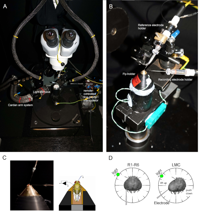
| Cardan arm system | Self-designed and constructed | | Providing accurate x,y,z-positioning of the light stimuli |
|---|
| Peltier temperature control system | Self-designed and constructed | | |
|---|
| Faraday Cage | Self-constructed | | Electromagnetic noise shielding |
|---|
| Filamented Borosilicate Glass Capillaries | | Outer diameter: 1 mm | |
|---|
| Inner diameter: 0.5 - 0.7 mm | | | |
|---|
| Filamented Quartz Glass Capillaries | | Outer diameter: 1 mm | |
|---|
| Inner diameter: 0.5 - 0.7 mm | | | |
|---|
| Pipette Puller | Sutter Instrument Company | Model P-2000 laser Flaming/Brown Micropipette Puller | For borosilicate reference electrodes, use the preset program #11 (patch electrodes): Heat = 350;
Filament = 4; Velocity 36; Delay
= 200).1.2.1). For borosilicate recording electrodes, use the preset program #12 (this typically pulls good conventional sharps for photoreceptor recordings): Heat = 355; Filament = 4; Velocity 50; Delay = 225; Pull = 150. For LMC recordings, which require electrodes with finer tips, these values need to be adjusted. For pulling quartz capillaries, P-2000 manual suggests programs for
fine tipped microelectrodes. These programs’ preset parameters serve as useful starting points
for systematic modifications to generate electrodes with good penetration success and low recording noise. |
|---|
| Extracellular Ringer Solution for the reference electrode | Chemicals from Fisher Scientific | 10326390, NaCl 10010310, KCl
10147753, TES 10161800, CaCl2
10159872, MgCl2 10000430,
sucrose | See the recipe in the protocol section |
|---|
| 3 M KCl solution for filling the filamented recording microelectrode | Salts from Fisher Scientific | 10010310, KCl | |
|---|
| Petroleum jelly | Vaselin | | |
|---|