Method Article

An Electrophysiological Study of Retinogeniculate and Corticogeniculate Synapses in Mouse Brain Slices

July 8th, 2025

In This Article

Abstract

Loading...
$$\rightleftharpoonup{xx}$$ $$\longleftharp{xx}$$, $$\longrightharp{xx}$$,

Source: Chen, X., et al. Electrophysiological Investigations of Retinogeniculate and Corticogeniculate Synapse Function. J. Vis. Exp. (2019).

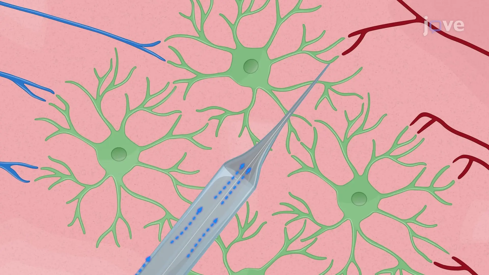
The video describes the electrophysiological recording of retinogeniculate and corticogeniculate synapses in mouse brain slices. The slices containing the dorsal lateral geniculate nucleus (dLGN) are placed in a recording chamber. A stimulating pipette is positioned on either the optic tract to stimulate retinal ganglion cells (RGCs) and investigate retinogeniculate synapses or the nucleus reticularis thalami to stimulate cortical neurons and investigate corticogeniculate synapses. Upon stimulation, the RGCs and cortical neurons release excitatory neurotransmitters, which produce synaptic currents in the LGN relay neurons. These currents are recorded using a patch-clamp recording pipette.

Protocol

Loading...
$$\rightleftharpoonup{xx}$$ $$\longleftharp{xx}$$, $$\longrightharp{xx}$$,

All procedures involving sample collection have been performed in accordance with the institute's IRB guidelines.

1. Solutions

  1. Dissection solution
    1. To reduce excitotoxicity, prepare a choline-based solution to be used during the dissection as presented here (in mM): 87 NaCl, 2.5 KCl, 37.5 choline chloride, 25 NaHCO3, 1.25 NaH2PO4, 0.5 CaCl2, 7 MgCl2, and 25 glucose. Prepare the dissection solution less than 1 week before the experiment.
  2. Recording solution
    1. Prepare an artificial cerebral spinal fluid (ACSF) soluti....

Access restricted. Please log in or start a trial to view this content.

Results

Loading...
$$\rightleftharpoonup{xx}$$ $$\longleftharp{xx}$$, $$\longrightharp{xx}$$,

Brain dissection process; anatomical diagrams and cutting setup in experiment; educational research.
Figure 1: Schemes representing the dissection protocol. (A) Horizontal view of the mouse skull, showing the position of the initial cuts. The first cut was performed along with the sagittal suture, followed by another two cuts along wit.......

Access restricted. Please log in or start a trial to view this content.

Disclosures

Loading...
$$\rightleftharpoonup{xx}$$ $$\longleftharp{xx}$$, $$\longrightharp{xx}$$,
No conflicts of interest declared.

Materials

List of materials used in this article
NameCompanyCatalog NumberComments
AmplifierHEKA ElektronikEPC 10 USB Double patch clamp amplifier
CaCl2EMSURE1.02382.1000
choline chlorideSigma-AldrichC1879-1KG
Confocal Laser Scanning MicroscopeLeica MicrosystemsTCS SP5
CsClEMSURE1.02038.0100
Cs-gluconateSelf-preparedSince there was no commercial Cs-gluconate, we prepared it by ourselves
D-600Sigma-AldrichM5644-50MG
D-APVBiotrendBN0085-100
Digital camera for microscope OOlympusXM10
EGTASERVA11290.02
GlucoseSigma-Aldrich49159-1KG
HEPESROTH9105.2
High Current Stimulus IsolatorWorld Precision InstrumentsA385
KClEMSURE1.04936.1000
MgCl2EMSURE1.05833.0250
MicromanipulatorsLuigs & NeumannSM7
MiroscopeOlympusBX51
mounting mediumThermoFisher ScientificP36930
NaClROTH3957.1
NaH2PO4EMSURE1.06346.1000
NaHCO3EMSURE1.06329.1000
PipetteHilgenberg 1807502
PullerSutterP-1000
razor bladePersonna 60-0138
Semiautomatic VibratomeLeica BiosystemsVT1200S
SR 95531 hydrobromideBiotrendAOB5680-10GABAA-receptor antagonist

Reprints and Permissions

Request permission to reuse the text or figures of this JoVE article

Request Permission

Tags

Retinogeniculate SynapsesPatch Clamp RecordingElectrophysiological RecordingOptic Tract StimulationNucleus Reticularis ThalamiDorsal Lateral Geniculate NucleusRelay NeuronsIntracellular Solution

Related Articles