Method Article

MicroRNA-Mediated Inhibition of Excitatory Postsynaptic Currents in Mouse Hippocampal Slices

July 8th, 2025

In This Article

Abstract

Loading...
$$\rightleftharpoonup{xx}$$ $$\longleftharp{xx}$$, $$\longrightharp{xx}$$,

Source: Thalhammer, A., et al. Combining Optogenetics with Artificial microRNAs to Characterize the Effects of Gene Knockdown on Presynaptic Function within Intact Neuronal Circuits. J. Vis. Exp. (2018)

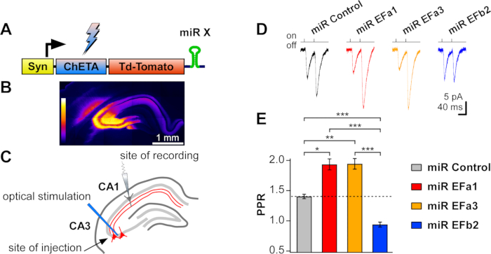
This video demonstrates microRNA-mediated inhibition of excitatory synaptic currents in mouse hippocampal brain slices. The CA3 neurons were infected with a recombinant viral vector harboring a construct for expressing a fluorescent reporter, a light-activated membrane channel, and a microRNA that downregulates the expression of voltage-gated calcium channels. Upon placing a slice in a recording chamber and patching a CA1 neuron with a recording pipette, light pulses were used to stimulate the light-activated channel, resulting in an ion influx and generating an action potential. The absence of voltage-gated calcium channels in CA3 neurons inhibits neurotransmitter release, causing inhibition of excitatory postsynaptic currents in the synaptically-connected CA1 neurons.
Protocol

Loading...
$$\rightleftharpoonup{xx}$$ $$\longleftharp{xx}$$, $$\longrightharp{xx}$$,

1. Assessing the Role of Presynaptic Proteins in Intact Neuronal Circuits by Targeted Stimulation of Knocked-down Neurons with Optogenetics

NOTE: The following protocol requires previous experience with electrophysiological recordings in acute brain slices and access to an electrophysiological setup.

  1. Stereotactically inject recombinant adeno-associated virus with mosaic serotype 1 and 2 (rAAV1/2) into the brain. Determine the stereotactic coordinates for the brain region of interest using stereotactic atlases for mouse or rat brain.
    1. Use a micropipette puller to pull injection micropipettes with long shanks of Ø 7–9 µm; clip the injection micropipettes on the shanks with scissors. Use a thin marker and graph paper to place calibrating marks on the injection micropipettes every 2 mm.
    2. Anesthetize the animal with isoflurane and fix it in the stereotactic apparatus.
    3. Keep the animal warm throughout the operation with a heating pad set at 37 °C.
    4. Protect the eyes with ocular lubricant.
    5. Shave the fur on the head with an electric razor.
    6. Spread povidone iodine on the shaved head using a cotton bud.
    7. Under a dissecting microscope, make a midline incision.
    8. Clean the skull surface with a cotton bud, so to make the bregma and lambda visible.
    9. Place the injection micropipette into its holder. Attach the holder to the stereotactic arm.
    10. Determine the x and y coordinates of the site of injection relative to the bregma and/or the lambda.
    11. Use a drill to thin the skull over the target area.
      NOTE: Use gentle circular movements and avoid drilling through the skull as this will damage the surface of the brain.
    12. If there is bleeding, absorb excessive blood with towel paper.
      NOTE: Excessive blood will make it difficult to determine correctly the z coordinate.
    13. Load ≤2 µL of virus into the injection micropipette by capillary action.
    14. Bring the injection micropipette to the x and y coordinates of the site of injection.
    15. Calculate the z coordinate from the dura and lower the pipette slowly into the brain.
    16. When the z coordinate is reached, wait 3 min to allow for tissue adjustment.
    17. Apply low positive pressure using a 1 mL syringe connected via a flexible tube to the back of the injection micropipette. Visually monitor the speed of ejection of the virus from the injection micropipette through the dissection microscope and using the calibrating marks as reference points.
      NOTE: Virus needs to be injected at slow rate (<100 nL/min) to avoid tissue damage.
    18. When injection is finished, wait 5 min, withdraw the injection micropipette by 0.2 mm, wait for another 5 min, to avoid backflow of the virus.
    19. Withdraw the injection micropipette slowly and completely from the brain and dispose of it into a container filled with bleach.
    20. Wet the skull with physiological solution and suture the skin with 3-4 stitches.
    21. Apply gentamicin ointment to the wound.
    22. Single-house the animal in a clean cage with food pellets and under a heat lamp till it fully recovers.
  2. ≥15 days post-injection, decapitate animals under deep isoflurane anesthesia.
  3. Prepare acute brain slices of the brain region of interest with a vibratome using gassed (95% O2, 5% CO2) ice-cold artificial cerebrospinal Fluid (aCSF) solution, containing (in mM): 123 NaCl, 1.25 KCl, 1.25 KH2PO4, 1.5 MgCl2, 1 CaCl2, 25 NaHCO3, 2 sodium pyruvate and 18 glucose (osmolarity adjusted to 300 mOsm). For example, if the aim is to analyze synaptic transmission from CA3 to CA1 pyramidal neurons, prepare sagittal slices of the hippocampal formation (350 µm thick).
    NOTE: From this step onwards work under low-light conditions to avoid activation of the optogenetic probe by environmental light.
  4. Let the slices recover for 30 min at 37 °C in the same aCSF in a chamber designed for holding brain slices. Keep the slices in the same brain slice chamber at room temperature till recording.
    NOTE: Using these conditions, slices can be maintained healthy for up to 6–8 h.
  5. Transfer one slice to a submerged recording chamber and superfuse it with 2 mL/min of the same aCSF used for recovery supplemented with 1.5 mM CaCl2 (total Ca2+: 2.5 mM) and without sodium pyruvate.
    NOTE: Slices are prepared and maintained in low concentration of Ca2+ (1 mM) to minimize toxicity. Recordings are performed in 2.5 mM Ca2+ to favor vesicle release.
  6. Check briefly the signal of the expressed fluorescent reporter (e.g. TdTomato; Figure 1B) to ascertain the localization and intensity of infection.
  7. Fill a patch electrode with an intracellular solution containing (in mM): 110 K-gluconate, 22 KCl, 5 NaCl, 0.5 ethylene glycol-bis(β-aminoethyl ether)-N,N,N′,N′-tetraacetic acid (EGTA), 3 MgCl2, 4 adenosine 5′-triphosphate magnesium salt (Mg-ATP), 0.5 Guanosine 5'-triphosphate trisodium salt (Na3-GTP), 20 K2-creatine phosphate, 10 4-(2-hydroxyethyl)-1-piperazineethanesulfonic acid buffer adjusted with KOH (HEPES-KOH, pH 7.28, 290 mΩ).
    NOTE: Use patch electrode with a pipette resistance of 5–6 mΩ.
  8. Under infrared illumination, reach tight-seal whole-cell configuration from a neuron receiving synaptic inputs from the infected neurons. For example, if CA3 pyramidal neurons were infected, patch pyramidal neurons in the proximal to medial tract of the CA1 region (Figure 1C).
    NOTE: Series resistance can be left uncompensated, but it should be constant and low (≤20 MΩ).
  9. Use pharmacology to isolate the synaptic currents under investigation. For example, if the aim is to investigate excitatory synaptic transmission, block inhibitory synaptic transmission with 10 µM bicuculline.
  10. Evoke synaptic currents, for example, excitatory postsynaptic currents (EPSCs), using a 473 nm blue laser coupled to an optical fiber (Ø ≤250 µm) positioned on the somata of the infected neurons (e.g. CA3 pyramidal neurons).
    1. Adjust stimulation length to a minimum, to reduce the possibility of evoking more than one action potential per light pulse. If using ChETA, set it to 2 ms.
    2. Adjust stimulation strength of the laser to yield small but clearly detectable synaptic currents (≤50 pA peak amplitude for EPSCs recorded at a holding potential of -70 mV between CA3 and CA1 pyramidal neurons; Figure 1D). If using ChETA and an optical fiber of 250 µm in diameter, stimulation strengths of 1–3 mW at fiber exit should be suitable.
      NOTE: if laser strength cannot be regulated, use neutral density filters.
    3. Shine the 473 nm laser light on the somata of the infected neurons, but not on their axons. For example, shine the light on CA3 somata, and away from the Schaffer collaterals, to avoid direct depolarization of the axons.
    4. Apply tetrodotoxin (TTX; 0.5 µM), a blocker of sodium channels, to the sample to confirm the channels are action potential driven.
    5. In different recordings, apply a selective blocker to the synaptic current under investigation to confirm the stimulation is selective.
      NOTE: For example, apply 2,3-dioxo-6-nitro-7-sulfamoyl-benzo[f]quinoxaline (NBQX, 10 µM) to α-amino-3-hydroxy-5-methyl-4-isoxazolepropionic acid (AMPA)-type glutamate receptor-mediated EPSCs.
    6. Compare optically and electrically evoked synaptic currents in the same acute brain slice in response to repetitive stimulation (≥2 stimuli at ≤20 Hz). A similar degree of synaptic facilitation or depression should be observed between electrical and optical stimulation when using a control microRNA (miR), thus suggesting that similar cellular mechanisms are activated by the two types of stimulations.
      NOTE: The above steps are necessary to ensure that optical stimulation does not induce a direct depolarization of presynaptic boutons, thus bypassing some mechanisms of synaptic transmission.

Access restricted. Please log in or start a trial to view this content.

Results

Loading...
$$\rightleftharpoonup{xx}$$ $$\longleftharp{xx}$$, $$\longrightharp{xx}$$,

Synaptic transmission analysis; diagram of optical stimulation; miR expression; electrophysiology data.
Figure 1. Assessing the role of Cav2.1[EFa] and Cav2.1[EFb] in the native hippocampus by targeted stimulation of knocked-down neurons with optogenetics. (A) Scheme of the expression cassette of the rAAV construc...

Access restricted. Please log in or start a trial to view this content.

Disclosures

Loading...
$$\rightleftharpoonup{xx}$$ $$\longleftharp{xx}$$, $$\longrightharp{xx}$$,
No conflicts of interest declared.

Materials

List of materials used in this article
NameCompanyCatalog NumberComments
Brain slices PrechamberHarvard ApparatusBSC-PC
CCD camera-based imagerBio-RadChemiDoc™ MP
Cell Culture reagentsLife Technologies
DrillForedomK.1030
EGTASigmaE4378
Gentamicin ointmentLocal pharmacy
GlucoseSigmaG7021
HEPESSigma54459
Injection micropipettesNarishigeGD1
Inorganic salts & detergentsSigma
K2-creatine phosphateCalbiochem237911
KGluconateFluka60245
LaserLaserglow TechnologiesLRS-0473-GFM-00100-03
MembraneAmershamProtran™ 0.2 µm NC
MgATPSigmaA9187
Micropipette holderNarishigeIM-H1
Micropipette pullerNarishigePC-100
Na3GTPSigmaG8877
NaPyruvateSigmaP5280
Ocular lubricantLocal pharmacyLacrigel
Povidone iodineLocal pharmacyBetadine
Stereotactic apparatusWPI
TetrodotoxinTocris1069Toxic
VibratomeLeicaVT1200S

Reprints and Permissions

Request permission to reuse the text or figures of this JoVE article

Request Permission

Tags

MicroRNA InhibitionOptogenetic StimulationViral Vector ExpressionVoltage Gated Calcium ChannelsPatch Clamp RecordingLight Activated ChannelsBrain Slice PreparationSynaptic Current Measurement

Related Articles