Method Article

Optogenetic Stimulation and Electrophysiological Recording of Synaptic Events in Rat Brain Slices

July 8th, 2025

In This Article

Abstract

Loading...
$$\rightleftharpoonup{xx}$$ $$\longleftharp{xx}$$, $$\longrightharp{xx}$$,

Source: Kinnavane, L., et al. Ex Vivo Optogenetic Interrogation of Long-Range Synaptic Transmission and Plasticity from Medial Prefrontal Cortex to Lateral Entorhinal Cortex. J. Vis. Exp. (2022)

This video demonstrates the procedure for studying synaptic efficiency in the lateral entorhinal cortex (LEC) by optogenetically stimulating medial prefrontal cortex (mPFC) axons and recording the responses of pyramidal neurons in the LEC. This method combines patch-clamp recordings with synaptic activation by light stimulation to study synaptic plasticity.
Protocol

Loading...
$$\rightleftharpoonup{xx}$$ $$\longleftharp{xx}$$, $$\longrightharp{xx}$$,

All procedures involving animal samples have been reviewed and approved by the appropriate animal ethical review committee.

1. Electrophysiology and optogenetic stimulation

  1. Identification of target cell.
    1. Place the slice into a submerged recording chamber at 34 °C perfused with artificial cerebrospinal fluid (aCSF) at a rate of 2 mL/min by a peristaltic pump. Immobilize the slice using a slice anchor.
    2. Under the low magnification (4x) objective of a widefield microscope using oblique infrared illumination, navigate to lateral entorhinal cortex (LEC) layer 5. Measure the distance from the pial surface to the required layer.
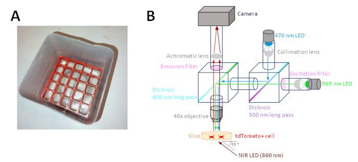
      NOTE: Oblique infrared illumination was achieved by positioning a near-infra-red LED approximately 3 mm below the recording chamber coverslip at an angle of ~55° to the plane of the coverslip (Figure 1B). Differential interference contrast is an alternative and commonly used imaging technique for slice electrophysiology.
    3. Change to a high-magnification water immersion objective (40x) and identify pyramidal neurons. Mark the position of the cell on the computer monitor with tape.
      NOTE: The pyramidal neurons have a roughly triangular morphology with prominent apical dendrites projecting toward the pial surface of the slice (Figure 2A). Cell health can be assessed by the absence of a condensed, visible nucleus and inspection of the plasma membrane which should appear smooth. It is unlikely that clear fluorescent labeling of axons by the opsin-fluorophore fusion protein will be visible when using a wide-field fluorescence microscope. To visualize axonal projections, perform immunohistochemistry post-hoc using a primary antibody against the opsin's fluorophore and amplify with a secondary antibody conjugated to a fluorophore of the same or similar wavelength.
  2. Formation of whole-cell patch clamp.
    1. Fabricate a borosilicate glass micropipette using a pipette puller and fill it with filtered intracellular recording solution (120 mM k-gluconate, 40 mM HEPES (N-2-hydroxyethylpiperazine-N'-2-ethanesulfonic acid), 10 mM KCl (potassium chloride), 2 mM NaCl (sodium chloride), 2 mM MgATP (magnesium adenosine triphosphate), 1 mM MgCl (magnesium chloride), 0.3 mM NaGTP (sodium guanosine triphosphate), 0.2 mM EGTA (Ethylene glycol tetraacetic acid), and 0.25% biocytin made in UPW (ultrapure water), pH 7.25, 285-300 mOsm). Place the filled micropipette in the electrode holder on the patch-clamp amplifier headstage, ensuring the electrode wire is in contact with the intracellular solution.
    2. Apply positive pressure by mouth by blowing hard into a mouthpiece (such as a 1 mL syringe with the plunger removed) connected by tubing to the electrode holder side port and maintain pressure by closing an in-line three-way valve. Raise the microscope objective such that a meniscus forms and insert the electrode into the meniscus until it can be seen on the microscope.
    3. Open the Seal Test window in WinLTP16 (or other acquisition software/oscilloscope), and with the amplifier in the voltage-clamp mode, apply a 5 mV square pulse to determine whether the pipette resistance is 3-6 MΩ.
    4. Approach and touch the identified cell with the pipette tip; this should result in an indentation in the cell membrane (Figure 2A; right panel) and a small increase in pipette resistance (0.1 MΩ).
    5. Release positive pressure and apply negative pressure by applying moderate suction at the mouthpiece; this should result in a vast increase in pipette resistance(>1000 MΩ). Pressure can now be left neutral. Apply negative pressure in a gradually increasing ramp until cell membrane ruptures resulting in whole-cell capacitance transients.
  3. Record optogenetically evoked synaptic events.
    1. Enter current-clamp configuration.
      NOTE: In most instances, long-range synaptic transmission is glutamatergic, therefore recording at membrane potentials close to the chloride reversal potential will best isolate AMPA receptor (AMPAR)-mediated transmission and minimize measurement of any feed-forward inhibition (FFI) evoked. Chloride reversal is dependent on the composition of intracellular recording solution and aCSF and can be calculated using the Goldman-Hodgkin-Katz equation; for the above solutions, this was -61.3 mV. Layer 5 LEC pyramidal neurons had an average resting membrane potential of -62 mV and, where necessary, were maintained at this potential by injection of constant current. Alternatively, cells can be voltage-clamped to the desired potential. To record long-range inhibitory projections16 or to record FFI, voltage-clamp at cation reversal potential to isolate GABAergic chloride conductance. When voltage-clamping neurons at membrane potentials above action potential threshold, a cesium-based intracellular solution containing voltage-gated sodium channel blockers is used to improve voltage-clamp and prevent initiation of action potentials (130 mM CsMeSO4 (cesium methanesulfonate), 10 mM HEPES, 8 mM NaCl, 5 mM QX 314 chloride, 4 mM MgATP, 0.5 mM EGTA, 0.3 mM NaGTP, 0.25% biocytin made in UPW, pH 7.25, 285-300 mOsm).
    2. Using data acquisition software, send transistor-transistor logic (TTL) signals to an LED (light emitting diode) driver to activate a mounted 470 nm LED. The mounted LED is directed into the microscope light path using filter cubes and appropriate optics (Figure 1B) to apply light pulses to the slice via the 40x objective to evoke optogenetic excitatory post-synaptic potentials (oEPSPs).
      NOTE: Light pulses can be applied perisomatically/over the dendrites, which will result in the activation of opsins in the axons and presynaptic bouton, or the investigator can move the objective to axons away from the recorded cell to avoid over-bouton stimulation. Maximal oEPSP amplitude depends on the strength of the synaptic projection, the efficacy of viral injections, and opsin used. oEPSPs can be titrated to the desired amplitude by varying light intensity and/or duration18; varying the duration of light pulses (typical duration between 0.2-5 ms at maximal LED power output, which results in 4.4 mW/mm light density19) gives more consistent oEPSP amplitudes than altering the power output of the LED.
    3. Investigate presynaptic release properties (change in voltage) by delivering trains of multiple light pulses with differing inter-stimulus intervals (Figure 2E); care should be taken while interpreting optogenetically evoked transmission.
    4. Investigate long-term plasticity either by repetitively evoking oEPSPs19 or application of ligands. Monitor oEPSP amplitude for 5-10 min to ensure stability before induction of plasticity, and then monitor until a stable amplitude is reached (typically 30-40 min).
      NOTE: Most current opsins are not capable of reliably evoking multiple action potentials at high frequencies, e.g., 100 stimuli delivered at 100 Hz, as is typically used to induce LTP.
    5. To confirm oEPSPs are monosynaptic, perform over-bouton activation of the transduced pathway by positioning the objective over the dendritic arbor and stimulating in the presence of 0.5 µM tetrodotoxin and 100 µM aminopyridine. Application of tetrodotoxin will abolish transmission if the responses are action potential-dependent, and subsequent inclusion of aminopyridine will partially restore transmission if the oEPSPs are generated monosynaptically19,20.
    6. To allow biocytin to fill the neuron, wait for at least 15 min after entering the whole-cell configuration. In voltage-clamp, monitor membrane capacitance and input resistance.
    7. Slowly withdraw the pipette along the approach angle away from the soma of the cell, observing the slow disappearance of capacitance transients and membrane current, indicating the re-sealing of the cell membrane and formation of an outside-out patch at the pipette tip. Note the orientation of the slice and the location of the cell(s) within the slice. Put the slice into PFA (paraformaldehyde) in a 24-well plate, incubate overnight at 4 °C, and then transfer to 0.1 M PB.
      ​NOTE: Slices can be stored for up to a week. If longer storage is required, change the PB (phosphate buffer) regularly or use PB containing sodium azide (0.02%-0.2% of sodium azide).

Access restricted. Please log in or start a trial to view this content.

Results

Loading...
$$\rightleftharpoonup{xx}$$ $$\longleftharp{xx}$$, $$\longrightharp{xx}$$,

Optical setup diagram: LED excitation, emission filters, camera for cell imaging; photonic study.
Figure 1: Slice collection chamber and optical configuration for visualized whole-cell recordings, optogenetic excitation, and identification of tdTomato-positive neurons. (A) The slice collection chamber14 is custom-made from a microcentr...

Access restricted. Please log in or start a trial to view this content.

Disclosures

Loading...
$$\rightleftharpoonup{xx}$$ $$\longleftharp{xx}$$, $$\longrightharp{xx}$$,
No conflicts of interest declared.

Materials

List of materials used in this article
NameCompanyCatalog NumberComments
3 way luer valveCole-ParmerWZ-30600-02
40x objectiveOlympusLUMPLFLN40XW
4-aminopyridineHello BioHB1073
4x objectiveOlympusPLN4X/0.1
Achromatic lensEdmund Optics49363Focusses visual spectrum and near-IR
Benchtop microcentrifugeBenchmark ScientificC1005*
BiocytinSigma-AldrichB4261
Borosillicate glass capillaryWarner InstrumentsG150F-6
CaCl2Sigma-AldrichC5670
Camera - Qimaging Retiga ElectroPhotometrics01-ELECTRO-M-14-C
CarbacholTocris2810
Chlorhexidine surgical scrubVetaseptXHG008
ClippersAndis22445AGC Super 2-Speed Detachable Blade Clipper
Collimation condenser lensThorLabsACL2520-A
CoverslipsFisher Scientific Ltd10011913
CryostatLeicaCM3050 S
CsMeSO4Sigma-AldrichC1426
Cyanoacrylate glueRapid Electronics Ltd84-4557
Data acquisition deviceNational InstrumentsUSB-6341 BNC
D-glucoseSigma-AldrichG8270
Dichroic mirror 500 nm long-passEdmund Optics69899
Dichroic mirror 600 nm long-passEdmund Optics69901
Dichroic mirror cubeThorLabsCM1-DCH/M
EGTAMillpore324626
Electrode holder with side portHEKA895150
Emission filterChroma59022m
Excitation filterChromaET570/20x
Eye gelDechraLubrithal
Fine paint brushScientific Laboratory SuppliesBRU2052
GuillotineWorld Precision InstrumentsDCAP
HEPESSigma-AldrichH3375
Hydrogen peroxide solutionSigma-AldrichH100930% (w/w)
IsofluraneHenry Schein988-3245
IsopentaneSigma-AldrichM32631
KClSigma-AldrichP3911
k-gluconateSigma-AldrichG4500
Kinematic fluorescence filter cubeThorLabsDFM1T1
LED driverThorLabsLEDD1B
MgATPSigma-AldrichA9187
MgClSigma-AldrichM2670
MgSO4Sigma-AldrichM7506
Microelectrode pullerSutter instrumentsP-87
Microinjection syringeHamilton7634-01/00
Microinjection syringe needleHamilton7803-05Custom specification: gauge 33, length 15mm, point style 4 - 12°
Microinjection syringe pumpWorld Precision InstrumentsUMP3T-1
Mounted blue LEDThorLabsM470L5
Na2HPO4.7H2OSigma-AldrichS9390
NaClSigma-AldrichS9888
NaGTPSigma-AldrichG8877
NaH2PO4Sigma-AldrichS0751
NaH2PO4.H2OSigma-AldrichS9638
NaHCO3Sigma-AldrichS5761
NIR LEDOSRAMSFH4550Used for refracted IR imaging of slice, differential interference contrast (DIC) optics is another commonly used method
ParaformaldehydeSigma-Aldrich158127
ParaformaldehydeSigma-AldrichP6148
Patch clamp amplifierMolecular Devices700A
Peristaltic pumpWorld Precision InstrumentsMinistar
Poly-L-lysine coated microscope slidesFisher Scientific Ltd23-769-310
Recording chamberWarner InstrumentsRC-26G
Slice anchorWarner InstrumentsSHD-26-GH/15
Stereotaxic holder for micro drillHarvard Apparatus75-1874
SucroseSigma-AldrichS0389
Surgical MicroscopeCarl ZeissOPMI 1 FR pro
Syringe filter for intracellular recording solutionThermo Scientific Nalgene171-0020
Upright fluorescence microscopeLeicaDM6 B
Solution
aCSF
sucrose cutting solution
PFA

Reprints and Permissions

Request permission to reuse the text or figures of this JoVE article

Request Permission

Tags

Patch ClampLight PulsesPyramidal NeuronsMedial Prefrontal CortexLateral Entorhinal CortexWhole Cell Configuration

Related Articles