All procedures involving animal models have been reviewed by the local institutional animal care committee and the JoVE veterinary review board.
1. Preparing the animal:
- Anesthetize the rat using an inhalant (isoflurane) at 1-2% delivered to effect or by injection (ketamine, xylazine, acepromazine). Dosage of injectable anesthesia is as follows: 95.0 mg/kg of ketamine, 10.0 mg/kg of xylazine, 0.075 mg/kg of acepromazine.
NOTE: We successfully use both inhalant and injectable anesthetic approaches when conducting this CMAP recording protocol, with no preference.
- The animal must be maintained at the appropriate anesthetic depth before the surgery is started, through the conclusion of surgery, and until post-operative buprenorphine has taken effect. Confirm proper anesthetization by brief toe pinch to determine that a withdrawal response is not elicited. Additionally, test the corneal reflex with a cotton swab. Throughout the surgical procedure, determine that heart rate and respiratory rate have not increased, which suggests that anesthetic depth has become too light.
- Apply vet ointment on the animal's eyes to prevent dryness while under anesthesia.
- The surgeon should wash her/his hands with a disinfectant (chlorhexidine scrub) before beginning surgery. The surgeon should wear sterile gloves, a facemask, and gown or clean lab coat. All instruments should be washed and autoclaved before surgery. Add a sterilization indicator strip inside the instrument kit before autoclaving at the level of the instruments to verify that sterilization is complete. The surgeon should verify that the entire line on the strip is black before beginning surgery. Tape the outside of the box with indicating autoclave tape as well. If surgery is to be performed on several animals on the same day, clean the instruments between surgeries and sterilize in a glass bead sterilizer. If necessary, sterilize both ends of the instrument in the glass bead sterilizer by placing the handle of the instrument in the sterilizer for the appropriate time, removing the instrument with a sterile hemostat, cooling it in a sterile field and then placing the functional end of the instrument into the glass bead sterilizer for the appropriate period.
- Once the animal is anesthetized, place it on surgical board with dorsal surface down and abdomen facing up so that tape can be used to secure animal's forelimbs to surgical board.
- Shave ventral surface caudally beginning at the base of the skull and ensure that the area around abdomen on either side of the midline is shaved depending on which hemi-diaphragm is being recorded. Apply povidone-iodine to the shaved surface of the skin.
2. Preparation for CMAP recordings:
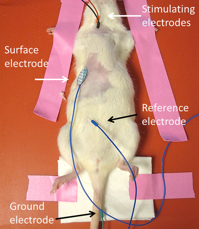
- Following animal preparation, place ground electrode subcutaneously into tail (Figure 1).
- Place the reference electrode subcutaneously into the contralateral lower abdominal region.
- Place conductive gel onto self-adhesive surface strip or disk recording electrode (dimensions of surface strip electrode: 0.5 x 3 cm), and then place surface electrode transversely along the unilateral costal margin.
- Place stimulating electrodes transcutaneously 0.5 cm apart lateral to trachea and superior to the clavicle. Insert the electrodes approximately 1.0 cm deep through the skin. Secure the electrodes by hand so that their location does not move with subsequent stimulations.
NOTE: Angle stimulating electrodes 90° relative to the skin at the site of insertion.
3. Compound Muscle Action Potential (CMAP) recordings:
- Obtain electrophysiological recordings using a stimulator/amplifier followed by computer assisted data analysis.
- The stimulus parameters of single pull stimulation should be 0.5 msec, 1.0 Hz supramaximal pulses. The amplitude of the stimulus should be between 6.0 and 8.0 V (Figure 2A).
NOTE: Within an experiment, use a stimulus intensity with the same amplitude across animals. The objective is to maximize the response amplitude by using the minimal amplitude of stimulation. It is important to note that the initial response that occurs at the time of stimulation is a stimulation artifact. In addition, care should be taken to obtain a CMAP response that is preceded by a stable/flat baseline after the stimulation artifact, which is then followed by the rapid CMAP response. To obtain such a response, it may be necessary to reposition the stimulation and/or recording electrodes.
- Wait 30 sec between stimulations and repeat 10 stimulations in a row to obtain the average response (Figure 2B-C). Measure the amplitude from baseline to peak (Figure 2A). Conduct phrenic nerve stimulation during the phase of the animal's respiratory cycle when the phrenic nerve/diaphragm is not activated. The procedure can be repeated on the same animal for the contralateral hemi-diaphragm.
- After the final CMAP recording session, perfuse the animal if diaphragm neuromuscular junction (NMJ) staining will be conducted. Conduct CMAP recordings repeatedly on the same animal approximately once every week (or at intervals of choice).
- Following survival CMAP recordings, allow animal to recover on a circulating warm water heating pad, and continuously monitor during recovery until awake and sternally recumbent. Do not return an animal that has undergone surgery to the company of other animals until fully recovered.
- Once sternally recumbent, monitor the animal every 12 hr for the first 48 hr (once daily thereafter). If the animal appears to be dehydrated (flaccid skin, listlessness), administer fluids (lactated ringers solution; subcutaneously; 1-2 ml per injection, with the sites rotated; 2 times per day) until post-surgical weight and fluid loss stabilizes. Provide animals with softened food in small dishes during the acute post-operative period if necessary to encourage eating if they cannot reach the wire bar lid feeder.
- Administer buprenorphine at a dose of 0.05 mg/kg before the end of surgery and then at 12 hr intervals for the first 24 hr (and dependent upon signs of pain/stress thereafter) via subcutaneous injection (site rotated). If signs of pain or distress are noticed thereafter, treat animals with buprenorphine. Signs indicative of pain/distress include lack of activity, vocalizations, lack of eating or drinking (dehydration), excessive weight loss, guarding, excessive gnawing or scratching at site of incision. Additional criteria used in determining pain and distress include unresponsiveness to extraneous stimuli, transient or unprovoked vocalization, labored or abnormal breathing, persistent reddish-brown naso-occular discharge, marked piloerection. Also monitor evidence of poor hygiene that would suggest ill animals. If the animal is still observed to be in distress after 72 hr of treatment (according to the pain/stress criteria outline above, and after receiving the analgesic protocol described above) euthanize the animal.