A subscription to JoVE is required to view this content. Sign in or start your free trial.

Method Article

Measuring the Contralateral Silent Period Induced by Transcranial Magnetic Stimulation

892 views

July 8th, 2025

In This Article

Abstract

Source: Rebello-Sanchez, I., et al. Measuring Contralateral Silent Period Induced by Single-Pulse Transcranial Magnetic Stimulation to Investigate M1 Corticospinal Inhibition. J. Vis. Exp. (2022).

This video demonstrates a technique for measuring the contralateral silent period (cSP) using transcranial magnetic stimulation. Position the patient's hand prone and place an electrode on the first dorsal interosseous (FDI) muscle. Then, place a force-measuring device between the thumb and index finger. Identify the primary motor cortex (M1) region of the brain. Maintain a standardized pressure on the force-measuring device and stimulate the M1 region to record the muscle activity, termed the motor-evoked potential (MEP), followed by the subsequent period of reduced muscle activity, termed cSP.
Protocol

All procedures involving human participants have been performed in compliance with the institutional, national, and international guidelines for human welfare and have been reviewed by the local institutional review board.

1. Experimental procedures (Figure 1)

  1. Select the muscle for positioning the electrodes.
    1. Ask the subject to put their hand over the table, in a prone position. Select the FDI muscle, localized between the first and second metacarpal osseous. To identify the FDI, ask the subject to abduct their index finger against resistance, keeping the rest of the hand still and laying on the tabl....

Access restricted. Please log in or start a trial to view this content.

Results

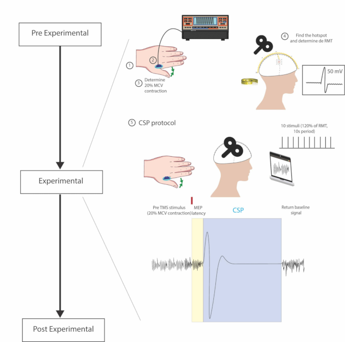
Transcranial magnetic stimulation (TMS) experimental protocol diagram depicting motor cortex activity.
Figure 1: Experimental steps. 1. Electrode placement on the belly of the FDI muscle 2. Positioning of the dynamometer between fingers. 3. Voluntary contraction of the target muscle to test the standardization of 20% MVC 4. Head measurements and TMS pu.......

Access restricted. Please log in or start a trial to view this content.

Disclosures

No conflicts of interest declared.

Materials

List of materials used in this article
NameCompanyCatalog NumberComments
Alcohol padsMedlinePreparation with 70% isopropyl alcohol
Conductive gelWeaver and CompanyUsed on the electrode
Echo PinchJTECH medical0902A302Digital dynamometer.
Mega-EMGSoterix MedicalNS006201Digital multiple channel EMG with built in software.
MEGA-TMS coilSoterix MedicalNS0632018 shaped TMS coil
Mega-TMS stimulatorSoterix Medical6990061Single Pulse TMS
Neuro-MEP.NETSoterix MedicalEMG software used to analyse the muscles eletrical activity.
Swim capKiefer

Tags

Motor Evoked PotentialFirst Dorsal InterosseousPrimary Motor CortexForce Measuring DeviceElectromyography RecordingHotspot IdentificationResting Motor ThresholdSuprathreshold Stimuli